Variations of Short Saphenous Vein and Great Saphenous Vein on Ultrasound Colour Doppler and Its Surgical Importance.
- Alka Agrawal , Professor and Head of Department , Department of Radiodiagnosis,MGMMC MYH indore (M.P).
- Gaurav Bhandari , Assistant Professor, Department of Radiodiagnosis,MGMMC MYH indore (M.P).
- Vivek Yonati , Associate Professor, Department of Radiodiagnosis,MGMMC MYH indore (M.P).
- Anjana Kumari , Junior Resident, Department of Radiodiagnosis,MGMMC MYH indore (M.P).
- Raj Bahor Patel , Junior Resident , Department of Radiodiagnosis,MGMMC MYH indore (M.P).
Article Information:
Abstract:
Background: Anatomical variations of the great saphenous vein (GSV) and small saphenous vein (SSV) influence venous haemodynamics and may contribute to varicose veins, persistent reflux, and recurrence after intervention. This study evaluated the prevalence of these variations using colour Doppler ultrasonography and assessed their surgical significance. Methods: A hospital-based cross-sectional observational study was conducted on 200 adults undergoing lower limb venous colour Doppler ultrasonography. The GSV and SSV were evaluated for anatomical variations, venous diameter, junctional anatomy, reflux, valve competence, perforator status, and their association with varicose veins. Statistical analysis was performed using SPSS version 23, with p<0.05 considered significant. Results: Varicose veins were identified in 90 (45.0%) patients. GSV and SSV variations were observed in 110 (55.0%) and 61 (30.5%) patients, respectively. Anterior accessory GSV and extrafascial GSV (31.0% each) were the most common GSV variants, while the vein of Giacomini (31.1%) was the commonest SSV variant. GSV variation (66.7% vs. 45.5%; p<0.001) and SSV variation (41.1% vs. 21.8%; p=0.038) were significantly associated with varicose veins. Increasing venous diameter, saphenofemoral and saphenopopliteal junction incompetence, perforator incompetence, and dilated calf deep veins also demonstrated significant associations with varicosity. Conclusion: Anatomical variations of the superficial venous system are common and significantly associated with varicose veins. Colour Doppler ultrasonography provides comprehensive preoperative venous mapping and should be routinely performed to optimize surgical planning and reduce disease recurrence.
Keywords:
Article :
INTRODUCTION:
Chronic venous disease (CVD) is one of the most common vascular disorders affecting the lower extremities and represents a major cause of morbidity worldwide. Varicose veins, the most frequent manifestation of CVD, affect a substantial proportion of adults and are associated with pain, edema, skin changes, reduced quality of life, and considerable healthcare expenditure. Despite advances in surgical and endovenous therapies, recurrence following treatment remains a significant clinical challenge, with unrecognized anatomical variations of the superficial venous system being an important contributing factor [1], [2].
The superficial venous system of the lower limb consists primarily of the great saphenous vein (GSV) and the small saphenous vein (SSV), which communicate with the deep venous system through perforating veins. Although these veins follow predictable anatomical pathways, considerable variations have been described in their origin, course, duplication, tributaries, diameter, and termination [3], [4]. Such variations may influence venous hemodynamics, predispose to valvular incompetence, and affect the development and progression of superficial venous insufficiency [5].
Recognition of these anatomical variants has become increasingly important with the widespread use of minimally invasive procedures such as endovenous laser ablation, radiofrequency ablation, and ultrasound-guided foam sclerotherapy. Failure to identify duplicated trunks, accessory veins, or atypical saphenofemoral and saphenopopliteal junctions may result in incomplete treatment, persistent reflux, and disease recurrence [6]. Furthermore, the variable relationship of the SSV with adjacent neurovascular structures, particularly the sural nerve, may increase the risk of iatrogenic injury during surgical interventions [7].
Duplex colour Doppler ultrasonography is the imaging modality of choice for evaluating superficial venous disorders because it provides real-time assessment of venous anatomy, valve competence, and reflux without exposing patients to ionizing radiation. In addition to identifying structural variations, it enables accurate preoperative venous mapping, thereby facilitating appropriate surgical planning and improving procedural outcomes. Beyond the management of varicose veins, detailed assessment of the GSV is valuable when it is considered for use as an autologous conduit in coronary artery bypass grafting and peripheral vascular reconstruction, where anatomical variations may influence conduit selection and surgical success [8], [9].
As the prevalence and pattern of superficial venous variations may differ across populations, region-specific data are essential for optimizing clinical decision-making. The present study was undertaken to evaluate anatomical variations of the great and small saphenous veins using colour Doppler ultrasonography and to assess their surgical relevance in the management of superficial venous disorders.
MATERIALS AND METHODS:
This hospital-based cross-sectional observational study was conducted in the Department of Radiodiagnosis, M.G.M. Medical College and M.Y. Hospital, Indore, Madhya Pradesh, India, after obtaining approval from the Institutional Scientific Review Board and the Institutional Ethics Committee. The study was carried out over a period of one year following ethical clearance. A total of 200 consecutive adult patients referred for lower limb colour Doppler ultrasonography during the study period were enrolled after providing written informed consent. Participants aged between 18 and 85 years who underwent lower limb venous Doppler examination were eligible for inclusion. Patients with previously diagnosed venous vasculopathy, a history of deep vein thrombosis, traumatic injury to the lower limb, prior venous surgery or sclerotherapy, or those unwilling to participate were excluded.
All ultrasound examinations were performed using a high-resolution real-time ultrasound system equipped with a linear-array transducer. Examinations were conducted according to a standardized scanning protocol by experienced radiologists. Before imaging, relevant clinical history and demographic information were recorded.
For evaluation of the great saphenous vein (GSV), patients were examined in the supine position with the lower limb slightly externally rotated to facilitate optimal visualization of the superficial venous system. Assessment of the small saphenous vein (SSV) was performed with the patient in the standing position and the knee flexed approximately 15°, allowing adequate venous distension and improved visualization of the saphenopopliteal junction.
Grey-scale and colour Doppler imaging were used to evaluate the anatomical characteristics of the superficial venous system. The GSV and SSV were assessed throughout their course for variations in formation, duplication, accessory veins, branching pattern, calibre, and termination. Particular attention was given to the anatomy of the saphenofemoral junction and saphenopopliteal junction, including their drainage patterns and tributaries. Venous diameter was measured at predefined anatomical levels, and colour Doppler was used to assess venous flow characteristics and valve competence where applicable. Anatomical variations identified during the examination were systematically documented (Figure 1).
The primary outcome was the prevalence and pattern of anatomical variations involving the GSV and SSV. Secondary outcomes included venous diameter, level of termination, drainage pattern of the saphenofemoral and saphenopopliteal junctions, colour flow characteristics, and their association with varicose veins.
Data were entered into Microsoft Excel and analysed using IBM SPSS Statistics software (version 23; IBM Corp., Armonk, NY, USA). Continuous variables are presented as mean ± standard deviation (SD), while categorical variables are expressed as frequencies and percentages. Associations between categorical variables were analysed using the Chi-square test or Fisher's exact test, as appropriate. A two-tailed p value of <0.05 was considered statistically significant.
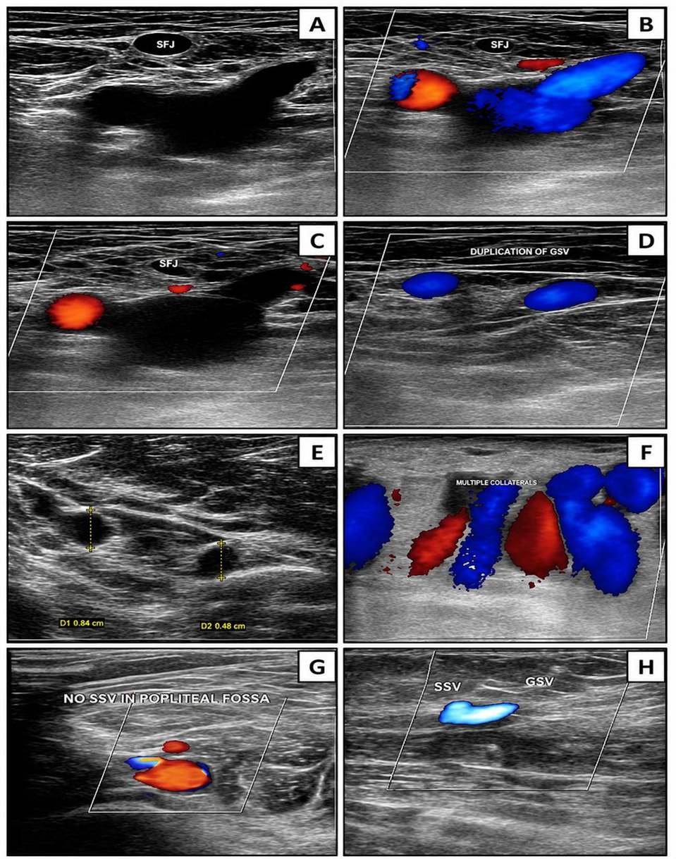
Figure 1. Ultrasonographic appearances of the superficial venous system: (A) Grayscale image of the saphenofemoral junction (SFJ); (B) Colour Doppler image of the saphenofemoral junction; (C) Colour Doppler demonstrating a competent saphenofemoral junction; (D) Colour Doppler image showing duplication of the great saphenous vein (GSV); (E) Grayscale image showing increased calibre of the great saphenous vein; (F) Colour Doppler image demonstrating multiple collateral veins along the course of the great saphenous vein; (G) Colour Doppler image showing absence of the saphenopopliteal junction (SPJ) with no small saphenous vein (SSV) identified in the popliteal fossa; (H) Colour Doppler image showing the small saphenous vein draining into the great saphenous vein at the medial aspect of the mid-leg.
RESULTS:
A total of 200 patients were included in the study. The largest proportion belonged to the 36–45-year age group (30.0%), followed by the 56–65-year (19.5%) and 46–55-year (18.0%) age groups. There was a marked male predominance, with 169 (84.5%) males and 31 (15.5%) females. Swelling (60.0%) was the most frequently reported clinical symptom, followed by visible dilated veins (48.0%), skin changes (15.0%), and ulceration (9.0%). Overall, 90 patients (45.0%) had varicose veins, while 110 (55.0%) showed no evidence of varicosity (Table 1).
Table 1. Baseline Demographic and Clinical Characteristics of the Study Population (N = 200)
|
Variable |
Category |
n (%) |
|
Age group (years) |
18–25 |
7 (3.5) |
|
26–35 |
34 (17.0) |
|
|
36–45 |
60 (30.0) |
|
|
46–55 |
36 (18.0) |
|
|
56–65 |
39 (19.5) |
|
|
>65 |
24 (12.0) |
|
|
Sex |
Male |
169 (84.5) |
|
Female |
31 (15.5) |
|
|
Clinical symptoms* |
Swelling |
120 (60.0) |
|
Visible veins |
96 (48.0) |
|
|
Skin changes |
30 (15.0) |
|
|
Ulcer |
18 (9.0) |
|
|
Varicose vein status |
Present |
90 (45.0) |
|
Absent |
110 (55.0) |
|
|
*Multiple responses permitted. |
||
Anatomical variations of the superficial venous system were identified in 110 patients (55.0%) for the great saphenous vein (GSV) and in 61 patients (30.5%) for the small saphenous vein (SSV), whereas combined GSV and SSV variations were observed in 9 patients (4.5%). Among GSV variations, the anterior accessory saphenous vein and an extrafascial course were the most common (31.0% each), followed by duplication (17.3%), circumflex GSV (14.5%), and posterior accessory saphenous vein (6.3%). Within the SSV, the vein of Giacomini was the most frequent variation (31.1%), followed by an extrafascial course (22.9%) and mid-thigh termination (21.3%) (Table 2).
Table 2. Distribution of Anatomical Variations of the Great and Small Saphenous Veins (N = 200)
|
Vein |
Anatomical variation |
n |
% |
|
Overall prevalence |
GSV variation |
110 |
55.0 |
|
SSV variation |
61 |
30.5 |
|
|
Combined GSV + SSV variation |
9 |
4.5 |
|
|
Great saphenous vein (n=110) |
Anterior accessory saphenous vein |
34 |
31.0 |
|
Extrafascial course |
34 |
31.0 |
|
|
Duplication |
19 |
17.3 |
|
|
Circumflex GSV |
16 |
14.5 |
|
|
Posterior accessory saphenous vein |
7 |
6.3 |
|
|
Small saphenous vein (n=61) |
Vein of Giacomini |
19 |
31.1 |
|
Extrafascial SSV |
14 |
22.9 |
|
|
Mid-thigh termination |
13 |
21.3 |
|
|
Upper-thigh termination |
8 |
13.1 |
|
|
Lower-thigh termination |
3 |
4.9 |
|
|
Hypoplastic SSV |
3 |
4.9 |
|
|
Duplication |
1 |
1.6 |
GSV variation was observed in 66.7% of patients with varicose veins compared with 45.5% of those without varicosity (p < 0.001). Similarly, SSV variation was more frequent in patients with varicose veins than in those without varicosity (41.1% vs. 21.8%; p = 0.038).
Combined GSV and SSV variations were identified in 7.8% of patients with varicosity and 1.8% of those without varicosity (p = 0.08) (Table 3).
Table 3. Association Between Anatomical Variations and Varicose Veins
|
Variable |
Varicose n (%) |
Non-varicose n (%) |
p value |
|
GSV variation present |
60 (66.7) |
50 (45.5) |
<0.001 |
|
GSV variation absent |
30 (33.3) |
60 (54.5) |
|
|
SSV variation present |
37 (41.1) |
24 (21.8) |
0.038 |
|
SSV variation absent |
53 (58.9) |
86 (78.2) |
|
|
Combined GSV + SSV variation present |
7 (7.8) |
2 (1.8) |
0.08 |
|
Combined variation absent |
83 (92.2) |
108 (98.2) |
Increasing GSV and SSV diameters were associated with a higher frequency of varicose veins. All patients with a GSV diameter of 6–10 mm at the upper thigh, lower thigh, and mid-leg levels had varicosity. Similarly, 76.9% of patients with an SSV diameter of 3–6 mm had varicose veins (Table 4).
Table 4. Association Between Saphenous Vein Diameter and Varicosity
|
Site |
Diameter (mm) |
Total (n) |
Varicose n (%) |
Non-varicose n (%) |
|
Upper-thigh GSV |
1–3 |
11 |
2 (18.2) |
9 (81.8) |
|
3–6 |
166 |
65 (39.2) |
101 (60.8) |
|
|
6–10 |
23 |
23 (100) |
0 (0) |
|
|
Lower-thigh GSV |
1–3 |
43 |
6 (14.0) |
37 (86.0) |
|
3–6 |
146 |
73 (50.0) |
73 (50.0) |
|
|
6–10 |
11 |
11 (100) |
0 (0) |
|
|
Mid-leg GSV |
1–3 |
112 |
32 (28.6) |
80 (71.4) |
|
3–6 |
81 |
51 (63.0) |
30 (37.0) |
|
|
6–10 |
7 |
7 (100) |
0 (0) |
|
|
Mid-leg SSV |
1–3 |
161 |
60 (37.3) |
101 (62.7) |
|
3–6 |
39 |
30 (76.9) |
9 (23.1) |
Doppler ultrasonography demonstrated significant associations between varicosity and SFJ incompetence, SPJ incompetence, GSV and SSV perforator incompetence, dilated calf deep veins, and the presence of GSV tributaries. Among patients with varicose veins, GSV reflux was observed in 60 (66.7%) patients, SSV reflux in 37 (41.1%), and dilated superficial veins in all 90 (100%) patients (Table 5).
Table 5. Doppler Ultrasonographic Findings and Their Association with Varicose Veins
|
Parameter |
Total (n) |
Varicose n (%) |
Non-varicose n (%) |
p value |
|
SFJ incompetence |
28 |
28 (31.1) |
0 (0.0) |
<0.001 |
|
SFJ competence |
172 |
62 (68.9) |
110 (100) |
|
|
SPJ formation present |
138 |
59 (65.6) |
79 (71.8) |
<0.001* |
|
SPJ formation absent |
62 |
31 (34.4) |
31 (28.2) |
|
|
SPJ incompetence† |
12 |
12 (13.3) |
0 (0.0) |
<0.001 |
|
SPJ competence† |
126 |
47 (52.2) |
79 (71.8) |
|
|
GSV tributaries present |
193 |
86 (95.6) |
107 (97.3) |
0.030 |
|
GSV tributaries absent |
7 |
4 (4.4) |
3 (2.7) |
|
|
GSV perforator incompetence |
44 |
44 (48.9) |
0 (0.0) |
<0.001 |
|
GSV perforator competence |
156 |
46 (51.1) |
110 (100) |
|
|
SSV perforator incompetence |
31 |
31 (34.4) |
0 (0.0) |
<0.001 |
|
SSV perforator competence |
169 |
59 (65.6) |
110 (100) |
|
|
Dilated calf deep veins |
31 |
31 (34.4) |
0 (0.0) |
<0.001 |
|
Normal calf deep veins |
169 |
59 (65.6) |
110 (100) |
|
|
GSV reflux on Colour Doppler |
60 |
60 (66.7) |
— |
— |
|
SSV reflux on Colour Doppler |
37 |
37 (41.1) |
— |
— |
|
Dilated superficial veins |
90 |
90 (100) |
— |
— |
|
† SPJ valve status was assessed only in patients with an identifiable saphenopopliteal junction. |
||||
DISCUSSION:
The present study evaluated the anatomical variations of the great saphenous vein (GSV) and small saphenous vein (SSV) using colour Doppler ultrasonography and assessed their relationship with varicose veins. Accurate identification of these variations is clinically important because superficial venous anatomy influences venous haemodynamics, treatment planning, and the risk of recurrence after surgery or endovenous procedures. Duplex colour Doppler remains the investigation of choice as it provides both anatomical and functional assessment of the superficial, perforator, and deep venous systems.
Most participants in the present study were middle-aged, with a mean age of 48 ± 15.28 years, and the highest proportion belonged to the 36–45-year age group. This finding is consistent with the known increase in chronic venous disease with advancing age due to progressive venous wall degeneration and valvular dysfunction [1], [10]. A male predominance was also observed, similar to previous Indian studies by Agale et al. and Sanjay et al. [11], [12], possibly reflecting occupational exposure to prolonged standing and healthcare-seeking patterns. Swelling and prominent veins were the commonest presenting complaints, while skin pigmentation and ulceration were less frequent, indicating that most patients presented during the earlier stages of chronic venous insufficiency [1].
GSV variations were identified in 55.0% of patients and SSV variations in 30.5%, confirming that superficial venous anatomy is highly variable. The most common GSV variations were the anterior accessory saphenous vein, extrafascial course, duplication, and circumflex GSV, whereas the vein of Giacomini, extrafascial SSV, and variable thigh termination were the predominant SSV variants. These findings are comparable with previous anatomical and ultrasonographic studies, which also reported accessory veins, duplication, and variable SSV termination as common anatomical variants [13–16]. Such variations are clinically significant because they may act as persistent reflux pathways if left untreated.
Patients with GSV and SSV variations showed a higher frequency of varicose veins, although varicosities were also observed in individuals with normal venous anatomy, indicating that venous disease is multifactorial and also depends on valvular incompetence, perforator dysfunction, and venous hypertension [14], [17]. Increasing GSV and SSV diameter was associated with a greater prevalence of varicosity, with all patients having a GSV diameter of 6–10 mm demonstrating varicose veins. Similar observations have been reported previously, where larger venous calibre correlated with reflux severity and disease progression [18], [19].
Junctional incompetence, perforator reflux, and dilated deep calf veins demonstrated a strong association with varicose veins, reinforcing the central role of venous reflux in the pathophysiology of chronic venous disease [20], [21]. Overall, the findings highlight the importance of comprehensive preoperative duplex mapping of the GSV, SSV, junctions, perforators, venous diameter, and deep venous system to optimize treatment planning and reduce postoperative recurrence.
The study was limited by its single-centre, cross-sectional design and lack of postoperative follow-up. Therefore, larger multicentre prospective studies are needed to validate these findings and assess their long-term clinical significance.
CONCLUSION:
This study demonstrates that anatomical variations of the great and small saphenous veins are common and are significantly associated with varicose veins. Great saphenous vein variations were more frequent than small saphenous vein variations and, together with increased venous diameter, junctional reflux, and perforator incompetence, were strongly associated with venous insufficiency. Colour Doppler ultrasonography proved to be a reliable modality for identifying these anatomical and functional abnormalities. Routine preoperative venous mapping can improve diagnostic accuracy, facilitate individualized treatment planning, reduce disease recurrence, and optimize surgical and endovenous outcomes in patients with superficial venous disorders.
REFERENCES:
1. Meissner MH, Gloviczki P, Bergan J, et al. Primary chronic venous disorders. J Vasc Surg. 2007;46 Suppl S:54S-67S.
2. Rabe E, Pannier F. Clinical, aetiological, anatomical and pathological classification (CEAP): gold standard and limits. Phlebology. 2012;27 Suppl 1:114-118.
3. Rai N, Nair S, Thanduri N, Joshi R. Variations of great saphenous vein: a cadaveric study in central Indian population. Int J Res Med Sci. 2017;5(7):2883-2886.
4. Ekin EE, Yildiz HK. The importance of great saphenous vein types and segmental aplasia in venous reflux. J Clin Ultrasound. 2017;45(6):332–336.
5. Veselá M, Beneš M, Dostálová G, Linhart A, Kachlík D. Variations of the saphenopopliteal junction: an ultrasonography study, systematic review and meta-analysis. Angiology. 2024;75(6):527–535.
6. Motwani JG, Topol EJ. Aortocoronary saphenous vein graft disease: pathogenesis, predisposition, and prevention. Circulation. 1998;97(9):916-931.
7. Meissner MH. Lower extremity venous anatomy. Semin Intervent Radiol. 2005;22(3):147-156.
8. Orhurhu V, Chu R, Xie K, et al. Management of Lower Extremity Pain from Chronic Venous Insufficiency: A Comprehensive Review. Cardiol Ther. 2021;10(1):111-140.
9. de Moraes Silva MA, Nakano LC, Cisneros LL, Miranda F Jr. Balneotherapy for chronic venous insufficiency. Cochrane Database Syst Rev. 2019;8(8):CD013085.
10. Laurikka JO, Sisto T, Tarkka MR, Auvinen O, Hakama M. Risk indicators for varicose veins in forty- to sixty-year-olds in the Tampere varicose vein study. World J Surg. 2002;26(6):648-651.
11. Agale SV, Kulkarni MV, Agale GV. Primary varicose veins: clinicopathological study with emphasis on histological changes. J Clin Diagn Res. 2013;7(9):1946–1948.
12. Sanjay P, Woodward A. A study of clinical profile of varicose vein disease and its management at a regional hospital in India. Indian J Surg. 2014;76(4):265–268.
13. Muhlberger D, Morandini L, Brenner E. Venous valves and major superficial tributary veins near the saphenofemoral junction. J Vasc Surg. 2009;49(6):1562–1569.
14. Caggiati A, Bergan JJ, Gloviczki P, et al. Nomenclature of the veins of the lower limbs: an international interdisciplinary consensus statement. J Vasc Surg. 2002;36(2):416-422.
15. Delis KT, Knaggs AL, Khodabakhsh P. Prevalence, anatomic patterns, valvular competence, and clinical significance of the Giacomini vein. J Vasc Surg. 2004;40(6):1174–1183.
16. Labropoulos N, Leon M, Nicolaides AN, Giannoukas AD, Volteas N, Chan P. Superficial venous insufficiency: correlation of anatomic extent of reflux with clinical symptoms and signs. J Vasc Surg. 1994;20(6):953–958.
17. Cavezzi A, Labropoulos N, Partsch H, et al. Duplex ultrasound investigation of the veins in chronic venous disease of the lower limbs — UIP Consensus Document. Part II. Anatomy. Eur J Vasc Endovasc Surg. 2006;31(3):288–299.
18. Sai Harathi M, Mohanty SS, Mohapatra CKR, Mohapatra M, Swain BM, Reddy MR, et al. Association between the venous reflux and diameter of great saphenous vein on colour Doppler in patients with varicose veins. Eur J Cardiovasc Med. 2025;15(6):408-414.
19. Hajiahmadi S, Elhaie M, Nikkhah A, et al. Predictive value of great and small saphenous vein diameters for venous reflux in lower extremity chronic venous insufficiency. J Ultrasound. 2026;29:75–83.
20. Darke SG, Vetrivel S, Foy DM, Smith S, Baker S. A comparison of duplex scanning and continuous wave Doppler in the assessment of primary and uncomplicated varicose veins. Eur J Vasc Endovasc Surg. 1997;14(6):457–461.
21. Ting ACW, Cheng SWK, Wu LLH, Cheung GCY. Perforator vein incompetence is an independent predictor of recurrent varicose veins following saphenous vein surgery. J Vasc Surg. 2006;43(5):1012–1017.