Clinical And Radiological Outcome Of Distal End Radius Fractures Treated With Five K-Wire Fixation: A Prospective Cohort Study.
- Mohammed Anas , Senior Resident, Department of Orthopaedics, Bangalore Medical College and Research Institute, Bengaluru, Karnataka, India.
- Suman S Patil , Post Graduate Dept of Orthopaedics Bangalore Medical College and Research Institute. Bangalore.
- Sharath Kumar P V , Professor Department of Orthopaedics Bangalore Medical College and Research Institute. Bangalore.
Article Information:
Abstract:
Background: Objective: Conventional crossed Kirschner (K)-wire fixation of the distal radius restores coronal and sagittal alignment but provides little rotational stability, because the wires converge near the fracture and behave as a single implant. The five K-wire technique adds two radio-ulnar wires to counter this. We assessed the functional and radiological outcome of distal end radius fractures treated with five K-wire fixation. Methods: In a prospective cohort study conducted at a tertiary teaching hospital between May 2023 and October 2024, 27 adults with AO/OTA type 2R3A–2R3C distal radius fractures underwent closed reduction and five K-wire fixation in a standardised sequence (radial styloid, Lister’s tubercle, distal radio-ulnar, medial corner and proximal radio-ulnar wires). The radio-ulnar wires were removed at 4 weeks and the remaining wires 2 weeks later. Range of motion and the Cooney modification of the Green and O’Brien score were recorded at 6, 12 and 24 weeks; radiographs at 24 weeks were graded by the Sarmiento modification of the Lidström criteria. Paired t-tests compared serial functional scores. Results: Mean age was 62.1 ± 10.1 years (range 45–82); 17 participants (63.0%) were women and 18 (66.7%) sustained a fall on an outstretched hand. All 27 participants completed 24 weeks of follow-up. Wrist flexion improved from 36.1 ± 5.1° at 6 weeks to 48.7 ± 8.2° at 24 weeks, dorsiflexion from 26.1 ± 4.2° to 40.6 ± 7.3°, supination from 37.8 ± 4.9° to 55.0 ± 9.9° and pronation from 26.7 ± 5.4° to 52.6 ± 8.0°. The Cooney score rose from 76.1 ± 9.9 at 6 weeks to 78.1 ± 8.2 at 12 weeks (mean difference 2.04; P < 0.001) and to 81.5 ± 8.6 at 24 weeks (mean difference 3.33; P < 0.001). Radiological grading was excellent in 5 (18.5%), good in 13 (48.1%) and fair in 9 (33.3%); no result was poor. Pin-tract infection occurred in 3 patients (11.1%) and resolved with oral antibiotics and pin-site care. Mean hospital stay was 2.3 days. Conclusion: Five K-wire fixation achieved progressive and statistically significant recovery of wrist motion and function over 24 weeks, with satisfactory radiological alignment, a short hospital stay and a low complication rate. It is a low-cost, reproducible option for distal radius fractures, particularly where implant cost or medical comorbidity limits open fixation.
Keywords:
Article :
INTRODUCTION:
Fractures of the distal end of the radius are among the commonest injuries in orthopaedic practice, accounting for a substantial share of all adult fractures and showing a characteristic bimodal age distribution [1]. Younger patients typically sustain them through high-energy trauma, whereas older patients — predominantly women with osteoporotic bone — sustain them after low-energy falls on an outstretched hand [1,2]. Because the distal radius carries the majority of the axial load across the wrist, malunion translates directly into pain, reduced grip strength and restricted forearm rotation, so the functional stakes of treatment are high [2].
The governing principle of treatment is accurate reduction followed by immobilisation that holds that reduction until union. There is, however, no consensus on how best to achieve it. Closed reduction and cast application remains widely used, but redisplacement within the cast is common in unstable and comminuted patterns [3]. Closed reduction with percutaneous K-wire fixation is one of the standard alternatives: it is inexpensive, technically undemanding and the least morbid of the operative options, and it yields better radiological outcomes than casting alone for extra-articular fractures [4]. Larger randomised evidence comparing percutaneous wires with volar locking plates has found no clinically important difference in patient-reported function at five years, which keeps wire fixation a defensible primary choice in appropriately selected fractures [5]. Long-term series of closed reduction and percutaneous pinning likewise report durable function [6].
The principal mechanical shortcoming of conventional pinning is rotational. When two or three wires are driven across the fracture they converge and cross at or near the fracture line, so the construct behaves as a single wire and resists rotation poorly. Comparative mechanical testing of different percutaneous pin configurations confirms that stiffness depends strongly on the number and divergence of the wires rather than on pinning per se [7]. Rotational laxity permits late settling of the metaphysis, and loss of radial length is a recognised determinant of a poorer functional result.
The five K-wire (five-pin) technique was developed to address exactly this weakness. Three wires stabilise the radial, intermediate and medial columns, and two additional wires transfix the radio-ulnar articulation proximally and distally. The radio-ulnar wires resist rotation and hold radial length constant during union, while retaining the advantages of a closed, low-cost, day-care procedure that can be performed under regional anaesthesia and avoids a plaster slab, allowing early mobilisation. Published series of the technique report functional outcomes superior to conventional K-wire fixation and comparable to volar plating [8,9,10].
Reports of the five-pin technique nevertheless remain few, are mostly small, and rarely track both function and radiographic alignment prospectively across the whole first six months. We therefore assessed the clinical and radiological outcome of distal end radius fractures treated with five K-wire fixation in a consecutive prospective cohort, with serial assessment at 6, 12 and 24 weeks.
MATERIALS AND METHODS:
Study design and setting
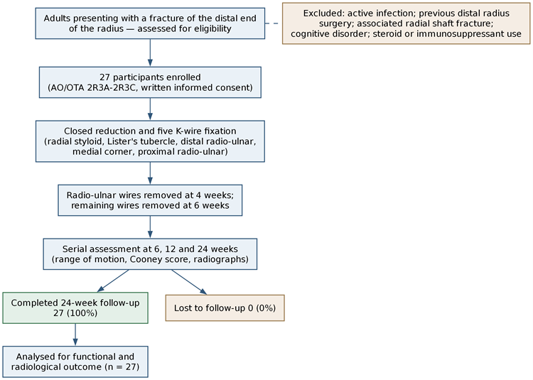
This was a prospective cohort study conducted in the Department of Orthopaedics of Bangalore Medical College and Research Institute, Bengaluru, and its affiliated hospitals, between May 2023 and October 2024. Reporting follows the STROBE recommendations for cohort studies. The study flow is summarised in Figure 1.
Participants
Consecutive patients presenting to the outpatient and inpatient services with a fracture of the distal end of the radius were screened. Adults older than 18 years with an AO/OTA type 2R3A to 2R3C fracture [11] who consented to participate and could attend a minimum of six months of follow-up were eligible; coexisting hypertension, diabetes mellitus, hypothyroidism, obesity, ischaemic heart disease or anaemia did not preclude inclusion. Patients were excluded if there was evidence of active infection, previous surgery on the distal radius, an associated radial shaft fracture, or a cognitive disorder or current treatment with steroids or immunosuppressants. Fractures were additionally classified by the Frykman system [12].
Surgical technique and postoperative care
After pre-anaesthetic evaluation, closed reduction was performed under image intensification and five K-wires were inserted in a fixed sequence: (i) a radial styloid wire passed from lateral to medial to fix the lateral column; (ii) a Lister’s tubercle wire passed dorsal to volar and lateral to medial to fix the intermediate column; (iii) a distal radio-ulnar wire passed transversely in the ulno-radial direction just below the articular surface to hold the articular reduction; (iv) a medial corner wire passed dorsal to volar and medial to lateral, entered between the fourth and fifth extensor compartments, to fix the medial column; and (v) a proximal radio-ulnar wire passed in the ulno-radial direction 5 cm proximal to the wrist joint to provide rotational stability. Each wire was introduced through a 1 cm incision after blunt dissection, with care to protect the superficial branch of the radial nerve and the extensor tendons.
No plaster slab was applied. Patients received perioperative antibiotics, pin-site care, early mobilisation and supervised physiotherapy. The proximal and distal radio-ulnar wires were removed at 4 weeks and the remaining three wires 2 weeks later. Length of hospital stay, operative time and complications were recorded.
Outcome measures
Functional outcome was assessed with the Cooney modification of the Green and O’Brien scoring system [13], which grades pain, functional status, range of motion, the flexion–extension arc and grip strength out of 100, and with the Disabilities of the Arm, Shoulder and Hand (DASH) questionnaire [15]. Range of motion of the shoulder, elbow, wrist (dorsiflexion, palmar flexion, radial and ulnar deviation) and forearm (supination, pronation) was measured with a goniometer at 6, 12 and 24 weeks.
Radiographs of the wrist were obtained in the immediate postoperative period and at 6, 12 and 24 weeks. Radial height, radial inclination, volar tilt, ulnar variance and any intra-articular step were measured, and the overall radiological result at 24 weeks was graded by the Sarmiento modification of the Lidström criteria [14], which grades loss of volar tilt, loss of radial deviation and radial shortening as excellent, good, fair or poor.
Sample size
The sample size was based on the series of Vipin et al. [9], in which 90% of patients achieved a good to excellent outcome six months after wire removal. Using n=z_α^2 pq/d^2 with a 95% confidence level and 12% absolute precision, n=(〖1.96〗^2×90×10)/〖12〗^2=24. Allowing for a 10% dropout rate, the required sample was 26.4, rounded to 27.
Statistical analysis
Data were compiled in a spreadsheet and analysed with jamovi. Continuous variables are summarised as mean ± standard deviation with median and range, and categorical variables as counts and percentages. Serial Cooney scores were compared with paired t-tests, with Cohen’s d reported as the effect size. A two-sided P value below 0.05 was considered statistically significant.
RESULTS:
Participant characteristics
Twenty-seven patients were enrolled and all 27 completed the full 24 weeks of follow-up, with no losses (Figure 1). The mean age was 62.1 ± 10.1 years (median 64; range 45–82) and 17 participants (63.0%) were women. A fall on an outstretched hand accounted for 18 injuries (66.7%) and road traffic accidents for 9 (33.3%); the mechanism was direct in every case. Twenty-three patients (85.2%) presented within one week of injury. Diabetes mellitus and hypertension were each present in 14 patients (51.9%), cardiovascular disease in 5 (18.5%) and asthma in 4 (14.8%). Baseline characteristics are given in Table 1.
Pain was universal at presentation; swelling was present in 25 patients (92.6%) and deformity in 26 (96.3%). The surrounding skin was normal in 12 patients (44.4%), with contusion, oedema and micro-abrasion each seen in 5 (18.5%). Two patients (7.4%) had associated injuries. By AO/OTA classification, 17 fractures (63.0%) were type 2R3B3, 6 (22.2%) type 2R3A3, 3 (11.1%) type 2R3C2 and 1 (3.7%) type 2R3B2; by the Frykman system, type IV was commonest (7 patients, 25.9%).
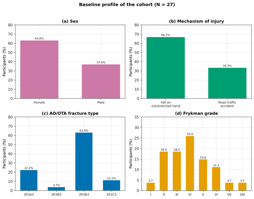
Figure 2. Baseline profile of the cohort (N = 27): sex, mechanism of injury, AO/OTA fracture type and Frykman grade.
Operative details and complications
Twenty-two procedures (81.5%) were performed under regional block and 5 (18.5%) under general anaesthesia. Reduction was achieved without difficulty in 23 patients (85.2%). Pin-tract infection developed in 3 patients (11.1%) and settled with oral antibiotics and regular pin-site dressing. There were no cases of excessive intraoperative or postoperative bleeding, deep wound infection, neurovascular injury or postoperative palsy. The mean hospital stay was 2.3 days (range 1–5), with 14 patients (51.9%) discharged after two days. All wires were removed by 6 weeks, and all fractures united.
Range of motion
Every patient had a full range of shoulder and elbow motion at both 6 and 12 weeks. Wrist and forearm motion improved steadily across the three assessments (Table 2, Figure 3). Mean wrist flexion rose from 36.1 ± 5.1° at 6 weeks to 43.1° at 12 weeks and 48.7 ± 8.2° at 24 weeks, and dorsiflexion from 26.1 ± 4.2° to 32.6° and 40.6 ± 7.3°. Forearm rotation showed the largest gains: supination increased from 37.8 ± 4.9° to 55.0 ± 9.9° and pronation from 26.7 ± 5.4° to 52.6 ± 8.0° over the same interval. Radial deviation improved from 5.7 ± 1.8° to 9.1 ± 2.0° and ulnar deviation from 10.0 ± 2.8° to 11.1 ± 2.1°.
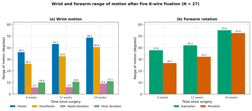
Figure 3. Mean wrist and forearm range of motion at 6, 12 and 24 weeks after five K-wire fixation (N = 27).
Functional outcome
The Cooney score improved at each interval (Table 3). The mean score was 76.1 ± 9.9 at 6 weeks, 78.1 ± 8.2 at 12 weeks and 81.5 ± 8.6 at 24 weeks. The 6-to-12-week gain was statistically significant (mean difference 2.04, standard error 0.482; t(26) = −4.23; P < 0.001; Cohen’s d = −0.81), as was the 12-to-24-week gain (mean difference 3.33, standard error 0.706; t(26) = −4.72; P < 0.001; Cohen’s d = −0.91). The narrowing standard deviation between 6 and 12 weeks indicates increasingly consistent recovery across the cohort.
Radiological outcome
At 24 weeks the mean volar tilt was 0.2°, with 12 wrists (44.4%) at 0°, 8 (29.6%) at +5° and 7 (25.9%) at −5°. Mean radial height was 8.5 mm and mean radial inclination 18.0°, with 24 wrists (88.9%) between 17° and 20°. Mean ulnar variance was −0.5 mm, most commonly −1 mm (10 wrists, 37.0%). An intra-articular step of 1 mm was present in 14 wrists (51.9%), while 9 (33.3%) had no step and 4 (14.8%) had a 2 mm step; no wrist had a step exceeding 2 mm (Table 4).
By the Sarmiento modification of the Lidström criteria, the overall radiological result was excellent in 5 patients (18.5%), good in 13 (48.1%) and fair in 9 (33.3%). No patient had a poor result, so 18 of 27 wrists (66.7%) achieved a good or excellent grade.
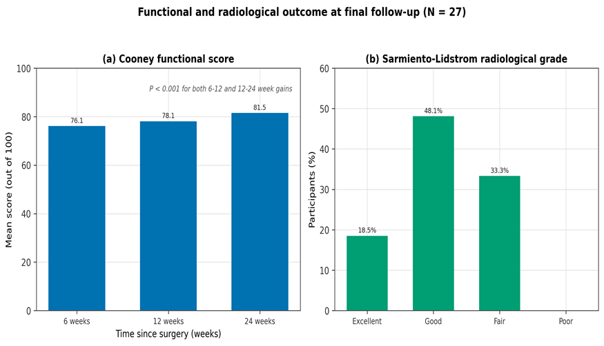
Figure 4. Functional and radiological outcome: (a) mean Cooney score at 6, 12 and 24 weeks; (b) Sarmiento radiological grade at 24 weeks (N = 27).
TABLES
Table 1. Baseline demographic, injury and fracture characteristics (N = 27).
|
Characteristic |
Category |
n (%) |
|
Age (years) |
Mean ± SD |
62.1 ± 10.1 |
|
Age (years) |
Median (range) |
64 (45–82) |
|
Sex |
Female |
17 (63.0) |
|
Sex |
Male |
10 (37.0) |
|
Mechanism of injury |
Fall on outstretched hand |
18 (66.7) |
|
Mechanism of injury |
Road traffic accident |
9 (33.3) |
|
Time to presentation |
Within 1 week |
23 (85.2) |
|
Time to presentation |
More than 1 week |
4 (14.8) |
|
Comorbidity |
Diabetes mellitus |
14 (51.9) |
|
Comorbidity |
Hypertension |
14 (51.9) |
|
Comorbidity |
Cardiovascular disease |
5 (18.5) |
|
Comorbidity |
Asthma |
4 (14.8) |
|
Comorbidity |
Previous fracture |
2 (7.4) |
|
Surrounding skin |
Normal |
12 (44.4) |
|
Surrounding skin |
Contused |
5 (18.5) |
|
Surrounding skin |
Oedematous |
5 (18.5) |
|
Surrounding skin |
Micro-abrasion |
5 (18.5) |
|
Associated injuries |
Present |
2 (7.4) |
|
AO/OTA type |
2R3A3 |
6 (22.2) |
|
AO/OTA type |
2R3B2 |
1 (3.7) |
|
AO/OTA type |
2R3B3 |
17 (63.0) |
|
AO/OTA type |
2R3C2 |
3 (11.1) |
|
Frykman grade |
I |
1 (3.7) |
|
Frykman grade |
II |
5 (18.5) |
|
Frykman grade |
III |
5 (18.5) |
|
Frykman grade |
IV |
7 (25.9) |
|
Frykman grade |
V |
4 (14.8) |
|
Frykman grade |
VI |
3 (11.1) |
|
Frykman grade |
VII |
1 (3.7) |
|
Frykman grade |
VIII |
1 (3.7) |
|
Anaesthesia |
Regional block |
22 (81.5) |
|
Anaesthesia |
General anaesthesia |
5 (18.5) |
Table 2. Wrist and forearm range of motion during follow-up (N = 27; degrees, mean ± SD).
|
Movement |
6 weeks |
12 weeks |
24 weeks |
|
Wrist flexion |
36.1 ± 5.1 |
43.1 |
48.7 ± 8.2 |
|
Wrist dorsiflexion |
26.1 ± 4.2 |
32.6 |
40.6 ± 7.3 |
|
Radial deviation |
5.7 ± 1.8 |
5.9 |
9.1 ± 2.0 |
|
Ulnar deviation |
10.0 ± 2.8 |
10.6 |
11.1 ± 2.1 |
|
Forearm supination |
37.8 ± 4.9 |
42.0 |
55.0 ± 9.9 |
|
Forearm pronation |
26.7 ± 5.4 |
32.2 |
52.6 ± 8.0 |
12-week values are means derived from the recorded frequency distributions; standard deviations were not reported at that interval.
Table 3. Cooney (modified Green and O’Brien) functional score and paired comparisons (N = 27).
|
Interval |
Mean ± SD |
Median |
Comparison |
Mean difference (SE) |
t (df = 26) |
P |
Cohen’s d |
|
6 weeks |
76.1 ± 9.9 |
75 |
— |
— |
— |
— |
— |
|
12 weeks |
78.1 ± 8.2 |
75 |
6 vs 12 weeks |
2.04 (0.482) |
−4.23 |
< 0.001 |
−0.81 |
|
24 weeks |
81.5 ± 8.6 |
80 |
12 vs 24 weeks |
3.33 (0.706) |
−4.72 |
< 0.001 |
−0.91 |
Table 4. Radiographic parameters at 24 weeks (N = 27).
|
Parameter |
Distribution, n (%) |
Mean |
|
Volar tilt |
−5°: 7 (25.9); 0°: 12 (44.4); +5°: 8 (29.6) |
0.2° |
|
Radial height |
6 mm: 3 (11.1); 7 mm: 4 (14.8); 8 mm: 6 (22.2); 9 mm: 8 (29.6); 10 mm: 3 (11.1); 11 mm: 3 (11.1) |
8.5 mm |
|
Radial inclination |
14–16°: 3 (11.1); 17°: 6 (22.2); 18°: 7 (25.9); 19°: 6 (22.2); 20°: 5 (18.5) |
18.0° |
|
Ulnar variance |
−2 mm: 5 (18.5); −1 mm: 10 (37.0); 0 mm: 8 (29.6); +1 mm: 2 (7.4); +2 mm: 2 (7.4) |
−0.5 mm |
|
Intra-articular step |
0 mm: 9 (33.3); 1 mm: 14 (51.9); 2 mm: 4 (14.8) |
0.8 mm |
|
Sarmiento–Lidström grade |
Excellent: 5 (18.5); Good: 13 (48.1); Fair: 9 (33.3); Poor: 0 (0.0) |
— |
DISCUSSION:
In this prospective cohort of 27 adults with distal radius fractures treated by five K-wire fixation, wrist and forearm motion and the Cooney functional score improved significantly at every interval up to 24 weeks, two-thirds of wrists achieved a good or excellent radiological grade, and the only complication was superficial pin-tract infection in three patients. The absence of any poor radiological result, and the maintenance of radial inclination between 17° and 20° in 88.9% of wrists, support the mechanical rationale for the technique.
That rationale is rotational. Conventional crossed pinning stabilises the coronal and sagittal planes but leaves the construct rotationally lax, because converging wires that cross at the fracture behave as a single implant. Mechanical testing of five different percutaneous pin configurations has shown that construct stiffness rises with the number and divergence of wires rather than with pinning alone [7], which is consistent with our observation that adding two radio-ulnar wires preserved radial length: mean ulnar variance at 24 weeks was −0.5 mm and mean radial height 8.5 mm, without the late collapse that limits conventional pinning. Because radial shortening is one of the three components of the Sarmiento–Lidström grade, holding length is also what drove the radiological results.
Our functional findings sit comfortably alongside the small published experience of the five-pin technique. Abdallah et al. compared the five-pin construct directly with conventional K-wire fixation and found the five-pin approach a safe, cost-effective method giving better pain, mobility and stability [8]. Vipin et al., in 20 patients, attributed the benefit specifically to the two additional ulno-radial wires, which preserve radial height and permit early mobilisation of the wrist and fingers [9]. Bhasme et al., in 30 patients, concluded that the technique gives functional outcomes better than conventional K-wire fixation and comparable to volar plating [10]. Our cohort reproduces that pattern in an older population than most of these series.
Compared with alternative fixation strategies, our results are unsurprising but reassuring. The five-year follow-up of the DRAFFT randomised trial found no difference in patient-reported function between percutaneous wires and volar locking plates for dorsally displaced fractures [5], and Mishra et al. similarly reported comparable functional outcomes for external fixation and volar plating in unstable fractures [21]. Randomised comparisons of augmented external fixation against percutaneous pinning and casting [17], and of external fixation with optional percutaneous wires against casting [20], have likewise failed to show a decisive advantage for the more invasive construct. Against casting alone, however, percutaneous fixation is clearly better radiologically in extra-articular fractures [4], and redisplacement remains the weakness of closed treatment [3]. Szyluk et al., in 112 patients, found percutaneous K-wire fixation to give good results across AO types A2, A3, B1, B2, C1 and C2 [16] — a spectrum that encompasses the fractures treated here. Snow et al. reported that interfocal wiring restores function without needing to over-correct position [18]. Taken together, the literature suggests that once reduction is held, the choice of implant matters less than commonly assumed, which strengthens the case for the cheapest and least invasive construct that will hold it.
Two features of our cohort deserve comment. First, at a mean of 62.1 years our patients were older than those in most five-pin series, and 63.0% were women — a distribution consistent with the osteoporotic, low-energy pattern that dominates distal radius fractures in older populations [1]. In precisely this group, Arora et al. showed that operative fixation in patients over 70 confers radiological but not always functional superiority over non-operative care [19], which makes a low-morbidity day-care construct particularly attractive. Second, avoiding a plaster slab allowed immediate mobilisation, and every patient had regained full shoulder and elbow motion by 6 weeks; shoulder–hand stiffness, historically a major source of morbidity after distal radius fracture, did not occur. The mean hospital stay of 2.3 days reflects the day-care nature of the procedure.
The 11.1% pin-tract infection rate is the expected trade-off of percutaneous fixation and is in keeping with published rates; all three cases were superficial and resolved with oral antibiotics and pin-site care, without wire removal or deep infection. This complication is the price of the technique’s advantages, and it is a low one.
STRENGTHS AND LIMITATIONS
The main strengths are the prospective design, a standardised and explicitly sequenced surgical technique, complete follow-up of all 27 participants to 24 weeks, and parallel functional and radiographic assessment using validated instruments. Several limitations temper the findings. This was a single-arm cohort without a comparator, so the comparisons with conventional pinning, plating and external fixation are indirect and drawn from the literature rather than from randomised contemporaneous controls. The sample was small and drawn from a single centre, which limits precision and generalisability. Outcome assessment was not blinded and was performed by the treating team, which risks observer bias in both goniometry and radiographic measurement. Follow-up ended at 24 weeks, so late collapse, post-traumatic radiocarpal arthritis and the durability of the functional gains beyond six months remain unknown. Grip strength was recorded only as a component of the composite Cooney score rather than as a separate dynamometric measurement, and DASH scores, although collected, are not reported here as a distribution. Finally, the cohort was skewed towards partial articular fractures (63.0% type 2R3B3), so these results should not be extrapolated to highly comminuted complete articular injuries, in which bridging constructs may still be preferable
CONCLUSION:
Five K-wire fixation produced progressive, statistically significant improvement in wrist and forearm motion and in the Cooney functional score over 24 weeks, with two-thirds of wrists achieving a good or excellent Sarmiento–Lidström grade, no poor radiological results, a mean hospital stay of 2.3 days and superficial pin-tract infection as the only complication. By adding two radio-ulnar wires to the conventional three-column construct, the technique addresses the rotational instability and late collapse that limit standard percutaneous pinning while preserving its low cost, short learning curve and day-care feasibility. It is a sound option for distal end radius fractures, especially in older or comorbid patients in whom implant cost or operative morbidity argues against open fixation. Randomised comparison against conventional pinning and volar plating, with longer follow-up, is the logical next step.
REFERENCES:
1. Alffram PA, Bauer GC. Epidemiology of fractures of the forearm. A biomechanical investigation of bone strength. J Bone Joint Surg Am. 1962;44-A:105-114.
2. Ikpeze TC, Smith HC, Lee DJ, Elfar JC. Distal radius fracture outcomes and rehabilitation. Geriatr Orthop Surg Rehabil. 2016;7(4):202-205.
3. Handoll HH, Madhok R. Closed reduction methods for treating distal radial fractures in adults. Cochrane Database Syst Rev. 2003;(1):CD003763.
4. Venkatesh RB, Maranna GK, Narayanappa RK. A comparative study between closed reduction and cast application versus percutaneous K-wire fixation for extra-articular fracture distal end of radius. J Clin Diagn Res. 2016;10(2):RC05-RC09.
5. Costa ML, Achten J, Rangan A, Lamb SE, Parsons NR. Percutaneous fixation with Kirschner wires versus volar locking-plate fixation in adults with dorsally displaced fracture of distal radius: five-year follow-up of a randomized controlled trial. Bone Joint J. 2019;101-B(8):978-983.
6. Glickel SZ, Catalano LW, Raia FJ, Barron OA, Grabow R, Chia B. Long-term outcomes of closed reduction and percutaneous pinning for the treatment of distal radius fractures. J Hand Surg Am. 2008;33(10):1700-1705.
7. Muratli SK, Uzun B, Celik S. Comparison of five percutaneous pinning methods for unstable extra-articular distal radius fractures: a mechanical study using sawbones. Jt Dis Relat Surg. 2021;32(1):51-58.
8. Abdallah KN, Salama AM, El-Adawy AM, Gaber AM. Assessment of five pin technique versus conventional Kirschner wire fixation for management of distal radial fractures. Egypt J Hosp Med. 2022;89(2):7457-7465.
9. Vipin R, Rengarajan N, Manoharan M, Kesavan K. A novel 5-pin fixation for distal radius fractures and its functional assessment. J Orthop Joint Surg. 2021;3(1):13-17.
10. Bhasme VK, Shettar VG, Battur MN. Functional outcome analysis of fixation of distal radius fractures using ‘five pin technique’. Int J Contemp Med Res. 2018;5(9):I14-I18.
11. Meinberg EG, Agel J, Roberts CS, Karam MD, Kellam JF. Fracture and Dislocation Classification Compendium-2018. J Orthop Trauma. 2018;32(Suppl 1):S1-S170.
12. Frykman G. Fracture of the distal radius including sequelae: shoulder-hand-finger syndrome, disturbance in the distal radio-ulnar joint and impairment of nerve function. A clinical and experimental study. Acta Orthop Scand. 1967;Suppl 108:3-153.
13. Cooney WP, Bussey R, Dobyns JH, Linscheid RL. Difficult wrist fractures. Perilunate fracture-dislocations of the wrist. Clin Orthop Relat Res. 1987;(214):136-147.
14. Sarmiento A, Pratt GW, Berry NC, Sinclair WF. Colles’ fractures. Functional bracing in supination. J Bone Joint Surg Am. 1975;57(3):311-317.
15. Hudak PL, Amadio PC, Bombardier C. Development of an upper extremity outcome measure: the DASH (disabilities of the arm, shoulder and hand). Am J Ind Med. 1996;29(6):602-608.
16. Szyluk K, Jasinski A, Koczy B, Widuchowski W, Widuchowski J. Results of operative treatment of unstable distal radius fractures using percutaneous K wire fixation. Ortop Traumatol Rehabil. 2007;9(5):511-519.
17. Harley BJ, Scharfenberger A, Beaupre LA, Jomha N, Weber DW. Augmented external fixation versus percutaneous pinning and casting for unstable fractures of the distal radius: a prospective randomized trial. J Hand Surg Am. 2004;29(5):815-824.
18. Snow M, Kelly M, Jeyam M, Fahmy N. Function versus position: a randomized controlled trial of interfocal Kirschner wiring of unstable distal radial fractures. Eur J Trauma Emerg Surg. 2007;33(1):81-86.
19. Arora R, Gabl M, Gschwentner M, Deml C, Krappinger D, Lutz M. A comparative study of clinical and radiologic outcomes of unstable Colles type distal radius fractures in patients older than 70 years: nonoperative treatment versus volar locking plating. J Orthop Trauma. 2009;23(4):237-242.
20. Kreder HJ, Agel J, McKee MD, Schemitsch EH, Stephen D, Hanel DP. A randomized, controlled trial of distal radius fractures with metaphyseal displacement but without joint incongruity: closed reduction and casting versus closed reduction, spanning external fixation, and optional percutaneous K-wires. J Orthop Trauma. 2006;20(2):115-121.
21. Mishra AK, Sud AD, Prasad M, Kaul R, Singh CM. A comparative study of functional outcome of external fixation and volar plating in unstable distal radius fractures. Int J Res Orthop. 2019;5(2):326-334.