Morphometric study of pedicles of typical and atypical lumbar vertebra.
- Shilpa M , Assistant professor, Department of Anatomy, KRIMS, KARWAR
- Madhukar KT , Professor and Head of Department, Department of orthopaedics, KRIMS, KARWAR
- Arun Jose , 2nd year postgraduate, Department of Orthopedics, KRIMS KARWAR.
Article Information:
Abstract:
Background: Detail knowledge of the lumbar pedicle morphology is necessary for the treatment of lumbar instability by transpedicular screw fixation in order to avoid risk complication with adjacent vital structure and fracture of pedicle cortex. Objective: To study height, width and interpedicular distance of typical and atypical lumbar vertebra. Materials and Methods: 80 dry human lumbar vertebra of undetermined gender and age were selected for the study.65 were typical lumbar vertebra and 15 were atypical. The various parameters listed below were measured with the help of digital vernier caliper. Results: The various parameters like height, width on the right and left side of pedicles were measured and interpedicular distance was also measured. Comparison between typical and atypical vertebra was done. In Typical vertebra the mean height was 21mm and mean width was 16.28mm and the interpedicular distance 21.8mm. In Atypical vertebra the mean height was 20.8mm and mean width was 22.76mm and the interpedicular distance 21.6mm. Conclusion: There is significant correlation between height of typical and atypical vertebrae, whereas the width of the pedicles in the atypical vertebra is comparatively more than the typical vertebra. This data can also help the surgeons for performing safe transpedicular surgeries.
Keywords:
Article :
INTRODUCTION:
The vertebral column is composed of 33 vertebrae and their vertebral discs. The vertebral column forms the central axis made up of Cervical-7, thoracic-12, lumbar-5, sacral-5 and coccygeal-4. The vertebral column forms the central axis and gives main support to the bones and muscles1.The pedicle of lumbar vertebra is strong and large. This helps in placement of screws through them2. Transpedicular screw fixation of spine has developed as a very successful method of spinal fixation. The fixation of lumbar spine is needed for various spinal problems like fracture in lumbar spine, resection of tumours in vertebral bodies, gross spondylolisthesis and lumbar instabilities3. Utmost care needs to be taken during pedicle screw placement to prevent potential damage to neural structures that closely approximate the bony pedicle4. Lumbar vertebral pedicles are strong and large which enables fixing of screws through them. This is currently used for spinal fixation for various spinal problems. The pedicles of lumbar vertebrae are used for placing screws through them. Dimensions of lumbar vertebrae are of utmost importance in diagnosing lower back pain and lumbar spinal stenosis5.
MATERIALS AND METHODS:
The present study was carried out on 80 dry lumbar vertebrae of undetermined age and sex collected from the department of Anatomy of Karwar Institute of Medical Sciences, Karwar. The lumbar vertebrae were divided into two categories, 65 Typical and 15 Atypical vertebrae.Vertical height and width of the pedicle was measured on both right and left side of the typical and atypical vertebrae using digital Vernier calliper. Interpedicular distance was also calculated on both the typical and atypical lumbar vertebrae. Statistical analysis of the collected data was done by the statistician. Mean and range of each side was calculated and tabulated.
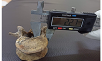
Measurement of width of lumbar pedicle
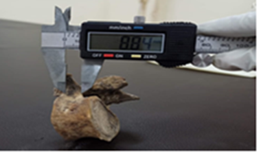
Measurement of length of lumbar pedicle
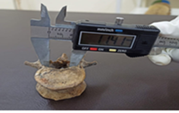
Measurement of length of interpedicular distance
RESULTS:
Typical Lumbar Vertebra(L1 –L4)
|
Height Right |
(mms) Left |
Width Right |
(mms) Left |
Interpedicular Distance (mms) |
|
12.02 |
12.8 |
15.04 |
15.06 |
18.06 |
|
13.6 |
12.2 |
17.6 |
18.02 |
20.8 |
|
14.8 |
16.7 |
19.6 |
19.9 |
27.8 |
|
16.03 |
15.9 |
10.3 |
11.02 |
14.3 |
|
16.5 |
17.9 |
13.7 |
12.6 |
23.3 |
|
17.11 |
19.8 |
15.3 |
15.6 |
19.8 |
|
18.3 |
18.5 |
20.08 |
20.4 |
22.03 |
|
18.4 |
21.2 |
15.7 |
16.1 |
19.4 |
|
18.76 |
19.7 |
13.7 |
13.8 |
22.7 |
|
18.8 |
18.7 |
14.6 |
14.04 |
13.8 |
|
19.17 |
18.7 |
15.8 |
17.3 |
24.3 |
|
20.1 |
19.7 |
13.5 |
14.1 |
22.6 |
|
20.3 |
20.1 |
11.8 |
13.2 |
20.7 |
|
20.3 |
21.04 |
13.7 |
13.2 |
19.2 |
|
20.4 |
20.7 |
18.4 |
17.5 |
26.04 |
|
20.6 |
21.4 |
14.6 |
15.7 |
25.4 |
|
20.64 |
20.4 |
15.8 |
17.2 |
24.3 |
|
20.9 |
20.7 |
20.16 |
19.6 |
22.6 |
|
20.6 |
21.4 |
18.2 |
18 |
23.4 |
|
20.64 |
20.4 |
14.8 |
15.1 |
20.2 |
|
20.9 |
20.7 |
19.7 |
20.2 |
18.4 |
|
21.5 |
20.5 |
11.7 |
12.3 |
26.4 |
|
22.02 |
21.7 |
25.3 |
24.8 |
26.2 |
|
22.2 |
21.6 |
18.6 |
17.9 |
25.3 |
|
22.3 |
23.7 |
14.8 |
14.4 |
17.4 |
|
22.3 |
24.4 |
15.6 |
14.9 |
16.9 |
|
23.6 |
22.3 |
17.8 |
18.3 |
20.3 |
|
23.4 |
22.6 |
14.3 |
15.2 |
14.2 |
|
23.6 |
22.5 |
16.5 |
16.1 |
14.2 |
|
23.9 |
22.9 |
14.4 |
13.8 |
16.2 |
|
24.01 |
23.8 |
20.3 |
20.1 |
23.4 |
|
24.4 |
24.1 |
16.7 |
16.2 |
22.3 |
|
24.9 |
23.8 |
15.8 |
15.3 |
21.7 |
|
27.6 |
26.5 |
17.4 |
17.1 |
19.9 |
|
29.5 |
28.8 |
14.6 |
14.1 |
20.3 |
|
20.4 |
21.2 |
17.4 |
16.8 |
25.3 |
|
16.7 |
16.2 |
15.6 |
14.9 |
24.2 |
|
27.4 |
26.4 |
18.3 |
18.1 |
26.1 |
|
24.2 |
23.2 |
15.6 |
15.4 |
19.8 |
|
18.06 |
17.9 |
14.4 |
14.1 |
17.2 |
|
19.7 |
18.9 |
17.8 |
17.2 |
23.3 |
|
22.1 |
21 |
14.7 |
14.3 |
18.7 |
|
24.01 |
23.3 |
13.5 |
13.2 |
22.2 |
|
23.4 |
22.7 |
17.7 |
16.9 |
17.8 |
RESULTS
Atypical Lumbar Vertebra
|
Height Right |
(mms) Left |
Width Right |
(mms) Left |
Interpedicular Distance (mms) |
|
19.4 |
19.6 |
22.7 |
23.1 |
23.5 |
|
19.9 |
19.7 |
22.7 |
23.1 |
19.8 |
|
20 |
21.3 |
23.2 |
22.9 |
24.4 |
|
21.9 |
20.7 |
21.5 |
20.8 |
21.8 |
|
22.7 |
23.3 |
24.4 |
24.1 |
22.4 |
|
18.6 |
19.2 |
21.6 |
22.1 |
23.6 |
|
21.6 |
20.9 |
23.9 |
23.2 |
24.2 |
|
18.8 |
18.2 |
19.2 |
18.7 |
22.8 |
|
20.2 |
20 |
18.4 |
17.6 |
24.1 |
|
18.8 |
18.2 |
19.4 |
18.9 |
26.1 |
|
19.2 |
19.1 |
21.2 |
20.8 |
21.1 |
|
17.6 |
17.4 |
19.3 |
19.7 |
25.2 |
|
18.8 |
18.2 |
22.2 |
22.4 |
23.3 |
|
19.2 |
19 |
23.4 |
23.1 |
21.4 |
|
PEDICLE |
Lumbar vertebra |
Parameter |
Mean (mm) |
Range(mm) |
|
Typical
|
Height
|
21 |
15.2-21.4
|
|
|
Width |
16.28 |
12.4-22.6 |
||
|
Interpedicular Distance |
19.6 |
13.7-25.6 |
||
|
Atypical |
Height |
22.8 |
21.4-26.7 |
|
|
Width |
22.7 |
20.5-25.2 |
||
|
Interpedicular Distance |
21.6 |
16.3-24.4 |
DISCUSSION:
One of the most common complaints amongPatients worldwide is low backache and it is amongst the top musculoskeletal symptoms observed. The pedicle is the strongest part of lumbar vertebra. It acts as a strut to transmit forces between the body and neural arch1.Lumbar vertebral pedicles are strong and large which enables fixing of screws through them.This is currently used for spinal fixation for various spinal problems2. A thorough knowledge of the variations in the size of the pedicles is extremely important when pedicle screws are used3.
In Kalpana Ramachandran et al1 the pedicles dimensions which include the mean height & width on the right and left side in typical vertebrae were 14.9 mm & 11mm and 14.7 mm & 11.1 mm while the mean height & width on the right side and left side in atypical vertebrae are 21 mm & 18 mm and 20 mm & 18.8 mm respectively. Singel et al2 found that the pedicle width of the lumbar segment increased progressively from:
L1 to L4 and increased abruptly at L5 level.
In a study done by Chandini et.al5 in typical lumbar vertebrae the mean height of the pedicle on both right and left side were more compared to the width, but in atypical vertebrae the mean height and width were almost the same.
In another study by Aruna & Rajeswari et al6, the range for breadth for typical lumbar vertebrae was 4.5 – 20 mm and range for the height was 11.5 mm- 20 mm & in atypical vertebra the range for breadth was 13-22 mm and height was 10- 16 mm respectively.
Chawla et al7, conducted the study on 30 dry L3 lumbar vertebrae and found that the average height of the pedicle was 14.0 ±1.1 mm on right side and the average height of the pedicle on left side was 14.1 ± 1.0 mm.The mean width of the pedicle was 8.7 ± 1.4 mm on the right side and 8.7 ± 1.7 mm on the left side.
In our present study it was found that there is increase in the height, width and interpedicular distance in atypical vertebra (L5) than typical vertebrae. In our study,we found the mean measurement 21 at L1 and 22.8at L5, mean interpedicular distance being 21.6mm
Lumbar vertebral pedicles are strong and large which enables fixing of screws through them.This is currently used for spinal fixation for various spinal problems8. The pedicles of lumbar vertebrae are used for placing screws through them. In this, the screw is inserted through the posterior aspect of the pedicle in to the body of the lumbar vertebra anteriorly. For this the screw required to be inserted is determined by the minimum diameter of the pedicle. Hence diameters of the pedicles are of utmost importance in pre-operative planning9.
The present study has found significant differences in various dimensions of lumbar vertebral pedicles at various levels (L1 – L5), that is from L1 to L5 both height and Widths of pedicle went on increasing and were more than the typical vertebrae. If there is mismatchbetween the size of the screw and the pedicle, it may lead to cortex perforation of pedicle, fracture pedicle or loosening of the screw, which may result in serious implications. Hencefor pedicle screw fixation the height and width of pedicles play an important role in decidingthe choice of the screw.
CONCLUSION:
Thus, according to the above discussion, the present study concludes that there is always an increase in width of lumbar pedicles proceeding from L1 to L5 levels and the width being maximum at L5 level to enable in weight transmission.
These differences have critical implication for spinal surgeons to perform safe operations in this area. There is an increase in use of pedicle screws and so there is great emphasis of studies on the dimensions of pedicles of lumbar vertebrae. These differences have critical implication for spinal surgeons to perform safe operations in this area. There is an increase in use of pedicle screws and so there is great emphasis of studies on the dimensions of pedicles of lumbar vertebrae in different populations.
REFERENCES:
1. Kalpana Ramachandra,Durgadevi Ganesan. A Morphometric study of Lumbar Vertebrae and its clinical significance.Int j med and applied sciences. 2015;4(1):189-193.
2. Singel TC, Patel MM, Gohil DV. A study of width and height of lumbar pedicles in Saurashtra region.J. Anat .Soc. India. 2004; 53(1): 4-9.
3. Suranjana Banik, Ajita Rajkumari.Morphometric analysis of lumbar vertebrae and its clinical significance.Int J Anat Res 2019, 7(2.1):6381-86.
4. Alam MM, Waqas m et al. Lumbar Morphometry: A study of Lumbar Vertebrae from a Pakistani Population using Computed Tomography scans. Asian Spine J.2014;8 (4):421-426.
5. Chandni G, Poonam K, Aravind KP, Antony SD. Morphometric study of Pedicles in Lumbar Vertebraeand its Clinical Significance. Journal of Surgical Academia. 2014; 4 (1): 37-40.
6. Arna N, Rajeshwari T.A study of Lumbar pedicle size in south Indians. Anatomica, Karnataka. 2011;5(2) 69 -73
7. Chawla K, Sharma M, Abhaya A . Kochhar S. Morphometry of the lumbar pedicle in West India.Eur J Anat. 2011; 15(3):155- 161.
8. Soumya Philipose, Santhosh Kuriakose et al. “Morphometric Study of Pedicles of Lumbar Vertebrae in Southern India”. Journal of Evidence based Medicine and Healthcare; Volume 2, Issue 39, Sept 28, 2015: 6182-6191.
9. Bin L, Yong H, Bi z, Mo S, Zhi-Min G. Pedicle screw piercer with warning device – A technique to increase accuracy of pedicle screw placement. A cadaveric Study. Indian Journal of Orthopaedics.2014;48 (6) : 545-549.
10. Matuoka CM, Junior RB. Anatomical study of Lumbar vertebral pedicles and adjacent neural structures. Acta Ortop Bras. 2002; 10 (3): 25-34.
11. Amonoo-Kuofi HS. Age related variations in the horizontal and vertical diameters of the pedicles of Lumbar spine. Journal of Anatomy.1995; 186 : 321-328.
12. Jadhav AS, Katti AS, Herekar NG, Jadhav SB.Osteological study of lumbar vertebrae in Western Maharashtra population. Journal of the Anatomical Society of India 2013;62(1):10-16.