CLINICOPATHOLOGICAL CHARACTERISTICS AND IMMUNOHISTOCHEMICAL PROFILE OF CARCINOMA BREAST PATIENTS IN A TERTIARY CARE CENTRE: A CROSS-SECTIONAL STUDY.
- MURALI S V , Assistant Professor, Department of General Surgery, PES University Institute of Medical Sciences and Research, Electronic City, Bengaluru, Karnataka, India, Pincode – 560100.
- SRIVACHAN S SHAPUR , Assistant Professor, Department of General Surgery, PES University Institute of Medical Sciences and Research, Electronic City, Bengaluru, Karnataka, India, Pincode – 560100.
- RADHA SURASETTY ANGADI , Assistant Professor, Department of Pathology, Basaveshwara Medical College & Hospital, Medehalli, Chitradurga, Karnataka, India, Pincode – 577501
- RAGHAVENDRA D , Assistant Professor, Department of Community Medicine, Sri Chamundeshwari Medical College, Hospital & Research Institute, Channapatna, Bengaluru South, Karnataka, India, Pincode – 562160.
Article Information:
Abstract:
Introduction: Breast cancer is a leading cause of cancer-related morbidity and mortality among women, with rising incidence in India. Histopathological classification and immunohistochemical assessment of ER, PR, and HER2/neu are essential for diagnosis, prognosis, and treatment selection, while triple-negative breast cancer represents an aggressive subtype. This study therefore evaluated histological patterns, receptor expression, TNBC prevalence, and age-related variations among patients attending a tertiary care centre. Material and Methods: This hospital-based cross-sectional study included 50 consecutive women with FNAC-confirmed breast carcinoma after ethical approval and informed consent. Clinical and tumour characteristics were recorded using a structured proforma. Surgical specimens underwent histopathological classification and immunohistochemical assessment of ER, PR, and HER2/neu. Data were analysed using SPSS; categorical variables were summarized as frequencies and percentages, and associations were tested using Chi-square or Fisher’s exact test, with p<0.05 considered significant. Results: Among 50 women with breast carcinoma, 42.0% were aged 41–50 years, 78.0% presented with a breast lump, 78.0% had upper outer quadrant involvement, and 62.0% had Stage IIB disease. IDC-NST was the predominant subtype (72.0%). ER, PR, and HER2/neu positivity were 56.0%, 54.0%, and 38.0%, respectively, while triple-negative breast cancer occurred in 32.0%. ER and PR positivity increased significantly with age (p=0.040 and p=0.050), whereas TNBC was confined to women aged ≤50 years (p=0.002). Conclusion: The study characterized the clinicopathological and receptor profile of breast carcinoma, identifying IDC-NST as predominant and triple-negative disease in nearly one-third of patients. ER and PR expression increased with age, whereas triple-negative breast cancer was concentrated among younger women, thereby fulfilling all stated objectives.
Keywords:
Article :
INTRODUCTION:
Breast cancer is the most commonly diagnosed malignancy and the leading cause of cancer-related mortality among women worldwide, contributing substantially to the global cancer burden.1,2 In India, the incidence of breast cancer has risen progressively over the past few decades, particularly in urban regions, where changing reproductive behaviour, increasing life expectancy, lifestyle modifications, and improved access to diagnostic facilities have contributed to enhanced disease detection.3,4 Despite considerable advances in screening strategies, diagnostic techniques, and therapeutic interventions, a significant proportion of patients continue to present with clinically advanced disease, emphasizing the importance of comprehensive evaluation of tumour characteristics that have prognostic and therapeutic relevance.5-7
Histopathological examination continues to serve as the gold standard for confirming the diagnosis of breast carcinoma and classifying tumours into distinct histological subtypes that differ in morphology, biological behaviour, and clinical outcome.8-10 In addition, immunohistochemical (IHC) evaluation of estrogen receptor (ER), progesterone receptor (PR), and human epidermal growth factor receptor-2 (HER2/neu) has become an indispensable component of routine pathological assessment, as these biomarkers are fundamental for molecular classification, prognostication, and the selection of endocrine and HER2-targeted therapies.9,10 Triple-negative breast cancer (TNBC), defined by the absence of ER, PR, and HER2 expression, represents a biologically aggressive subtype characterized by a higher likelihood of recurrence, limited targeted treatment options, and comparatively poorer clinical outcomes.11-13 Several investigators have also examined the relationship between clinicodemographic characteristics, particularly patient age, and hormone receptor expression; however, the reported associations have remained variable across different populations.14-16
Although the histopathological spectrum and immunohistochemical profiles of breast carcinoma have been extensively investigated, considerable geographic and population-specific variations continue to exist.10,14 Moreover, comprehensive data integrating histological subtypes, ER, PR, HER2/neu status, triple-negative breast cancer, and age-related receptor expression from Indian tertiary care centres remain relatively limited. Such region-specific evidence is essential for improving prognostic stratification, optimizing individualized treatment strategies, and understanding the clinicopathological characteristics of breast carcinoma within the local population.3,4 Therefore, this study aimed to describe the histopathological spectrum, determine receptor expression, estimate the prevalence of triple-negative breast cancer, and assess the association between age at presentation and immunohistochemical receptor status.
MATERIALS AND METHODS:
This cross-sectional study was conducted over a period of 18 months, including a 6-month follow-up period, in the Department of General Surgery at ESIC Medical College Hospital and Post Graduate Institute of Medical Sciences and Research (ESIC-MH & PGIMSR), Rajajinagar, Bengaluru, after obtaining approval from the Institutional Ethics Committee. The study aimed to evaluate the clinicopathological characteristics and immunohistochemical profile of carcinoma breast. A total of 50 female patients with clinically palpable breast lumps and fine needle aspiration cytology (FNAC)-confirmed carcinoma breast were included. All eligible patients presenting during the study period were enrolled after obtaining written informed consent.
Female patients aged 20–80 years with palpable breast lumps and FNAC-confirmed carcinoma breast were included in the study. Patients with benign breast diseases, recurrent carcinoma breast, and male breast carcinoma were excluded. A detailed clinical history was obtained using a structured proforma, followed by thorough clinical examination. Baseline demographic details, presenting complaints, clinical findings, tumour characteristics, and staging information were recorded systematically.
All patients underwent routine laboratory investigations, including complete blood count, urine analysis, random blood sugar, renal function tests, and electrocardiography. Disease evaluation was performed using appropriate radiological and pathological investigations, including ultrasonography and/or mammography of the breast, chest radiography, ultrasonography of the abdomen and pelvis, liver function tests, serum alkaline phosphatase estimation, bone imaging whenever indicated, positron emission tomography/computed tomography (PET/CT) or oncological CT in selected patients, and trucut/core biopsy where appropriate. Histopathological examination of the postoperative specimen confirmed the diagnosis and determined the histological subtype.
Immunohistochemical evaluation was performed on postoperative tumour specimens to determine estrogen receptor (ER), progesterone receptor (PR), and human epidermal growth factor receptor-2 (HER2/neu) status. Tumours negative for ER, PR, and HER2/neu were classified as triple-negative breast cancer. Histopathological subtype, receptor expression, and age-wise distribution of immunohistochemical markers were analysed to evaluate clinicopathological characteristics.
The collected data were entered into Microsoft Excel and analysed using SPSS software version 26. Categorical variables, including clinicopathological characteristics, histopathological subtypes, and ER, PR, HER2/neu, and triple-negative receptor status, were summarised as frequencies and percentages. Fisher’s exact test was used to assess the association of age group with ER, PR, HER2/neu, and triple-negative breast cancer status. A p-value of <0.05 was considered statistically significant.
RESULTS:
Among the 50 study participants, the majority were aged 41–50 years (42.0%) and presented with a breast lump as the sole complaint (78.0%). The left breast (60.0%) and upper outer quadrant (78.0%) were most commonly affected. Most tumours measured 2–5 cm (92.0%), with N1 nodal involvement observed in 78.0% of patients. Clinically, Stage IIB disease was the predominant presentation (62.0%), followed by Stage IIA (22.0%). (Table 1)
Table 1: Baseline clinicopathological characteristics of the study participants (N = 50)
|
Variable |
Category |
Frequency (N) |
Percentage (%) |
|
Age group |
30 to 40 years |
10 |
20.0% |
|
41 to 50 years |
21 |
42.0% |
|
|
51 to 60 years |
12 |
24.0% |
|
|
61 to 70 years |
6 |
12.0% |
|
|
71 to 80 years |
1 |
2.0% |
|
|
Presenting complaint |
Breast lump only |
39 |
78.0% |
|
Breast lump with pain |
5 |
10.0% |
|
|
Breast lump with nipple discharge |
3 |
6.0% |
|
|
Breast lump with skin changes |
3 |
6.0% |
|
|
Side involved |
Left |
30 |
60.0% |
|
Right |
20 |
40.0% |
|
|
Tumour location |
Central quadrant |
2 |
4.0% |
|
Lower inner quadrant |
3 |
6.0% |
|
|
Lower outer quadrant |
4 |
8.0% |
|
|
Upper inner quadrant |
2 |
4.0% |
|
|
Upper outer quadrant |
39 |
78.0% |
|
|
Tumour size |
2 to 5 cm |
46 |
92.0% |
|
>5 cm |
4 |
8.0% |
|
|
Clinical nodal status |
N0 |
11 |
22.0% |
|
N1 |
39 |
78.0% |
|
|
Clinical stage |
Stage IIA |
11 |
22.0% |
|
Stage IIB |
31 |
62.0% |
|
|
Stage IIIA |
2 |
4.0% |
|
|
Stage IIIB |
3 |
6.0% |
|
|
Stage IV |
3 |
6.0% |
Invasive ductal carcinoma–No Special Type (IDC-NST) was the predominant histopathological subtype, accounting for 72.0% of cases. ER and PR positivity were observed in 56.0% and 54.0% of patients, respectively, while HER2/neu positivity was noted in 38.0%. Triple-negative breast cancer constituted 32.0% of the study population, indicating that approximately one-third of patients lacked expression of all three therapeutic receptors. (Figure 1 & 2)
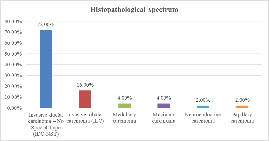
Figure 1: Histopathological spectrum of carcinoma breast among the study participants (N = 50)
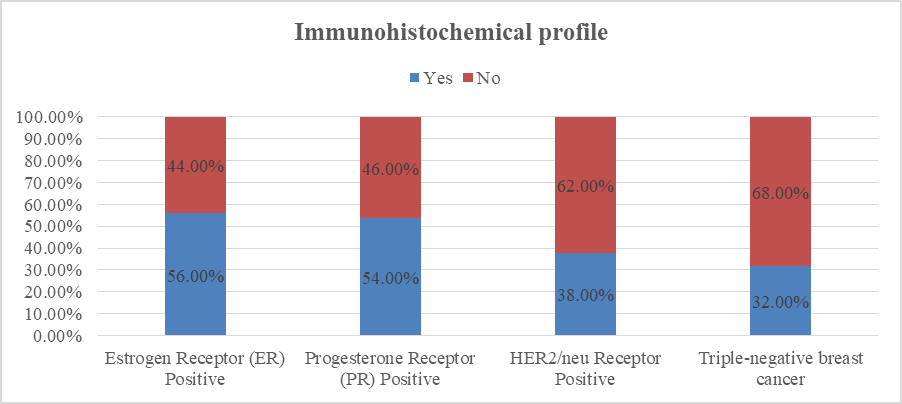
Figure 2: Immunohistochemical profile of carcinoma breast among the study participants (N = 50)
ER positivity varied significantly across the age groups (p = 0.040). It was observed in 40.0% of participants aged 30–40 years and 38.1% aged 41–50 years. A higher proportion of ER-positive tumours was seen among those aged 51–60 years and 61–70 years (83.3% each). The single participant aged 71–80 years was ER positive. (Table 2)
Table 2: Association between age group and estrogen receptor status among the study participants (N = 50)
|
Age group |
ER positive, n (%) |
ER negative, n (%) |
p-value# |
|
30 to 40 years |
4 (40.0%) |
6 (60.0%) |
0.040* |
|
41 to 50 years |
8 (38.1%) |
13 (61.9%) |
|
|
51 to 60 years |
10 (83.3%) |
2 (16.7%) |
|
|
61 to 70 years |
5 (83.3%) |
1 (16.7%) |
|
|
71 to 80 years |
1 (100.0%) |
0 (0.0%) |
# Fisher’s exact test; * Statistically significant at <0.05
PR positivity varied across age groups, with statistical significance (p = 0.050). PR positivity was observed in 50.0% of participants aged 30–40 years and 33.3% aged 41–50 years. It was highest among those aged 51–60 years (83.3%). The only participant aged 71–80 years was PR positive. (Table 3)
Table 3: Association between age group and progesterone receptor status among the study participants (N = 50)
|
Age group |
PR positive, n (%) |
PR negative, n (%) |
p-value# |
|
30 to 40 years |
5 (50.0%) |
5 (50.0%) |
0.050* |
|
41 to 50 years |
7 (33.3%) |
14 (66.7%) |
|
|
51 to 60 years |
10 (83.3%) |
2 (16.7%) |
|
|
61 to 70 years |
4 (66.7%) |
2 (33.3%) |
|
|
71 to 80 years |
1 (100.0%) |
0 (0.0%) |
# Fisher’s exact test; * Statistically significant at <0.05
HER2/neu positivity did not differ significantly across the age groups (p = 0.754). HER2/neu positivity was observed in 50.0% of participants aged 30–40 years, 42.9% aged 41–50 years, 25.0% aged 51–60 years, and 33.3% aged 61–70 years, while none of the participants aged 71–80 years were HER2/neu positive. (Table 4)
Table 4: Association between age group and HER2/neu receptor status among the study participants (N = 50)
|
Age group |
HER2/neu positive, n (%) |
HER2/neu negative, n (%) |
p-value# |
|
30 to 40 years |
5 (50.0%) |
5 (50.0%) |
0.754 |
|
41 to 50 years |
9 (42.9%) |
12 (57.1%) |
|
|
51 to 60 years |
3 (25.0%) |
9 (75.0%) |
|
|
61 to 70 years |
2 (33.3%) |
4 (66.7%) |
|
|
71 to 80 years |
0 (0.0%) |
1 (100.0%) |
# Fisher’s exact test
Table 5: Association between age group and triple-negative breast cancer among the study participants (N = 50)
|
Age group |
Triple-negative, n (%) |
Non-triple-negative, n (%) |
p-value# |
|
30 to 40 years |
5 (50.0%) |
5 (50.0%) |
0.002* |
|
41 to 50 years |
11 (52.4%) |
10 (47.6%) |
|
|
51 to 60 years |
0 (0.0%) |
12 (100.0%) |
|
|
61 to 70 years |
0 (0.0%) |
6 (100.0%) |
|
|
71 to 80 years |
0 (0.0%) |
1 (100.0%) |
# Fisher–Freeman–Halton exact test; * Statistically significant at <0.05
Triple-negative breast cancer showed a statistically significant association with age (p = 0.002). It was present in 50.0% of participants aged 30–40 years and 52.4% of those aged 41–50 years. No triple-negative cases were observed among participants older than 50 years. These findings indicate that triple-negative breast cancer was concentrated predominantly among younger participants. (Table 5)
DISCUSSION:
Given the limited regional evidence on histopathological patterns and receptor profiles of breast carcinoma, this hospital-based cross-sectional study was conducted in the Department of General Surgery of a tertiary care teaching hospital after ethics approval and written informed consent. Fifty consecutive women with FNAC-confirmed breast carcinoma were enrolled using convenience sampling. Demographic, clinical, tumour, nodal, and staging details were recorded using a structured proforma. Following definitive surgery, resected specimens underwent histopathological classification and immunohistochemical assessment of ER, PR, and HER2/neu. Triple-negative breast cancer was identified by the absence of all three receptors.
Breast carcinoma most commonly affected women aged 41–50 years (42.0%), followed by 51–60 years (24.0%), 30–40 years (20.0%), 61–70 years (12.0%), and 71–80 years (2.0%), with an age range of 20–80 years. Yadav P et al.17 similarly reported a 41–50-year peak (38.0%; mean age 45.58 years), while Upadhyay AK et al.18 and Jagadeesh N et al.19 reported median ages of 49 and 48 years, respectively. In contrast, Sagar S et al.20 observed a 51–60-year peak (53.0%; mean 52.2 years), and Wemimo RM et al.21 reported a mean age of 52.1±12.1 years. Overall, Indian women presented earlier than Western women.
A palpable breast lump was the predominant presenting symptom in the present study (78.0%), followed by pain (10.0%), nipple discharge (6.0%), and skin changes (6.0%), consistent with Yadav P et al.17 (breast lump 98.0%, pain 22.0%, nipple discharge 10.0%, ulceration 6.0%), Upadhyay AK et al.18 (74.0%, 12.0%, 3.0%, and 6.0%, respectively), and Wemimo RM et al.21 (breast lump 92.9%, breast deformity 69.0%, ulceration 18.6%). Left-sided disease predominated (60.0%), whereas Yadav P et al.17 reported right-sided predominance (66.0%), Jagadeesh N et al.19 observed nearly equal right (50.0%) and left (48.0%) involvement with 2.0% bilateral disease, and Upadhyay AK et al.18 reported 1.4% bilateral cases.
The upper outer quadrant was most frequently involved (78.0%), similar to Yadav P et al.17 (71.43%), Jagadeesh N et al.19 (70.0%), and Sagar S et al.20 (67.0%). Most tumours measured 2–5 cm (92.0%), compared with 68.0% reported by Sagar S et al.20 and 71.43% by Yadav P et al.17, whereas Wemimo RM et al.21 found >5 cm tumours in 75.9%. Clinically, N1 nodal disease was present in 78.0%, compared with nodal positivity of 62.0% in Yadav P et al.17, 57.8% in Wemimo RM et al.21, 66.0% pathological N1 disease in Jagadeesh N et al.19, and 85.0% pathological nodal positivity in Sagar S et al.20 Stage IIB was the commonest presentation (62.0%), similar to Sagar S et al.20 (41.0% Stage IIB) and Yadav P et al.17 (66.0% Stage II), whereas Upadhyay AK et al.18 (48.08% Stage III) and Jagadeesh N et al.19 (52.0% Stage III) reported more advanced disease, highlighting persistent delays in diagnosis despite increasing detection of Stage II disease in tertiary care settings.
Invasive ductal carcinoma–No Special Type was the predominant histopathological subtype, accounting for 72.0% of cases, followed by invasive lobular carcinoma in 16.0%, medullary carcinoma in 4.0%, mucinous carcinoma in 4.0%, neuroendocrine carcinoma in 2.0%, and papillary carcinoma in 2.0%. IDC predominance was also reported by Wemimo RM et al.21 (94.7%), Jagadeesh N et al.19 (92.0%), Upadhyay AK et al.18 (91.96%), Yadav P et al.17 (88.0%), and Sagar S et al.20 (81.0%). However, the 16.0% prevalence of invasive lobular carcinoma in the present study was distinctly higher than the 5.0%, 6.0%, and 1.71% reported by Sagar S et al.20, Yadav P et al.17, and Upadhyay AK et al.18, respectively, indicating possible regional histopathological variation.
ER positivity was observed in 56.0% of patients, comparable to Sagar S et al.20 (54.0%), Yadav P et al.17 (54.0%), and Upadhyay AK et al.18 (ER/PR positivity 56.2%), while Wemimo RM et al.21 reported lower ER positivity (36.3%). PR positivity was 54.0%, similar to Sagar S et al.20 (45.0%) and Yadav P et al.17 (44.0%), but higher than Wemimo RM et al.21 (28.3%). HER2/neu positivity was 38.0%, comparable with Wemimo RM et al.21 (41.6%), Yadav P et al.17 (32.0%), Sagar S et al.20 (32.0%), and Upadhyay AK et al.18 (30.7%), whereas Jagadeesh N et al.19 reported only 8.0%. Triple-negative breast cancer (TNBC) was identified in 32.0% of cases, compared with 26.93% by Upadhyay AK et al.18, 22.0% by Jagadeesh N et al.19, and 42.5% by Wemimo RM et al.21, confirming TNBC as a substantial aggressive subtype in developing populations.
Age was significantly associated with ER (p=0.040) and PR status (p=0.050). ER positivity increased from 40.0% at 30–40 years and 38.1% at 41–50 years to 83.3% in both the 51–60- and 61–70-year groups. PR positivity was 50.0%, 33.3%, and 83.3% in the 30–40-, 41–50-, and 51–60-year groups, respectively. Yadav P et al.17 reported similar age-related increases in ER and PR positivity (p<0.0001), while Sagar S et al.20 found greater receptor expression among older women (p<0.001); however, Saleh et al. reported no significant age-related variation in PR. HER2/neu showed no significant association with age (p=0.754), with positivity ranging from 25.0% to 50.0%, contrary to Yadav P et al.17 (p<0.0001) and Sagar S et al.20 (p<0.001), who linked HER2 positivity with younger age. TNBC was significantly associated with age (p=0.002), affecting 50.0% of women aged 30–40 years and 52.4% aged 41–50 years, but none above 50 years, consistent with Jagadeesh N et al.19, Sagar S et al.20, and Wemimo RM et al.21
Strengths and limitations
The major strengths of this study include its comprehensive evaluation of both clinicopathological characteristics and immunohistochemical receptor profile in histopathologically confirmed breast carcinoma, standardized assessment of ER, PR, and HER2/neu status, and analysis of age-wise receptor distribution, which directly addressed all study objectives. The study also provides valuable baseline data from a tertiary care centre. However, the findings should be interpreted considering certain limitations, including the relatively small sample size (n=50), single-centre cross-sectional design, inclusion of only female patients with palpable breast lumps, lack of long-term survival or treatment outcome assessment, and limited generalizability to the wider population.
CONCLUSION:
The study demonstrated that breast carcinoma most commonly presented in women aged 41–50 years, predominantly as Stage IIB disease, with invasive ductal carcinoma–No Special Type being the principal histopathological subtype. ER, PR, and HER2/neu positivity were observed in 56%, 54%, and 38% of patients, respectively, while triple-negative breast cancer accounted for 32%. ER and PR expression increased significantly with age, whereas HER2/neu status showed no age-related association. Triple-negative breast cancer was significantly concentrated among women aged 30–50 years, thereby fulfilling the stated clinicopathological and immunohistochemical objectives.
REFERENCES:
1. Filho AM, Laversanne M, Ferlay J, Colombet M, Piñeros M, Znaor A, et al. The GLOBOCAN 2022 cancer estimates: data sources, methods, and a snapshot of the cancer burden worldwide. International journal of cancer. 2025 Apr 1;156(7):1336-46.
2. Bray F, Laversanne M, Sung H, Ferlay J, Siegel RL, Soerjomataram I. Global cancer statistics 2022: GLOBOCAN estimates of incidence and mortality worldwide for 36 cancers in 185 countries. CA Cancer J Clin. 2024;74(3):229–63.
3. Asthana S, Chauhan S, Labani S. Breast and cervical cancer risk in India: an update. Indian J Public Health. 2014;58(1):5–10.
4. Malvia S, Bagadi SA, Dubey US, Saxena S. Epidemiology of breast cancer in Indian women. Asia Pac J Clin Oncol. 2017;13(4):289–95.
5. Kumar V, Abbas AK, Aster JC. Robbins and Cotran pathologic basis of disease. 11th ed. Philadelphia: Elsevier; 2023.
6. Waks AG, Winer EP. Breast cancer treatment: a review. JAMA. 2019;321(3):288–300.
7. Gradishar WJ, Moran MS, Abraham J, Abramson V, Aft R, Agnese D, et al. NCCN Clinical Practice Guidelines in Oncology: breast cancer. Version 5.2025. Plymouth Meeting (PA): National Comprehensive Cancer Network; 2025.
8. WHO Classification of Tumours Editorial Board. Breast tumours. 5th ed. Lyon: International Agency for Research on Cancer; 2019. (WHO classification of tumours series; vol. 2).
9. Allison KH, Hammond MEH, Dowsett M, McKernin SE, Carey LA, Fitzgibbons PL, et al. Estrogen and progesterone receptor testing in breast cancer: ASCO/CAP guideline update. J Clin Oncol. 2020;38(12):1346–66.
10. Ellis IO, Schnitt SJ, Sastre-Garau X, Bussolati G, Tavassoli FA, Eusebi V, et al. Invasive breast carcinoma. In: WHO Classification of Tumours Editorial Board. Breast tumours. 5th ed. Lyon: International Agency for Research on Cancer; 2019.
11. Wolff AC, Hammond MEH, Allison KH, Harvey BE, Mangu PB, Bartlett JMS, et al. Human epidermal growth factor receptor 2 testing in breast cancer: ASCO/CAP clinical practice guideline focused update. Arch Pathol Lab Med. 2018;142(11):1364–82.
12. Harbeck N, Penault-Llorca F, Cortes J, Gnant M, Houssami N, Poortmans P, et al. Breast cancer. Nat Rev Dis Primers. 2019;5(1):66.
13. Foulkes WD, Smith IE, Reis-Filho JS. Triple-negative breast cancer. N Engl J Med. 2010;363(20):1938–48.
14. Rakha EA, Reis-Filho JS, Ellis IO. Basal-like breast cancer: a critical review. J Clin Oncol. 2008;26(15):2568–81.
15. Ghosh J, Gupta S, Desai S, Shet T, Radhakrishnan S, Suryavanshi P, et al. Estrogen, progesterone and HER2 receptor expression in breast cancer in India: a retrospective analysis of 798 tumours. Breast Cancer Res Treat. 2011;127(3):695–702.
16. Goldhirsch A, Winer EP, Coates AS, Gelber RD, Piccart-Gebhart M, Thürlimann B, et al. Personalizing the treatment of women with early breast cancer: highlights of the St Gallen International Expert Consensus on the primary therapy of early breast cancer 2013. Ann Oncol. 2013;24(9):2206–23.
17. Yadav P, Rathore SS, Verma V, Yadav MK, Sangwan Y. Immunohistochemical profile of carcinoma breast and its association with clinicopathological features: a single institutional experience. Int Surg J. 2022;9(4):828–34.
18. Upadhyay AK, Prakash A. Clinicopathological profile of breast cancer at a tertiary cancer center in Jharkhand, India: a descriptive cohort study. Cureus. 2023;15(6).
19. Jagadeesh N, Babu BC, Raja CDK. Clinicopathological spectrum of breast carcinoma with emphasis on hormone receptor patterns: a two-year institutional study. Eur J Cardiovasc Med. 2025;15(4):375–83.
20. Sagar S, Maida ML, Singh D, Bhardwaj G. Immunohistochemical markers in carcinoma breast: evaluation and correlation with clinicopathological features. Int J Med Pharm Res. 2025:637–46.
21. Wemimo RM, Eziagu UB, Sulaiman OA, Abiodun AE, Idowu NA, Ayinde AA, et al. Immunohistochemical and clinicopathological characteristics of invasive breast carcinoma in Nigeria. Oman Med J. 2023;38(5).