Diagnostic Utility of High-Resolution Ultrasonography in Clinically Suspected Lateral Epicondylitis: A Prospective Observational Study.
- Alka Agrawal , Professor,Department Of Radiodiagnosis, M.G.M Medical College & M.Y Hospital, Indore .
- Saurabh Atram , Professor,Department Of Radiodiagnosis, M.G.M Medical College & M.Y Hospital, Indore.
- Priyal Chouhan , Assistant Professor,Department Of Radiodiagnosis, M.G.M Medical College & M.Y Hospital, Indore.
- Sarvesh Shyamkumar Arya , Junior Resident,Department Of Radiodiagnosis, M.G.M Medical College & M.Y Hospital, Indore.
Article Information:
Abstract:
Background: Lateral epicondylitis is a common cause of lateral elbow pain, and its diagnosis is primarily based on clinical examination. High-resolution ultrasonography (HRUS) provides objective assessment of tendon morphology and vascularity and may improve diagnostic accuracy. This study evaluated the diagnostic utility of HRUS in clinically suspected lateral epicondylitis.Materials and Methods: This prospective observational study included 70 consecutive patients with clinically suspected lateral epicondylitis. Both symptomatic and contralateral asymptomatic elbows (140 elbows) were examined using gray-scale, colour Doppler, and power Doppler ultrasonography. Tendon thickness, echogenicity, fibrillar pattern, tears, calcifications, cortical irregularity, peritendinous fluid, ligament abnormalities, and neovascularization were assessed. Diagnostic performance was evaluated using sensitivity, specificity, positive predictive value (PPV), negative predictive value (NPV), and overall accuracy.Results: Gray-scale ultrasonography demonstrated significantly higher frequencies of tendon thickening, abnormal echogenicity, loss of fibrillar pattern, tendon tears, calcifications, cortical irregularity, and peritendinous fluid in symptomatic elbows than in contralateral controls (all p<0.001, except tendon tears, p=0.002, and lateral collateral ligament tears, p=0.004). Neovascularization was detected in 51.4% of symptomatic elbows (p<0.001). Combined gray-scale and colour Doppler ultrasonography showed the highest diagnostic performance with a sensitivity of 80.43%, specificity of 75.00%, PPV of 86.05%, NPV of 66.67%, and overall accuracy of 78.57%.Conclusion: High-resolution ultrasonography is a reliable, non-invasive imaging modality for evaluating clinically suspected lateral epicondylitis. Gray-scale ultrasonography effectively detects structural tendon abnormalities, while Doppler imaging provides complementary information on vascularity. Combined assessment improves diagnostic performance and supports accurate diagnosis, disease grading, and treatment planning.
Keywords:
Article :
INTRODUCTION:
Lateral epicondylitis, commonly known as tennis elbow, is one of the most frequent causes of lateral elbow pain in adults. It is characterized by pain and tenderness over the lateral epicondyle, which are typically aggravated by activities involving repetitive wrist extension, forearm supination, or forceful gripping. Although traditionally considered an inflammatory disorder, current evidence suggests that lateral epicondylitis is primarily a degenerative tendinopathy resulting from repetitive mechanical stress at the origin of the common extensor tendon, particularly the extensor carpi radialis brevis (ECRB).1,2
The condition affects approximately 1–3% of the general population, with the highest incidence reported between 35 and 50 years of age. Men and women are affected almost equally. Despite its association with racquet sports, most patients are non-athletes and develop the condition due to repetitive occupational or recreational activities involving the forearm and wrist. Although many cases resolve with conservative treatment, persistent symptoms may lead to chronic pain, reduced grip strength, and functional impairment.3,4
Diagnosis is primarily based on clinical history and physical examination. Patients usually present with localized pain over the lateral epicondyle that worsens during resisted wrist extension or gripping activities. Provocative clinical tests such as Cozen's test, Mill's test, and Maudsley's test are commonly used to reproduce symptoms and support the diagnosis. However, these tests have variable diagnostic accuracy and cannot reliably differentiate lateral epicondylitis from other causes of lateral elbow pain, including radial tunnel syndrome, ligament injuries, or intra-articular disorders.5,6 Imaging plays an important role when the clinical diagnosis is uncertain or when structural tendon abnormalities need to be assessed. Plain radiographs are mainly useful for excluding alternative bony pathologies but are frequently normal in patients with lateral epicondylitis.
Magnetic resonance imaging provides excellent soft-tissue evaluation but is relatively expensive and less readily available. In contrast, high-resolution ultrasonography is a cost-effective, readily accessible, and dynamic imaging modality that allows detailed assessment of tendon morphology. Gray-scale ultrasonography can demonstrate tendon thickening, loss of normal fibrillar pattern, hypoechogenicity, calcifications, cortical irregularity, and partial tendon tears, while colour and power Doppler imaging can detect increased vascularity associated with active disease.
Previous studies have reported moderate to high sensitivity and high specificity of ultrasonography for diagnosing lateral epicondylitis, supporting its value in routine clinical practice.7–9. Considering these advantages, high-resolution ultrasonography has emerged as an important adjunct to clinical examination by providing objective evaluation of tendon pathology and facilitating early diagnosis. Therefore, the present study was undertaken to evaluate the role of high-resolution ultrasonography in clinically suspected cases of lateral epicondylitis.
The study also compares ultrasonographic findings with clinical assessment and the contralateral asymptomatic elbow to determine its diagnostic utility in improving the evaluation and management of this common condition.
MATERIALS AND METHODS:
This prospective observational study was conducted in the Department of Radiodiagnosis, M.G.M. Medical College and M.Y. Hospital, Indore, Madhya Pradesh, India, after obtaining approval from the Institutional Scientific Review Board (ISRB) and the Institutional Ethics Committee. The study was carried out over a period of one year from the date of ethical clearance.
A total of 70 consecutive patients with clinically suspected lateral epicondylitis were enrolled in the study. Both elbows of each participant were evaluated, with the asymptomatic contralateral elbow serving as an internal control, resulting in the assessment of 140 elbows. Written informed consent was obtained from all participants before inclusion in the study. Patients aged 18 years or older presenting with pain or tenderness over the lateral aspect of the elbow along with at least one positive clinical provocation test (Cozen's test, Mill's test, or Maudsley's test) were included.
Patients with inflammatory or infective arthritis, neuromuscular disorders, traumatic injuries involving the elbow joint, or those younger than 18 years were excluded. All ultrasonographic examinations were performed using a high-resolution ultrasound system equipped with a high-frequency linear transducer. Patients were examined in the sitting position with the elbow flexed to approximately 90° and the forearm comfortably supported on the examination table.
The common extensor tendon origin at the lateral humeral epicondyle was systematically evaluated in both longitudinal and transverse planes. Imaging parameters, including depth, focus, and gain, were optimized to obtain clear visualization of the superficial musculoskeletal structures (Figure 1).
Gray-scale ultrasonography was used to assess the common extensor tendon for abnormalities including hypoechogenicity, loss of normal fibrillar architecture, tendon thickening, partial or complete tendon tears, intratendinous calcifications, peritendinous fluid collection, and cortical irregularity of the lateral humeral epicondyle. Colour Doppler and power Doppler examinations were subsequently performed to evaluate intratendinous or peritendinous vascularity.
Doppler settings were optimized for the detection of low-velocity blood flow by appropriately adjusting the pulse repetition frequency and gain to minimize background noise and motion artefacts. The ultrasonographic findings of the symptomatic elbow were compared with those of the contralateral asymptomatic elbow in each patient. Clinical findings, including the results of provocative tests, were also recorded to evaluate the diagnostic performance of ultrasonography in clinically suspected lateral epicondylitis.
The collected data were entered into Microsoft Excel and analyzed using the Statistical Package for the Social Sciences (SPSS) software (IBM SPSS Statistics, version 23.0; IBM Corp., Armonk, NY, USA). Continuous variables were expressed as mean ± standard deviation (SD), whereas categorical variables were presented as frequencies and percentages. Comparisons between symptomatic and contralateral asymptomatic elbows were performed using appropriate statistical tests based on the nature of the data.
Diagnostic performance was evaluated by calculating sensitivity, specificity, positive predictive value (PPV), negative predictive value (NPV), and overall diagnostic accuracy. Agreement between gray-scale ultrasonography and Doppler findings was assessed using Cohen's kappa (κ) statistic. A p-value of <0.05 was considered statistically significant.
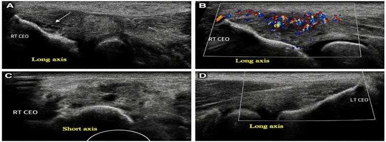
Figure 1. Ultrasonographic findings in right-sided severe lateral epicondylitis. (A) Long-axis view showing thickening of the right common extensor tendon (CET), measuring 6.8 mm, with diffuse hypoechogenicity and loss of the normal fibrillar pattern. An irregular hypoechoic area involving the middle and deep fibres at the entheseal attachment suggests an intrasubstance tear. Cortical irregularity of the underlying lateral epicondyle is also present. (B) Colour Doppler image demonstrating increased vascularity within the right CET, consistent with neovascularization. (C) Short-axis view of the right CET showing focal, rounded hypoechoic areas suggestive of micro-intrasubstance tears. (D) Long-axis view of the contralateral left CET showing normal morphology. Overall findings are consistent with right-sided severe lateral epicondylitis.
RESULTS:
A total of 70 patients with clinically suspected lateral epicondylitis were included in the study. The highest proportion of patients belonged to the 26–35 years and 46–55 years age groups (27.1% each), followed by the 36–45 years age group (25.7%). Females constituted 55.7% of the study population. Domestic workers represented the largest occupational category (42.9%), followed by manual and technical workers (28.6%). Most participants were right-hand dominant (78.6%), and the right elbow was the predominantly affected side (78.6%). More than half of the patients presented during the subacute phase (51.4%), while 42.9% had chronic symptoms. Tenderness was present in all patients (100%), whereas provocative clinical tests were positive in 65.7% and grip weakness was observed in 30.0% of cases (Table 1).
Table 1. Demographic and Clinical Characteristics of the Study Population (N = 70)
|
Characteristic |
Category |
n (%) |
|
Age group (years) |
18–25 |
6 (8.6) |
|
26–35 |
19 (27.1) |
|
|
36–45 |
18 (25.7) |
|
|
46–55 |
19 (27.1) |
|
|
56–65 |
8 (11.4) |
|
|
Sex |
Female |
39 (55.7) |
|
Male |
31 (44.3) |
|
|
Occupational category |
Domestic work |
30 (42.9) |
|
Manual/technical work |
20 (28.6) |
|
|
Sports/fitness activities |
9 (12.9) |
|
|
Student/desk-based work |
9 (12.9) |
|
|
Other occupations |
2 (2.9) |
|
|
Dominant hand |
Right |
55 (78.6) |
|
Left |
15 (21.4) |
|
|
Duration of symptoms |
Acute (<1 month) |
4 (5.7) |
|
Subacute (1–6 months) |
36 (51.4) |
|
|
Chronic (>6 months) |
30 (42.9) |
|
|
Clinically affected side |
Right |
55 (78.6) |
|
Left |
15 (21.4) |
|
|
Clinical findings |
Tenderness present |
70 (100.0) |
|
Positive provocative test |
46 (65.7) |
|
|
Grip weakness |
21 (30.0) |
Gray-scale ultrasonography demonstrated a wide spectrum of tendon abnormalities in symptomatic elbows, whereas all contralateral asymptomatic elbows showed normal findings. Mild tendon thickening was the most frequent abnormality (37.1%), with an overall mean tendon thickness of 4.20 ± 0.66 mm in symptomatic elbows compared with 3.54 ± 0.36 mm in controls.
Abnormal echogenicity was identified in 67.1% of cases, with focal hypoechoic areas being the most common pattern (28.6%). Deep fibre involvement was the predominant site of pathology (54.3%), followed by mid-fibre (24.3%) and superficial fibre involvement (21.4%).
Loss of the normal fibrillar pattern was present in 58.6% of symptomatic tendons, while intratendinous calcifications, cortical irregularity or bony spur formation, peritendinous fluid, and lateral collateral ligament tears were observed in 31.4%, 37.1%, 22.9%, and 11.4% of cases, respectively. No radial or posterior interosseous nerve involvement was detected (Table 2).
Table 2. Gray-Scale Ultrasonographic Findings in Symptomatic and Contralateral Asymptomatic Elbows
|
Ultrasonographic Parameter |
Category |
Cases n (%) |
Controls n (%) |
|
Common extensor tendon thickness |
Normal (<4.2 mm) |
29 (41.4) |
70 (100) |
|
Mild (4.2–5.0 mm) |
26 (37.1) |
0 |
|
|
Moderate (5.1–6.0 mm) |
12 (17.1) |
0 |
|
|
Severe (>6.0 mm) |
3 (4.3) |
0 |
|
|
Mean tendon thickness (mm) |
Mean ± SD |
4.20 ± 0.66 |
3.54 ± 0.36 |
|
Echogenicity |
Normal |
23 (32.9) |
70 (100) |
|
Diffuse hypoechogenicity |
18 (25.7) |
0 |
|
|
Focal hypoechoic areas |
20 (28.6) |
0 |
|
|
Linear hypoechoic areas |
9 (12.9) |
0 |
|
|
Predominant fibre involvement |
Deep fibres |
38 (54.3) |
0 |
|
Mid fibres |
17 (24.3) |
0 |
|
|
Superficial fibres |
15 (21.4) |
0 |
|
|
Loss of fibrillar pattern |
Present |
41 (58.6) |
0 |
|
Absent |
29 (41.4) |
70 (100) |
|
|
Intratendinous calcification |
Present |
22 (31.4) |
0 |
|
Absent |
48 (68.6) |
70 (100) |
|
|
Bony changes |
None |
44 (62.9) |
70 (100) |
|
Cortical irregularity |
18 (25.7) |
0 |
|
|
Bony spur |
8 (11.4) |
0 |
|
|
Peritendinous fluid |
Present |
16 (22.9) |
0 |
|
Absent |
54 (77.1) |
70 (100) |
|
|
Lateral collateral ligament tear |
Present |
8 (11.4) |
0 |
|
Absent |
62 (88.6) |
70 (100) |
|
|
Radial/Posterior interosseous nerve involvement |
Present |
0 |
0 |
|
Absent |
70 (100) |
70 (100) |
Tendon tears were identified in 12.9% of symptomatic elbows. Partial-thickness tears were more common than complete-thickness tears, accounting for 10.0% of cases, including intrasubstance tears (5.7%) and articular surface tears (4.3%), whereas complete-thickness tears were observed in only 2.9% of patients (Table 3).
Table 3. Types of Common Extensor Tendon Tear on Gray-Scale Ultrasonography
|
Type of Tear |
Cases n (%) |
Controls n (%) |
|
No tear |
61 (87.1) |
70 (100) |
|
Partial-thickness tear |
7 (10.0) |
0 |
|
• Intrasubstance tear |
4 (5.7) |
0 |
|
• Articular surface tear |
3 (4.3) |
0 |
|
Complete-thickness tear |
2 (2.9) |
0 |
Comparison between symptomatic and contralateral asymptomatic elbows demonstrated significantly higher frequencies of all major gray-scale ultrasonographic abnormalities in symptomatic elbows. Thickened common extensor tendon, abnormal echogenicity, loss of fibrillar pattern, intratendinous calcifications, cortical irregularity or bony spur formation, and peritendinous fluid were all significantly associated with lateral epicondylitis (all p<0.001). Tendon tears (p=0.002) and lateral collateral ligament tears (p=0.004) were also significantly more frequent in symptomatic elbows, while nerve involvement was absent in both groups (Table 4).
Table 4. Comparison of Gray-Scale Ultrasonographic Findings Between Symptomatic and Contralateral Elbows
|
Gray-scale USG Finding |
Cases n (%) |
Controls n (%) |
χ² |
p-value |
|
Thickened common extensor tendon (>4.2 mm) |
41 (58.6) |
0 |
60.2 |
<0.001 |
|
Abnormal echogenicity |
47 (67.1) |
0 |
73.6 |
<0.001 |
|
Loss of fibrillar pattern |
41 (58.6) |
0 |
60.2 |
<0.001 |
|
Tendon tear |
9 (12.9) |
0 |
9.7 |
0.002 |
|
Intratendinous calcification |
22 (31.4) |
0 |
26.1 |
<0.001 |
|
Cortical irregularity/Bony spur |
26 (37.1) |
0 |
32.8 |
<0.001 |
|
Peritendinous fluid |
16 (22.9) |
0 |
18.2 |
<0.001 |
|
Lateral collateral ligament tear |
8 (11.4) |
0 |
8.5 |
0.004 |
|
Nerve involvement |
0 |
0 |
— |
— |
|
Chi-square test; p <0.05 considered statistically significant. |
||||
Colour Doppler ultrasonography demonstrated neovascularization in 51.4% of symptomatic elbows, whereas none of the contralateral asymptomatic elbows showed Doppler vascularity, indicating a significant association with lateral epicondylitis (p<0.001). Based on ultrasonographic grading, moderate disease was the most common category (35.7%), followed by mild (21.4%) and severe (4.3%) grades, while 38.6% of symptomatic elbows demonstrated normal ultrasonographic findings (Tables 5 and 6).
Table 5. Doppler Ultrasonographic Findings
|
Parameter |
Cases n (%) |
Controls n (%) |
p-value |
|
Neovascularization present |
36 (51.4) |
0 |
<0.001 |
|
Neovascularization absent |
34 (48.6) |
70 (100) |
|
|
Chi-square test; p <0.05 considered statistically significant. |
|||
Table 6. Ultrasonographic Grading of Lateral Epicondylitis
|
Grade |
Cases n (%) |
Controls n (%) |
|
Normal |
27 (38.6) |
70 (100) |
|
Mild |
15 (21.4) |
0 |
|
Moderate |
25 (35.7) |
0 |
|
Severe |
3 (4.3) |
0 |
Among the evaluated imaging modalities, combined gray-scale and colour Doppler ultrasonography demonstrated the highest diagnostic performance, with a sensitivity of 80.43%, specificity of 75.00%, positive predictive value of 86.05%, negative predictive value of 66.67%, and an overall diagnostic accuracy of 78.57%. Gray-scale ultrasonography alone showed better diagnostic performance than either colour or power Doppler individually. Agreement analysis demonstrated an observed agreement of 77.1% between gray-scale and Doppler ultrasonography, with a Cohen's κ value of 0.52, indicating moderate agreement between the two techniques (Tables 7–9).
Table 7. Diagnostic Performance of Ultrasonography
|
Parameter |
Gray-scale USG |
Colour Doppler |
Power Doppler |
Combined Gray-scale + Colour Doppler |
|
Sensitivity (%) |
69.57 |
54.35 |
60.87 |
80.43 |
|
Specificity (%) |
66.67 |
54.17 |
62.50 |
75.00 |
|
Positive predictive value (%) |
80.00 |
69.44 |
75.68 |
86.05 |
|
Negative predictive value (%) |
53.33 |
38.24 |
45.45 |
66.67 |
|
Overall accuracy (%) |
68.57 |
54.29 |
61.43 |
78.57 |
Table 8. Cross-tabulation Between Gray-Scale and Doppler Ultrasonography
|
Gray-scale USG |
Doppler Positive |
Doppler Negative |
Total |
|
Positive |
30 |
10 |
40 |
|
Negative |
6 |
24 |
30 |
|
Total |
36 |
34 |
70 |
Table 9. Agreement Statistics Between Gray-Scale and Doppler Ultrasonography
|
Parameter |
Value |
|
Observed agreement |
77.1% |
|
Cohen's κ |
0.52 |
|
Interpretation |
Moderate agreement |
DISCUSSION:
Lateral epicondylitis is a common degenerative disorder of the common extensor tendon caused by repetitive mechanical stress rather than an acute inflammatory process. High-resolution ultrasonography has emerged as an important imaging modality because it provides detailed assessment of tendon morphology and vascularity in a rapid, non-invasive, and cost-effective manner. In the present study, the contralateral asymptomatic elbow was used as an internal control, allowing comparison of ultrasonographic findings while minimizing individual anatomical variation.
The majority of patients in the present study belonged to the middle-aged population, with the highest frequencies observed in the 26–35 and 46–55 year age groups. A slight female predominance was also observed. These findings are comparable with those reported by Connell et al. and Shrestha et al., who demonstrated that lateral epicondylitis is most frequently encountered during the fourth and fifth decades of life.10,11 The predominance of domestic workers and manual laborers in our study further supports previous observations by Silverstein et al. and Palmer et al., emphasizing the association between repetitive upper-limb activities and tendon degeneration.12,13
Gray-scale ultrasonography demonstrated characteristic structural abnormalities in symptomatic elbows. Tendon thickening, abnormal echogenicity, loss of fibrillar architecture, calcifications, cortical irregularity, and peritendinous fluid were significantly more frequent in affected elbows than in contralateral controls. Among these findings, abnormal echogenicity and loss of fibrillar pattern were the most common abnormalities, suggesting that disruption of the normal tendon architecture is an early feature of tendinosis. Similar observations have been reported by Miller et al., Tsai et al., and Draghi et al., highlighting the value of these sonographic features in diagnosing lateral epicondylitis.14–16.
Tendon tears were identified in a relatively small proportion of patients, with partial-thickness tears occurring more frequently than complete tears. Deep fibre involvement was the predominant pattern, corresponding to the extensor carpi radialis brevis tendon, which is recognized as the principal site of pathology in lateral epicondylitis. These findings are consistent with the anatomical and imaging observations described by Ricci et al. and Clarke et al.17,18.
Colour Doppler examination demonstrated neovascularization in approximately half of the symptomatic elbows, whereas no vascularity was detected in controls. Although Doppler imaging provides useful information regarding tendon vascularity, its diagnostic performance was lower than that of gray-scale ultrasonography when used alone. However, combining gray-scale and Doppler evaluation produced the highest sensitivity, specificity, positive predictive value, and overall diagnostic accuracy. These findings agree with previous reports by Park et al. and Clarke and Ahmad, who emphasized that Doppler imaging should complement rather than replace gray-scale assessment.18,19.
Overall, the findings of the present study demonstrate that high-resolution ultrasonography is a reliable imaging modality for evaluating clinically suspected lateral epicondylitis. Gray-scale ultrasonography effectively identifies structural tendon abnormalities, while Doppler imaging provides additional information regarding disease activity. The combined use of both techniques improves diagnostic performance and may assist in disease grading, treatment planning, and follow-up. The use of the contralateral asymptomatic elbow as an internal control further strengthened the assessment by providing patient-specific comparison, thereby improving the detection of subtle pathological changes.
Limitations
The present study has several limitations. First, the diagnostic performance of ultrasonography was evaluated using clinical suspicion as the reference standard rather than MRI or histopathological confirmation, which may have introduced verification bias. Second, this was a single-centre study with a predominance of domestic workers and manual laborers, potentially limiting the generalizability of the findings. Third, ultrasonography is inherently operator-dependent despite the use of a standardized imaging protocol. In addition, MRI comparison was not performed, preventing direct evaluation of diagnostic performance. Finally, the absence of longitudinal follow-up precluded assessment of ultrasonographic changes following treatment or during disease progression.
CONCLUSION:
The present study demonstrates that high-resolution ultrasonography is a valuable imaging modality for the evaluation of clinically suspected lateral epicondylitis. Gray-scale ultrasonography effectively identifies structural tendon abnormalities, while the addition of Doppler imaging improves diagnostic performance by detecting neovascularization. Comparison with the contralateral asymptomatic elbow enhances diagnostic confidence by accounting for individual anatomical variation. Ultrasonography complements clinical examination by providing objective assessment of tendon pathology, facilitating accurate diagnosis, disease grading, and treatment planning. Owing to its accessibility, real-time capability, and non-invasive nature, high-resolution ultrasonography should be considered a primary imaging tool in the routine evaluation and management of lateral epicondylitis.
REFERENCES:
1. Cutts S, Gangoo S, Modi N, Pasapula C. Tennis elbow: A clinical review article. J Orthop. 2019;17:203-207.
2. Vaquero-Picado A, Barco R, Antuña SA. Lateral epicondylitis of the elbow. EFORT Open Rev. 2017;1(11):391-397.
3. Shiri R, Viikari-Juntura E. Lateral and medial epicondylitis: role of occupational factors. Best Pract Res Clin Rheumatol. 2011;25(1):43-57.
4. Smidt N, van der Windt DA. Tennis elbow in primary care. BMJ. 2006;333(7575):927-928.
5. Kheiran A, Pandey A, Pandey R. Common tendinopathies around the elbow; what does current evidence say?. J Clin Orthop Trauma. 2021;19:216-223.
6. Ma KL, Wang HQ. Management of Lateral Epicondylitis: A Narrative Literature Review. Pain Res Manag. 2020;2020:6965381.
7. Weinreb JH, Sheth C, Apostolakos J, et al. Tendon structure, disease, and imaging. Muscles Ligaments Tendons J. 2014;4(1):66-73.
8. Hodgson RJ, O’Connor PJ, Grainger AJ. Tendon and ligament imaging. Br J Radiol. 2012;85(1016):1157-1172.
9. Bala E, Cetin O. Demonstrating the relationship of ultrasonographic parameters with disease activity and pain in lateral epicondylitis. Medicine (Baltimore). 2023;102(40):e35499.
10. Connell DA, Burke F, Coombes P, McNealy S, Freeman D, Pryde D. Sonographic examination of lateral epicondylitis. AJR Am J Roentgenol. 2001;176(3):777–82.
11. Shrestha D, Shrestha R, Sharma UK. Ultrasonographic evaluation of lateral epicondylitis. J Ultrason. 2021;21:e45–52.
12. Silverstein BA, Fine LJ, Armstrong TJ. Occupational risk factors. Am J Ind Med. 1987;11(3):343–58.
13. Palmer KT, Harris EC, Coggon D. Work-related epicondylitis. Occup Environ Med. 2007;64(2):133–9.
14. Miller TT. Imaging of elbow disorders. Radiol Clin North Am. 2002;40(2):241–54.
15. 15. 15. Tsai WC, Hsu CC, Chen CP. Ultrasonographic evaluation of tendinopathy. J Ultrasound Med. 2010;29(3):435–42.
16. Draghi F, Bortolotto C. Elbow ultrasound review. J Ultrasound. 2019;22(3):267–75.
17. Ricci V, Cocco G, Mezian K, et al. Anatomy and Sonographic Examination for Lateral Epicondylitis: EURO-MUSCULUS/USPRM Approach. Am J Phys Med Rehabil. 2023;102(4):300-307.
18. Clarke AW, Ahmad M. Imaging of elbow tendinopathy. Skeletal Radiol. 2018;47(3):313–25.
19. Park GY, Kwon DR, Lee SC. Doppler in tendinopathy. Ann Rehabil Med. 2013;37(6):823–30.