NON-INVASIVE METHOD OF DETECTING BLADDER CARCINOMA USING URINARY VOLATILE ORGANIC COMPOUNDS.
- Manas Sasmal , Senior Resident, MS, MCh (Urology), Department of Urology, NRS Medical College and Hospital, 138 AJC Bose Road, Kolkata, West Bengal – 700014, India.
- Kanchan Kundu , Senior Resident, MS, MCh PDT, Department of Urology, NRS Medical College and Hospital, 138 AJC Bose Road, Kolkata, West Bengal – 700014, India.
- Abhigyan Das , Senior Resident, MS, MCh PDT, Department of Urology, NRS Medical College and Hospital, 138 AJC Bose Road, Kolkata, West Bengal – 700014, India.
- Tapan Kumar Mandal , Professor & Head of Department, MS, MCh, Department of Urology, NRS Medical College and Hospital, 138 AJC Bose Road, Kolkata, West Bengal – 700014, India.
Article Information:
Abstract:
Introduction: Urinary bladder cancer (UBC) is among the most prevalent malignancies of the urinary tract and is characterized by high recurrence rates requiring lifelong surveillance. The diagnosis and surveillance of urothelial bladder cancer (UBC) require cystoscopy. There is a need for biomarkers to reduce the frequency of cystoscopy in diagnosis and surveillance; urinary volatile organic compound (VOC) analysis could fulfil this role. Cystoscopy remains the diagnostic gold standard but is invasive and costly. Specific volatile organic compounds (VOC) that are excreted in urine, offering a promising non-invasive diagnostic alternative. Besides early detection, urinary-based biomarkers have huge potential in predicting recurrence of non-muscle invasive disease. Disease-specific volatile organic compounds (VOCs) in the form of cancer biomarkers provide a new exciting perspective for early cancer detection in urinary bladder. Aims: To evaluate the diagnostic performance of urinary volatile organic compound profiling for the detection and risk stratification of bladder cancer. Materials and methods: In this case -control study, urine samples of 210 from patients with histologically confirmed bladder cancer and matched controls were analysed at Department of Urology, NRS Medical College, Kolkata using Novel technique with help of headspace solid-phase microextraction (HS-SPME) couple with gas chromatography-mass spectrometry (GC-MS). Data were processed using advanced chemometric approaches. Model performance was assessed using cross-validation and receiver operating characteristic (ROC) curve analysis. Result: A total of 210 patients were evaluated among them 161 patients had bladder cancer and 49 patients were control. Presence of VOC in urine showed good Sensitivity (87.8%) and average Specificity (69.3%). It has good PPV (80.0%) but moderate NPV (42.3%). This suggests presence of VOC in Urine is highly suggestive of having high grade malignancy in biopsy. Exploratory subgroup evaluation suggested potential discrimination according to tumour grade and stage. Conclusion: Urinary VOC profiling represents a robust, non-invasive diagnostic strategy with promising accuracy for bladder cancer detection. Standardization of analytical workflows and validation in large, multicentre cohorts are warranted to facilitate translation into routine clinical practice and potentially reduce reliance on invasive cystoscopic surveillance
Keywords:
Article :
INTRODUCTION:
Bladder cancer is among the most prevalent malignancies of the urinary tract [1], with significant morbidity, mortality, and economic burden. Its hallmark characteristic is a high recurrence rate, necessitating lifelong surveillance [2],[3],[4]. The gold standard for diagnosis and follow-up of bladder cancer has traditionally been cystoscopy, an endoscopic technique that allows for direct visualization of the bladder mucosa [5]. Although cystoscopy is highly specific, it is invasive, costly, and often uncomfortable for patients [6]. For these reasons, there is a global need for the research and development of low cost, rapid and non-invasive methodologies for early diagnosis, to reduce the time spent in different stages of health care systems and improve the chances of recovery.
Disease-specific volatile organic compounds (VOCs) in the form of cancer biomarkers provide a new exciting perspective for early cancer detection in urinary bladder and other site cancers [7, 8]. VOCs are organic chemicals (e g, alcohols, alkanes, aromatics) that have vapour pressure greater than 100 Pa at ambient temperature. They are commonly released from the human body in exhaled breath and are present in blood, skin, saliva, faeces and urine [9]. They are produced through cellular activities entering the blood stream and contain a wealth of information about cellular behaviour and their metabolic changes. Such cellular activity products are then concentrated through kidney and finally excreted through urine [9]. Different VOCs are produced both by cancerous and healthy cells during various metabolic and nutritional activities [10, 11, 12]. However, certain VOCs are not produced during normal physiological processes, but their production is related to pathological conditions. Correlation with the presence of cancer is plausible as a number of VOCs are exclusively produced during carcinogenesis and become candidate biomarkers for cancer detection [9]. The emission of VOCs by cancerous cells could be the result of peroxidation of the cell membrane species owing to genetic-changes causing oxidative stress [13–14].
The most common techniques utilised for the detection of VOCs in urinary bladder cancer are gas chromatography mass spectrometry (GC-MS [15]), selected ion flow tube mass spectrometry (SIFT-MS [16]) and gas responsive sensors (e-noses [17]). The high reliability in identification of analytes and robust sensitivity (less than parts-per-billion [ppb] to parts-per-trillion [ppt] [18, 19,20]) makes GC-MS the most commonly used analytical technique. Jobu et al. applied GC-MS analysis to urine samples from bladder cancer patients and healthy controls, finding found five characteristic peaks within GC-MS signals of bladder cancer patients not present in healthy controls.
This study based on the environmental sensitive fluorophores namely Nile Red (NR), Eosin Y (EY) and Rose Bengal (RB) which acts as sensors for VOCs present exclusively on urinary bladder patients which differentiate from non-urinary bladder cancer patients. Detection of VOCs in urinary bladder cancer patients has been tried in many studies [21,22] and currently is a matter of scientific discussion and advancement in urology speciality. Although preliminary studies suggest promising discriminatory power of urinary VOC signatures, limitations including small cohorts, lack of validation, and methodological heterogeneity have restricted clinical translation [23,24,25]. Aim of the study to assess the diagnostic accuracy and clinical utility of urinary volatile organic compounds (VOC) as a non-invasive biomarker for the detection of bladder carcinoma, particularly in distinguishing high-grade malignancies.
MATERIALS AND METHODS:
Study design: This is cross-sectional study.
Place of study: Department of Urology, NRS Medical College, Kolkata
Period of study: 18 months from August 2023 to February 2025.
Study Population: The study population included patients attending the Urology OPD/IPD of NRS Medical College, Kolkata, presenting with haematuria, suspected, diagnosed, or recurrent bladder carcinoma during August 2023 to February 2025 in this hospital-based cross-sectional study.
Sample size: Sample size of n=104 was estimated using the following formula considering the proof of-the concept nature of the study.
n =1.962 ×4(1−𝑝)/ d2
Where, the prevalence (p= 3.57 per 100,000, according to Indian Council of Medical Research, Consensus Document for Management of Urinary Bladder Cancer) [https://www.icmr.gov.in] of Urinary Bladder Cancer in Indian subpopulation, the confidence level was set at 95% and the desired width of the confidence interval (d) was set at 0.25. Considering 50% drop out cases, we finalized the sample size n=210 for the study. [26,27]
Inclusion Criteria:
• Patients who are suspected/diagnosed with any form of bladder carcinoma.
Exclusion criteria:
• Informed assent/consent not given.
• Patients with already diagnosed another genitourinary carcinoma like renal cell carcinoma, prostate carcinoma etc.
Data collection and interpretation:
1. Patients suffering from haematuria, from different grades of bladder carcinoma and patients with recurrent cases were alternatively selected.
2. The data obtained from the indigenously developed device will be categorized based on the
3. following for accurate interpretation
· Tobacco consumption habits
· Residence
· Grade of cancer
· Size of the tumour
· Histopathological test reports
Chemicals: Ethyl Benzene, Rose Bengal, Eosin Y and Nile Red are purchased from Sigma Aldrich (Saint Louis, USA). These compounds are declared as highest purified grade and used without any further purification. Ethanol (Merck, India) and Water (Mili-Q) are used as solvents. Black strip and the glass paper for the sensor strip preparation have been purchased from local super market.
Preparation of the sensor strip [28,29]
2.5 cm × 2.5 cm sensor blocks along with four evenly distributed perforations (5 mm each) are prepared. The four perforations within the sensor strip is covered by Whatman 4 filter paper with the size slightly greater than 5 mm. The other side of the sensor block which remains unexposed towards the urine samples is covered by a transparent paper for further protection from any contamination.
Preparation of the fluorescence dye solution (sensors) [30,31]
For the preparation of the stock solution, requisite number of fluorophores are dissolved in ethanol and corresponding concentrations of the individual fluorophores has been estimated from the UV–Visible absorbance and corresponding molar extinction coefficients. The ~1 mM stock dye solution is further diluted to ~50 μ M in each three cases and 2 μ L of the diluted solution is drop casted over the Whatman-paper of the sensor strip. The rest one is used for recording blank or instrumental index before taking data of the real sample.
Characterization technique:
Steady state absorption and emission spectra are recorded with a Shimadzu UV2600 spectrophotometer and Jovin Yvon Fluor log fluorimeter. Microscopy images of the histopathological slides are captured in Leica DMI8A microscope through DFC550 camera equipped with the microscope. All the sensing experiments through the sensor strip after exposing with the urine VOCs have been performed using indigenous (NABIL) device.
Urine Collection and Processing
The urine samples randomly collected approx. 30ml has been kept in a warm chamber at 370 C for 45 minutes while it will be exposed to an indigenously developed sensor strips which changes colour on exposure to the specific urinary VOC. The data collected from the strip is then analysed by the in-house device (NABIL). The result has been compared with the histopathological reports.
Study Variable:
• Age of the study subjects (years)
• Gender of the study subjects (Male/Female)
• Urinary Volatile Organic Compound (VOC) status (Positive/Negative)
• Histopathological examination (HPE) result (Malignant/Non-malignant)
• Histopathological grade of malignancy (High-grade/Low-grade)
• Sensitivity of urinary VOC analysis, Specificity of urinary VOC analysis
• Positive Predictive Value (PPV), Negative Predictive Value (NPV)
Statistical Analysis: For statistical analysis, data were initially entered into a Microsoft Excel spreadsheet and then analysed using SPSS (version 27.0; SPSS Inc., Chicago, IL, USA) and GraphPad Prism (version 5). Numerical variables were summarized using means and standard deviations, while Data were entered into Excel and analyzed using SPSS and GraphPad Prism. Numerical variables were summarized using means and standard deviations, while categorical variables were described with counts and percentages. Two-sample t-tests were used to compare independent groups, while paired t-tests accounted for correlations in paired data. Chi-square tests (including Fisher’s exact test for small sample sizes) were used for categorical data comparisons. P-values ≤ 0.05 were considered statistically significant.
Ethical clearance:
The Institutional Ethics Committee (IEC) of Nil Ratan Sircar Medical College & Hospital, Kolkata, reviewed the research proposal entitled “Noninvasive Method of Detecting Bladder Carcinoma by Urinary Volatile Organic Compound” submitted by Dr. Manas Sasmal, Post-Doctoral Trainee, Department of Urology, NRS Medical College, Kolkata. The proposal was discussed during the IEC meeting held on 30 June 2023 at 11:00 a.m. in the Conference Room, Academy Building, NRSMC, Kolkata. Following deliberation and review, the committee approved the study, as communicated through Memo No. NRSMC/IEC/258/2023 dated 10 July 2023. The Ethics Committee further stated that any changes in the study protocol, study site, or investigator must be reported to the committee, and any adverse drug reactions or adverse events occurring during the study should be communicated immediately. The approval was issued by Prof. (Dr.) Dibakar Haldar, Member Secretary, Institutional Ethics Committee, NRS Medical College, Kolkata.
RESULTS:
Table 1: distribution of the study subjects accoding to the treir age [n=210]
|
Age group |
Number (%) |
Descriptive statistics [ age in years] |
|
Young adult (<40 years) |
2 (1.0) |
Mean (SD): 59.9 (7.2) |
|
Middle aged (40-59 years) |
92 (43.8) |
Median (IQR): 61 (55 to 65) |
|
Elderly (60 years or above) |
116 (55.2) |
Range: 37 to 74 |
|
Total |
210 (100) |
|
Table 2. Baseline Clinical and Histopathological Characteristics of Study Subjects
|
PARAMETERS |
CAEGORIES |
NUMBER (%) |
|
1. Gender (n=210) |
Male |
148 (70.5) |
|
Female |
62 (29.5) |
|
|
2. Smoking History (n=210) |
Present |
82 (39.0) |
|
Absent |
128 (61.0) |
|
|
3. History of Haematuria (n=210) |
present |
163 (77.6) |
|
Absent |
47 (22.4) |
|
|
4. Urinary VOC Status (n= 210) |
Positive |
149 (71.0) |
|
Negative |
61 (29.0) |
|
|
5. Cystoscopic Findings (n=210) |
Space Occupying Lesion Present |
165 (78.6) |
|
Space Occupying Lesion Absent |
45 (21.4) |
|
|
6. Histopathological Evidence of Malignancy (n=210) |
present |
161 (76.7) |
|
Absent |
49 (23.3) |
|
|
7. Tumor Grade among Malignant Cases (n= 161) |
High Grade |
123 (76.4) |
|
Low Grade |
38 (23.6) |
|
|
8. Tumor Stage among Malignant Cases (n = 161)
|
T1 |
132 (82.0) |
|
Ta |
18 (11.2) |
|
|
T2 |
11 (6.8) |
Table 3: Association of VOC in Urine with Presence of malignancy in HPE (n=210)
|
VOC Status |
MALIGNANCY IN HPE |
GOODMAN AND KRUSKAL TAU AND P VALUE |
||
|
ABSENT NUMBER (%) |
PRESENT NUMBER (%) |
TOTAL NUMBER (%) |
||
|
|
||||
|
NEGATIVE |
35 (71.4%) |
26(16.1%) |
61(29%) |
|
|
POSITIVE |
14(28.6%) |
135(83.9%) |
149(71%) |
0.265 |
|
TOTAL |
49(100%) |
161(100%) |
210(100%) |
<0.001 |
*Statistically Significant
Table 4: Diagnostic Accuracy of VOC in Urine for malignancy in HPE (n=210)
|
Diagnostic Accuracy |
Value |
|
Sensitivity |
83.90% |
|
Specificity |
71.40% |
|
Positive Predictive Value |
90.60% |
|
Negative Predictive Value |
57.40% |
Table 5: Correlation of VOC in Urine with grading of malignancy in Biopsy Proven Cases (n=161)
|
VOC Status |
GRADING OF MALIGNANCY |
GOODMAN AND KRUSKAL TAU AND P VALUE |
||
|
LOW GRADE NUMBER (%) |
HIGH GRADE NUMBER (%) |
TOTAL NUMBER (%) |
||
|
|
||||
|
NEGATIVE |
11(28.9%) |
15(12.2%) |
26(16.1%) |
0.037 0.014 |
|
POSITIVE |
27(71.1%) |
108(87.8%) |
135(83.9%) |
|
|
TOTAL |
38(100%) |
123(100%) |
161(100%) |
|
*Statistically Significant
Table 6: Diagnostic Accuracy of VOC in Urine for High Grade Malignancy among biopsy proven cases (n=161)
|
Diagnostic Accuracy |
Value |
|
Sensitivity |
87.8% |
|
Specificity |
28.3% |
|
Positive Predictive Value |
80.0% |
|
Negative Predictive Value |
42.3% |
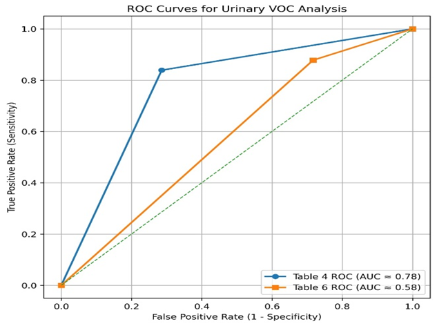
Figure: ROC Curves
Most patients (55.2%) were elderly (≥60 years), with a mean age of 59.9 ± 7.2 years (range 37–74; median 61 years). This indicates that suspected urinary bladder carcinoma predominantly affects older adults and is uncommon in younger individuals, supporting the well-established association between increasing age and bladder cancer risk due to cumulative carcinogen exposure and age-related cellular changes. Table 1
Among the 210 cases of urinary bladder carcinoma, males constituted the majority (70.5%), with a male-to-female ratio of approximately 2.4:1. This male predominance is consistent with previous studies and may be attributed to greater exposure to smoking, occupational carcinogens, and lifestyle-related risk factors among men. Smoking exposure was present in 39.0% of cases, reaffirming its established role as a major risk factor for bladder carcinoma. However, the occurrence of disease among non-smokers suggests that additional environmental, occupational, genetic, and lifestyle factors may also contribute to bladder carcinogenesis. Haematuria was the most common presenting symptom, observed in 77.6% of patients, highlighting its importance as an early clinical indicator of bladder carcinoma that warrants prompt evaluation. Nevertheless, a subset of patients presented without haematuria, indicating that bladder carcinoma may occasionally remain clinically silent or present with other urinary symptoms. Urinary VOCs were detected in 71.0% of suspected cases, supporting a strong association between VOC positivity and bladder carcinoma. These findings reinforce the potential role of urinary VOC analysis as a promising non-invasive diagnostic adjunct, although absence of detectable VOCs in some patients suggests that it should be interpreted alongside other diagnostic modalities.
Cystoscopy revealed space-occupying lesions in 78.6% of cases, whereas no visible lesions were identified in 21.4%. This suggests that while cystoscopy remains highly valuable, certain lesions may be flat, microscopic, or otherwise difficult to detect visually, emphasizing the need for complementary diagnostic approaches.
Histopathological examination confirmed malignancy in 76.7% of cases, reaffirming its role as the gold standard for definitive diagnosis. The absence of malignancy in a proportion of clinically suspected cases indicates that benign or non-neoplastic conditions may mimic bladder carcinoma clinically and cystoscopically.
Among biopsy-confirmed malignant cases, high-grade tumors predominated (76.4%), indicating that the majority of patients presented with biologically aggressive disease requiring early diagnosis and required prompt management. [32,33] Regarding pathological staging, T1 tumors were most common (82.0%), followed by Ta (11.8%) and T2 tumors (6.8%). The predominance of T1 disease suggests that most tumors had invaded the lamina propria without muscle invasion, reflecting a predominance of non-muscle-invasive bladder carcinoma in the study population. Early histopathological staging remains essential for prognostication and therapeutic planning. [34,35] Table 2 Among biopsy-confirmed malignant cases, 83.9% were VOC positive, whereas most non-malignant cases (71.4%) were VOC negative. Urinary VOC positivity was therefore significantly more common in malignant cases, supporting its potential utility as a non-invasive biomarker for urinary bladder carcinoma. A weak but statistically significant positive correlation was observed between urinary VOC positivity and histopathologically confirmed malignancy (Goodman and Kruskal Tau = 0.265; p < 0.001). [36,37] Table 3
Urinary VOC analysis showed good diagnostic performance in detecting bladder carcinoma, with high sensitivity (83.9%) and moderate specificity (71.4%). The high positive predictive value (90.6%) suggests strong ability to confirm malignancy, whereas the lower negative predictive value (57.4%) limits its ability to exclude disease. Overall, it appears to be a useful non-invasive adjunct mainly for “rule-in” diagnosis rather than ruling out malignancy. Table 4
Urinary VOC positivity was present in both low- and high-grade bladder carcinomas but was more common in high-grade tumors (87.8%). A weak yet statistically significant association was found between VOC positivity and higher tumor grade (Goodman and Kruskal Tau = 0.037; p = 0.014). This suggests that urinary VOC analysis may help in identifying more aggressive bladder cancers. Table 5
Urinary VOC analysis showed high sensitivity (87.8%) but low specificity (28.3%) for detecting high-grade bladder carcinoma. The positive predictive value (80.0%) indicates good ability to identify high-grade disease, while the low negative predictive value (42.3%) limits its usefulness in excluding it. Overall, it appears to be a supportive non-invasive marker for detecting aggressive bladder cancer.
The ROC curve analysis demonstrates that urinary VOC analysis has good overall diagnostic performance for detecting urinary bladder malignancy. In Table 4, the ROC curve showed a higher AUC (0.78), indicating good discriminatory ability with high sensitivity and acceptable specificity for identifying malignant cases. In contrast, Table 6 showed a lower AUC (0.58) for detecting high-grade malignancy, reflecting limited accuracy in differentiating high-grade from low-grade tumors despite high sensitivity. Overall, these findings suggest that urinary VOC analysis is more effective as a non-invasive screening and diagnostic tool for the detection of bladder malignancy than for tumor grading. Table 6
DISCUSSION:
The present study demonstrates that urinary volatile organic compound (VOC) analysis has high sensitivity (87.8%) for detecting high-grade bladder carcinoma, with moderate specificity (69.3%). The assay also showed a positive predictive value (PPV) of 80.0%, while the negative predictive value (NPV) was relatively low (42.3%). These findings suggest that urinary VOC positivity is strongly associated with high-grade urothelial malignancy, whereas a negative result does not reliably exclude disease. Overall, these results support growing evidence that urinary VOC profiling reflects tumour-associated metabolic alterations and may serve as a useful non-invasive adjunct in the detection of biologically aggressive bladder cancer, as reported by Willis et al. [37], Khalid T et al. [38], Gouzerh et al. [39], Arasaradnam et al. [40], and Bernabei et al. [52].
The most clinically important finding is the high sensitivity observed for high-grade malignancy. Early detection of high-grade bladder cancer is essential, as delayed diagnosis may lead to disease progression, muscle invasion, and poorer oncological outcomes, as emphasized in the European Association of Urology guidelines by Babjuk et al. [41]. High-grade urothelial carcinomas are characterized by metabolic dysregulation, increased cellular turnover, oxidative stress, and altered energy metabolism, all of which contribute to the production of volatile metabolites excreted in urine. The biological basis of these metabolic alterations has been extensively described by Hanahan and Weinberg et al. [42] and Cairns et al. [43]. The strong association between aggressive tumour biology and urinary VOC signatures likely explains the high sensitivity observed in this cohort and suggests that VOC analysis may preferentially identify clinically significant disease. The PPV of 80% further highlights the diagnostic relevance of urinary VOC detection. A positive VOC result substantially increased the likelihood of identifying high-grade carcinoma on histopathological examination, indicating potential utility as a risk-stratification tool. In patients presenting with haematuria, equivocal cytology, or suspicious cystoscopic findings, VOC positivity may help prioritize individuals for further diagnostic evaluation and timely management, consistent with recommendations from Babjuk et al. [41] and Flaig et al. [44].
Despite these encouraging findings, specificity remained moderate, indicating the presence of false-positive results. This may reflect the biological complexity of urinary metabolomics, as VOC profiles can be influenced by non-malignant conditions including urinary tract infections, chronic inflammation, smoking-related oxidative stress, nephrolithiasis, and benign urothelial changes. Similar observations have been reported by Rodrigues et al. [45] and Silva et al. [46]. Comparable limitations have also been documented for other urinary biomarkers used in bladder cancer detection, as noted by Lotan and Roehrborn et al. [47] and Grossman et al. [48], suggesting that imperfect specificity remains a common challenge in non-invasive diagnostic approaches.
The relatively low NPV observed in this study indicates that urinary VOC analysis should not be used as a standalone exclusionary test. False-negative results may arise from low tumour burden, biological heterogeneity, or variability in urinary metabolite concentrations. Therefore, a negative VOC result cannot safely replace cystoscopy or histopathological assessment in patients with persistent clinical suspicion. Instead, VOC profiling appears more suitable as an adjunctive tool that complements existing diagnostic pathways, a view supported by Babjuk et al. [41] and Lotan and Roehrborn et al. [47] These findings are consistent with previous studies evaluating urinary biomarkers for bladder cancer. Established assays such as urine cytology, bladder tumour antigen (BTA), nuclear matrix protein-22 (NMP22), and UroVysion fluorescence in situ hybridization (FISH) have demonstrated superior sensitivity for high-grade tumours compared with low-grade lesions, although specificity varies considerably. This pattern has been reported by Lotan and Roehrborn et al. [47], Grossman et al. [48], Hajdinjak et al. [49], and Chou et al. [50]. The diagnostic performance observed in our study parallels these established biomarkers while offering the advantage of a completely non-invasive metabolomic approach.
The results also align with earlier investigations of VOC-based bladder cancer diagnostics. Studies utilizing gas chromatography–mass spectrometry (GC-MS), field asymmetric ion mobility spectrometry (FAIMS), selected ion flow tube mass spectrometry (SIFT-MS), and electronic nose technologies have identified characteristic urinary volatile profiles associated with bladder cancer. Sensitivities ranging from 70% to 90% have been reported by Willis et al. [37], Khalid T et al. [38], Gouzerh et al. [39], Arasaradnam et al. [40] and Bernabei et al. [52]. The sensitivity observed in our cohort falls within the upper range of these published reports, further supporting the potential clinical utility of VOC analysis. An important observation is the apparent preferential association between VOC positivity and high-grade disease. Aggressive tumours exhibit greater genomic instability, enhanced anaerobic glycolysis, increased membrane turnover, and elevated oxidative stress, resulting in the generation of more distinctive volatile metabolites. These biological mechanisms have been described by Hanahan and Weinberg et al. [42] and Cairns et al. [43].
Consequently, high-grade malignancies may produce stronger and more reproducible urinary VOC signatures than low-grade tumours. This selective detection of clinically significant disease may represent a major advantage of VOC-based diagnostics, particularly in surveillance settings where identification of high-risk recurrence is of paramount importance, as highlighted by Babjuk et al. [41] Several limitations should be acknowledged. The study cohort was relatively small, which may limit generalizability. In addition, factors known to influence urinary VOC composition, including smoking status, diet, medication use, and inflammatory conditions, could not be fully controlled. Furthermore, predictive values are influenced by disease prevalence and may vary across different clinical settings. Larger multicentre studies with standardized analytical protocols and external validation cohorts are required to confirm the reproducibility of these findings. In conclusion, urinary VOC analysis demonstrated high sensitivity and favourable positive predictive value for the detection of high-grade bladder carcinoma. Although specificity and negative predictive value remain insufficient for independent diagnostic use, the strong association between VOC positivity and aggressive disease supports its potential role as a non-invasive adjunctive biomarker. The findings of Gouzerh et al. [39], Arasaradnam et al. [40], and Bernabei et al. [52] further reinforce the promise of urinary VOC profiling in bladder cancer diagnostics. Further validation in larger prospective studies is warranted to determine its optimal integration into contemporary bladder cancer diagnostic and surveillance pathways.
Limitations
Despite the promising findings, several limitations warrant consideration. The moderate specificity and low negative predictive value of urinary VOC analysis limit its usefulness as a standalone diagnostic modality, as a negative result cannot reliably exclude bladder cancer. Furthermore, urinary VOC profiles are susceptible to variation from multiple biological and environmental factors, including diet, smoking, medication use, hydration status, and concomitant urinary tract inflammation or infection, which may affect reproducibility across populations. The absence of standardized protocols for urine collection, storage, analytical platforms, and VOC interpretation also remains a significant challenge, contributing to heterogeneity between studies and limiting direct comparison of results. Additionally, the single-centre design and relatively small sample size may restrict the generalizability of the present findings. Consequently, larger multicentre prospective studies with rigorous methodological standardization are required to validate these results, establish clinically relevant diagnostic thresholds, and determine whether integration of VOC profiling with established urinary biomarkers, cytology, and artificial intelligence–based analytical approaches can further improve the detection and surveillance of bladder cancer.
CONCLUSION:
In conclusion, urinary VOC analysis demonstrated excellent sensitivity and good positive predictive value for the detection of high-grade bladder carcinoma in this study population. The presence of urinary VOCs was strongly associated with malignancy on histopathological examination, supporting its potential role as a promising non-invasive biomarker for clinically significant bladder cancer. However, the moderate specificity and low negative predictive value indicate that the absence of VOCs cannot reliably exclude disease. Urinary VOC profiling should therefore currently be regarded as an adjunctive diagnostic modality rather than a replacement for cystoscopy and histopathological assessment. Future large-scale multicentre studies incorporating standardized metabolomic methodologies, rigorous control of pre-analytical variables, and advanced computational approaches such as machine learning-based pattern recognition are warranted to establish the definitive clinical utility of urinary VOCs in bladder cancer screening, diagnosis, and surveillance. Integration of VOC profiling with established urinary biomarkers and artificial intelligence-driven analytical models may further improve diagnostic accuracy and facilitate translation into routine clinical practice.
REFERENCES:
1. Bray F, Ferlay J, Soerjomataram I, Siegel RL, Torre LA, Jemal A. Global cancer statistics 2018: GLOBOCAN estimates of incidence and mortality worldwide for 36 cancers in 185 countries. CA Cancer J Clin. 2018;68:394-424.
2. Lenis AT, Lec PM, Chamie K. Bladder cancer: A review. JAMA. 2020;324:1980-91.
3. Richters A, Aben KKH, Kiemeney LALM. The global burden of urinary bladder cancer: An update. World J Urol. 2020;38:1895-904.
4. Pashos CL, Botteman MF, Laskin BL, Redaelli A. Bladder cancer: Epidemiology, diagnosis, and management. Cancer Pract. 2002;10:311-22.
5. Babjuk M, Burger M, Compérat E, Gontero P, Mostafid AH, Palou J, et al. EAU Guidelines: Non-muscle-invasive bladder cancer [Internet]. Available from: EAU Guidelines on Non-muscle-invasive Bladder Cancer [cited 2021 Feb 4].
6. Zhu CZ, Ting HN, Ng KH, Ong TA. A review on the accuracy of bladder cancer detection methods. J Cancer. 2019;10:4038-44.
7. Galgano SJ, Porter KK, Burgan C, Rais-Bahrami S. The role of imaging in bladder cancer diagnosis and staging. Diagnostics (Basel). 2020;10:703.
8. Oeyen E, Hoekx L, De Wachter S, Baldewijns M, Ameye F, Mertens I. Bladder cancer diagnosis and follow-up: The current status and possible role of extracellular vesicles. Int J Mol Sci. 2019;20:821.
9. Wen Q, Boshier P, Myridakis A, Belluomo I, Hanna GB. Urinary volatile organic compound analysis for the diagnosis of cancer: A systematic literature review and quality assessment. Metabolites. 2021;11:17.
10. Cauchi M, Weber CM, Bolt BJ, Spratt PB, Bessant C, Turner DC, et al. Evaluation of gas chromatography mass spectrometry and pattern recognition for the identification of bladder cancer from urine headspace. Anal Methods. 2016;8:4037-46.
11. Janfaza S, Khorsand B, Nikkhah M, Zahiri J. Digging deeper into volatile organic compounds associated with cancer. Biol Methods Protoc. 2019;4.
12. Reingruber H, Pontel LB. Formaldehyde metabolism and its impact on human health. Curr Opin Toxicol. 2018;9:28-34.
13. Wishart DS, Feunang YD, Marcu A, Guo AC, Liang K, Vázquez-Fresno R, et al. HMDB 4.0: The human metabolome database for 2018. Nucleic Acids Res. 2018;46:D608-17.
14. Viant MR, Kurland IJ, Jones MR, Dunn WB. How close are we to complete annotation of metabolomes? Curr Opin Chem Biol. 2017;36:64-69.
15. Baldini C, Billeci L, Sansone F, Conte R, Domenici C, Tonacci A. Electronic nose as a novel method for diagnosing cancer: A systematic review. Biosensors (Basel). 2020;10:84.
16. Gut JM, Hegele A, Hofmann R, Boeselt T, Hattesohl A, Koczulla AR, et al. Non-invasive detection of bladder tumours through volatile organic compounds: A pilot study with an electronic nose. Anticancer Res. 2018;38:833-7.
17. Horstmann M, Steinbach D, Fischer C, Enkelmann A, Grimm MO, Voss A. PD25-03 an electronic nose system detects bladder cancer in urine specimen: First results of a pilot study. J Urol. 2015;193: e560-1.
18. Chou R, Gore JL, Buckley D, Fu R, Gustafson K, Griffin JC, et al. Urinary biomarkers for diagnosis of bladder cancer: A systematic review and meta-analysis. Ann Intern Med. 2015; 163:922-31.
19. Lima AR, Pinto J, Azevedo AI, Barros-Silva D, Jerónimo C, Henrique R, et al. Identification of a biomarker panel for improvement of prostate cancer diagnosis by volatile metabolic profiling of urine. Br J Cancer. 2019; 121:857-68.
20. Lima AR, Pinto J, Carvalho-Maia C, Jerónimo C, Henrique R, de Bastos ML, et al. A panel of urinary volatile biomarkers for differential diagnosis of prostate cancer from other urological cancers. Cancers (Basel). 2020; 12:2017.
21. Liu JF, Yuan BF, Feng YQ. Determination of hexanal and heptanal in human urine using magnetic solid phase extraction coupled with in-situ derivatization by high performance liquid chromatography. Talanta. 2015; 136:54-59.
22. Porto-Figueira P, Pereira J, Miekisch W, Câmara JS. Exploring the potential of NTME/GC-MS in the establishment of urinary volatomic profiles: Lung cancer patients as case study. Sci Rep. 2018; 8:13113.
23. Opitz P, Herbarth O. The volatilome—Investigation of volatile organic metabolites (VOM) as potential tumour markers in patients with head and neck squamous cell carcinoma (HNSCC). J Otolaryngol Head Neck Surg. 2018;47:42.
24. Silva CL, Perestrelo R, Silva P, Tomás H, Câmara JS. Implementing a central composite design for the optimization of solid phase microextraction to establish the urinary volatomic expression: A first approach for breast cancer. Metabolomics. 2019; 15:64.
25. Jiménez-Pacheco A, Salinero-Bachiller M, Iribar MC, López-Luque A, Miján-Ortiz JL, Peinado JM. Furan and p-xylene as candidate biomarkers for prostate cancer. Urol Oncol. 2018; 36:243. e21-243.e27.
26. Pua HL, Lerman J, Crawford MW, Wright JG. An evaluation of the quality of clinical trials in anesthesia. Anesthesiology. 2001; 95:1068-73. doi: 10.1097/00000542-200111000-00007. [DOI] [PubMed] [Google Scholar]
27. Villeneuve E, Mathieu A, Goldsmith CH. Power and sample size calculations in clinical trials from anesthesia journals. Anesth Analg. 1992;74: S337. [Google Scholar]
28. Porto-Figueira P, Pereira JAM, Câmara JS. Exploring the potential of needle trap microextraction combined with chromatographic and statistical data to discriminate different types of cancer based on urinary volatomic biosignature. Anal Chim Acta. 2018; 1023:53-63.
29. Pérez Antón A, Ramos ÁG, del Nogal Sánchez M, Pavón JLP, Cordero BM, Pozas ÁPC. Headspace-programmed temperature vaporization-mass spectrometry for the rapid determination of possible volatile biomarkers of lung cancer in urine. Anal Bioanal Chem. 2016; 408:5239-46.
30. Pinto, J.; Carpito, Amaro, F.; Lima, A.R.; Carvalho-Maia, C.; Martins, M.C.; Jeronimo, C.; Henrique, R.; de Lourdes Bastos, M.; Guedes de Pinho, P. Discovery of volatile biomarkers for bladder cancer detection and staging through urine metabolomics. Metabolites 2021, 11, 199. [Google Scholar] [CrossRef]
31. Zhu, S.; Corsetti, S.; Wang, Q.; Li, C.; Huang, Z.; Nabi, G. Opitical sensory arrays for the detection of urinary bladder cancer-related volatile organic compounds. J. Biophotonics 2019, 12, e201800165. [Google Scholar] [CrossRef] [Green Version]
32. Epstein JI, Amin MB, Reuter VR, et al. The World Health Organization/International Society of Urological Pathology consensus classification of urothelial (transitional cell) neoplasms of the urinary bladder. Bladder Consensus Conference Committee. Am J Surg Pathol 1998;22:1435-1448. https://doi.org/10.1097/00000478-199812000-00001. PMID: 9850170.10.1097/00000478-199812000-00001 [DOI] [PubMed] [Google Scholar]
33. WHO Classification of Toumours Editorial Board. Urinary and male genital tumours. Lyon (France): IARC Press; 2022. (WHO classification of tumours series, 5th ed.; vol. 8) [Google Scholar]
34. National Cancer Institute: SEER Cancer Stat Facts: Bladder Cancer. Bethesda, Md: National Cancer Institute. Available online. Last accessed January 31, 2025.
35. American Cancer Society: Cancer Facts and Figures 2025. Americal Cancer Society, 2025. Available online. Last accessed January16, 2025.
36. Arasaradnam RP, Westenbrink E, McFarlane MJ, et al. Differentiating bladder cancer patients from healthy controls using urinary volatile compounds. BJU Int. 2014;114(6):868–874.
37. Willis CM, Church SM, Guest CM, et al. Olfactory detection of human bladder cancer by dogs: proof of principle study. BMJ. 2004;329(7468):712.
38. Khalid T, White P, De Lacy Costello B, et al. Urinary volatile organic compounds for the detection of prostate cancer. PLoS One. 2015;10(11): e0143283.
39. Gouzerh F, Wallet F, Tachon G, et al. Analysis of volatile organic compounds in urine for cancer diagnosis: a review. Clin Biochem. 2020; 83:1–8.
40. Arasaradnam RP, Westenbrink E, McFarlane MJ, et al. Differentiating bladder cancer patients from healthy controls using urinary volatile compounds. BJU Int. 2014;114(6):868–874.
41. Babjuk M, Burger M, Compérat EM, et al. European Association of Urology Guidelines on Non-muscle-invasive Bladder Cancer (TaT1 and CIS). Eur Urol. 2022;81(1):75–94.
42. Hanahan D, Weinberg RA. Hallmarks of cancer: the next generation. Cell. 2011;144(5):646–674.
43. Cairns RA, Harris IS, Mak TW. Regulation of cancer cell metabolism. Nat Rev Cancer. 2011;11(2):85–95.
44. Flaig TW, Spiess PE, Agarwal N, et al. NCCN Guidelines Insights: Bladder Cancer, Version 2.2022. J Natl Compr Canc Netw. 2022;20(8):866–878.
45. Rodrigues D, Pinto J, Araújo AM, et al. Volatile metabolomic signature of bladder cancer and related pathways. J Breath Res. 2018;12(2):027105.
46. Silva CL, Passos M, Câmara JS. Investigation of urinary volatile organic metabolites as potential cancer biomarkers by solid-phase microextraction in combination with gas chromatography–mass spectrometry. Br J Cancer. 2011;105(12):1894–1904.
47. Lotan Y, Roehrborn CG. Sensitivity and specificity of commonly available bladder tumor markers versus cytology: results of a comprehensive literature review and meta-analyses. Urology. 2003;61(1):109–118.
48. Grossman HB, Messing E, Soloway M, et al. Detection of bladder cancer using a point-of-care proteomic assay. JAMA. 2005;293(7):810–816.
49. Hajdinjak T. UroVysion FISH test for detecting urothelial cancers: meta-analysis of diagnostic accuracy and comparison with urinary cytology testing. Urol Oncol. 2008;26(6):646–651.
50. Chou R, Gore JL, Buckley D, et al. Urinary biomarkers for diagnosis of bladder cancer: a systematic review and meta-analysis. Ann Intern Med. 2015;163(12):922–931.
51. Willis CM, Britton LE, Harris R, et al. Volatile organic compounds as biomarkers of bladder cancer: sensitivity and specificity using trained sniffer dogs. Cancer Biomark. 2011;8(3):145–153.
52. Bernabei M, Pennazza G, Santonico M, et al. A preliminary study on the possibility to diagnose urinary tract cancers by an electronic nose. Sensors Actuators B Chem. 2008;131(1):1–4.