PREVALENCE OF ANTIPHOSPHOLIPID ANTIBODIES IN PATIENTS OF SYSTEMIC LUPUS ERYTHEMATOSUS & CORRELATION WITH ORGAN INVOLVEMENT & PROGNOSIS.
- Jayeeta Bandyopadhyay , Senior DMO/ ADMIN, MD (Pathology), Department of Pathology, B. R Singh Hospital, Sealdah, Kolkata
- Manas Kumar Dutta , Associate professor, MS, FIAGES, Department of General Surgery, Medical College & Hospital, Kolkata.
Article Information:
Abstract:
Introduction: Systemic lupus erythematosus (SLE) is a chronic autoimmune disease characterized by multisystem involvement and the production of various autoantibodies. Among these, antiphospholipid antibodies (aPLs) are clinically important due to their association with thrombotic events, pregnancy morbidity, and variable patterns of organ involvement. The presence of aPLs in SLE patients may influence disease severity, clinical manifestations, and long-term prognosis. Identifying the prevalence of these antibodies and their correlation with organ involvement can help in risk stratification and early management of complications. Aim: To determine the prevalence of antiphospholipid antibodies in patients with systemic lupus erythematosus and evaluate their association with organ involvement and clinical prognosis. Materials and Methods: This was a prospective observational study conducted over a period of 1 year (January 2010 to January 2011) at the Institute of Post Graduate Medical Education & Research (IPGMER), Kolkata. The study was carried out in the Departments of Pathology and Rheumatology, including the rheumatology outpatient department and laboratory facilities.The study population consisted of 60 patients diagnosed with Systemic Lupus Erythematosus (SLE) who attended the rheumatology OPD of IPGMER. The study evaluated clinical manifestations, organ involvement, and their association with APLA positivity among SLE patients. Results: Among the 33 patients with nephropathy, 7 patients were APLA positive and 26 patients were APLA negative. In Class I lupus nephritis, no APLA positive cases were observed, whereas 7 APLA negative cases were present.In Class II lupus nephritis, 1 patient was APLA positive and 4 patients were APLA negative. In Class III lupus nephritis, no APLA positive cases were found, while 2 patients were APLA negative.The majority of cases belonged to Class IV lupus nephritis, with 16 patients in total (APLA positive: 6, APLA negative: 10). Class V lupus nephritis was observed in 3 patients, all of whom were APLA negative. Conclusion: Antiphospholipid antibodies are important immunological markers in systemic lupus erythematosus that may influence disease presentation and prognosis. Screening for aPLs in SLE patients can help identify individuals at higher risk for organ complications and guide preventive and therapeutic strategies.
Keywords:
Article :
INTRODUCTION:
Systemic Lupus Erythematosus (SLE) and Antiphospholipid Antibodies
The term lupus originates from the Latin word meaning “wolf,” as the destructive facial lesions caused by the disease were historically compared to wolf bites. Systemic lupus erythematosus (SLE) is a chronic autoimmune inflammatory disorder with a variable clinical spectrum ranging from mild disease to severe multisystem involvement affecting organs such as the kidneys, heart, lungs, nervous system, and blood cells. [1] Due to its diverse manifestations, diagnosis has evolved over time and is currently based on established classification criteria including the American College of Rheumatology (ACR) criteria. Early diagnosis and aggressive treatment have significantly improved outcomes by preventing organ damage and reducing morbidity. [2]
The early descriptions of lupus date back centuries. Hippocrates described ulcerative skin lesions, while later physicians including Rogerius Frugardi and Giovanni Manardi used the term lupus for destructive skin lesions. [3] In the 19th century, dermatologists Robert Willan and Thomas Bateman described characteristic cutaneous lesions, while Pierre Cazenave introduced the term lupus erythematosus in 1833. Ferdinand von Hebra described the classic butterfly-shaped malar rash, a hallmark of SLE. [4]
The understanding of lupus changed significantly with Moriz Kaposi’s description of systemic features such as fever, anemia, arthritis, lymphadenopathy, and internal organ involvement. Sir William Osler later introduced the term systemic lupus erythematosus after recognizing cardiac, pulmonary, and renal manifestations. Subsequent discoveries including the LE cell phenomenon by Malcolm Hargraves in 1948 and the identification of antinuclear antibodies established lupus as an autoimmune disease. [5]
Antiphospholipid antibodies (aPLs) are autoantibodies directed against phospholipid-binding plasma proteins and are strongly associated with thrombotic events and pregnancy complications. Although initially discovered during investigations of syphilis, these antibodies were later recognized as important markers in autoimmune disorders, particularly SLE. The term antiphospholipid syndrome (APS) describes the clinical condition characterized by vascular thrombosis, recurrent pregnancy loss, and the presence of persistent antiphospholipid antibodies. [6]
The history of aPL antibodies began with Wassermann’s discovery of a complement-fixing antibody in syphilis testing in 1906. Later, Pangborn identified cardiolipin as the antigen responsible for these reactions. Persistent false-positive syphilis tests were observed in autoimmune diseases, leading to further understanding of antiphospholipid antibodies. [7]
The lupus anticoagulant was first described by Conley and Hartman in 1952 in patients with prolonged coagulation tests. Despite the name, lupus anticoagulant is paradoxically associated with increased risk of thrombosis rather than bleeding. In 1983, Harris and colleagues developed sensitive assays for anticardiolipin antibodies, particularly ELISA techniques, which greatly improved diagnosis and research into APS. [8]
Further clinical observations by Dr. Graham Hughes demonstrated that patients with antiphospholipid antibodies developed thrombosis, neurological complications, and pregnancy morbidity. This led to the recognition of APS as a distinct clinical entity, occurring either secondary to SLE or independently as primary APS. [9]
Modern management of SLE involves immunomodulatory therapies including corticosteroids, antimalarials such as hydroxychloroquine, immunosuppressive drugs, and biological agents. APS management mainly depends on anticoagulation therapy to prevent recurrent thrombosis. Understanding antiphospholipid antibody positivity in SLE patients helps predict organ involvement, disease severity, and long-term prognosis. [10]
The aim of the study was to determine the prognostic value of antiphospholipid antibodies (APLA) and their prevalence in SLE patients by the following steps: Detection of anti-phospholipid antibodies in patients having diagnosed as SLE and to note organ involvement if any.Detection of anti-phospholipid antibodies in previously anti phospholipid antibody positive patients after 12 weeks. Follow up of the patients both positive & negative for antiphospholipid antibodies after 1 year to note the organ involvements in them.
MATERIALS AND METHODS:
Study design: Prospective observational study.
Study period: 1 year (January 2010- January 2011)
Study place: Institute of Post Graduate Medical Examination & Research, Department of Pathology & Department of Rheumatology (Including the outpatient department and laboratory), Kolkata.
Study Population: SLE Patients attending the rheumatology OPD of IPGMER.
Sample size: 60 SLE patients
Study Variables:
• Association Of Clinical Manifestations With Apla Positivity
• Association Of Thrombotic And Cardiovascular Manifestations With Apla Positivity
• Association Of Hematological Manifestations With Apla Positivity
• Association Of Nephropathy And Thyroid Abnormalities With Apla Positivity
• WHO classification of nephropathy
• Other Organ Involvements In Apla Positive And Apla Negative Cases
Inclusion Criteria
60 consecutive SLE patients attending Rheumatology outdoor will be included in the study. Diagnosis of SLE will be done following ACR criteria:
• Malar rash (fixed erythema ,flat or raised ,over the malar eminences ,tending to spare the nasolabial folds)
• Discoid rash (Erythematous raised patches withadherent keratotic scaling and follicular plugging ;atrophic scarring occurs in older lesions.
• Photosensitivity (Skin rash as a result of unusual reaction to sunlight t,by patient history or physician observation)
• Oral ulcers (Oral or nasopharyngeal ulceration ,usually painless ,observed by a physician)
• Arthritis (non erosive arthritis involving two or more peripheral joints ,characterised by tenderness,swelling or effusion)
Serositis:
• Pleuritis-convincing history of pleuritic pain or rub heard by a physician or evidence of pleural effusion or
• Pericarditis – Documented by ESG or rub or evidence of pericardial effusion
Renal disorders
• Persistent Proteinuria > 0.5g/day > 3 + if quantitation is not performed or
• Cellular casts – may be red blood cell, haemoglobin , granular, tubular, or mixed.
Neurologic disorder
• Seizures – in the absence of offending drugs or known metabolic derangements (e.g, uremia, acidosis or electrolyte imbalance) or
• Psychosis – in the absence of offending drugs known metabolic derangements (e.g, uremia, acidosis or electrolyte imbalance)
Hematologic disorder
• Hemolytic anemia – with reticulocytosis or
• Leukopenia - < 4000/mm3 or
• Lymphopenia - < 1500/ mm3 or
• Thrombocytopenia - < 100,000 / mm3 in the absence of offending drugs
Immunologic disorder
• Anti – DNA – antibody to native DNA in abnormal titer, or
• Anti – Sm – presence of antibody to Sm nuclear antigen, or
• Positive finding of APLA based on
• Abnormal serum concentration of IgG or IgM anticardiolipin antibodies
• Positive test result for lupus anticoagulant using a standard method or
• False – positive cerologic fluorescent treponemal antibody absorption test
ANA – Abnormal titer of ANA by immunofluorescence or equivalent assay at point in time and in the absence of drugs known to be associated with drug induced lupus syndrome.
Exclusion criteria
• Patients who did not give the consent
• Known procoagulant disorder
• Known hyperlipidemia
Statistical Analysis:
For statistical analysis data were entered into a Microsoft excel spreadsheet and then analyzed by SPSS (version 27.0; SPSS Inc., Chicago, IL, USA) and GraphPad Prism version 5. Data had been summarized as mean and standard deviation for numerical variables and count and percentages for categorical variables. Two-sample t-tests for a difference in mean involved independent samples or unpaired samples. Paired t-tests were a form of blocking and had greater power than unpaired tests. A chi-squared test (χ2 test) was any statistical hypothesis test wherein the sampling distribution of the test statistic is a chi-squared distribution when the null hypothesis is true. Without other qualification, 'chi-squared test' often is used as short for Pearson's chi-squared test. Unpaired proportions were compared by Chi-square test or Fischer’s exact test, as appropriate.
Explicit expressions that can be used to carry out various t-tests are given below. In each case, the formula for a test statistic that either exactly follows or closely approximates a t-distribution under the null hypothesis is given. Also, the appropriate degrees of freedom are given in each case. Each of these statistics can be used to carry out either a one-tailed test or a two-tailed test.
Once a t value is determined, a p-value can be found using a table of values from Student's t-distribution .If the calculated p-value is below the threshold chosen for statistical significance (usually the 0.10, the 0.05, or 0.01 level), then the null hypothesis is rejected in favour of the alternative hypothesis.
P-value ≤ 0.05 was considered for statistically significant.
RESULTS:
Table 1 : Association Of Clinical Manifestations With Apla Positivity
|
Clinical manifestations |
APLA positive |
APLA negative |
Total |
p value |
|
Stroke present |
1 |
0 |
1 |
0.267 |
|
Stroke absent |
15 |
44 |
59 |
|
|
Skin manifestations present |
7 |
18 |
25 |
1 |
|
Skin manifestations absent |
9 |
26 |
35 |
|
|
Serositis present |
4 |
8 |
12 |
0.716 |
|
Serositis absent |
12 |
36 |
48 |
|
|
Neuropsychiatric manifestations present |
3 |
3 |
6 |
0.328 |
|
Neuropsychiatric manifestations absent |
13 |
41 |
54 |
|
|
Optic neuritis present |
0 |
3 |
3 |
0.558 |
|
Optic neuritis absent |
16 |
41 |
57 |
|
|
Miscarriage present |
3 |
1 |
4 |
0.054 |
|
Miscarriage absent |
13 |
43 |
56 |
Table 2 : Association Of Thrombotic And Cardiovascular Manifestations With Apla Positivity
|
Manifestation |
APLA positive |
APLA negative |
Total |
p value |
|
Deep vein thrombosis (DVT) present |
3 |
0 |
3 |
0.016 |
|
DVT absent |
13 |
44 |
57 |
|
|
Hypertension present |
3 |
5 |
8 |
0.429 |
|
Hypertension absent |
13 |
39 |
52 |
|
|
Ischaemic heart disease (IHD) present |
0 |
0 |
0 |
NA |
|
IHD absent |
16 |
44 |
60 |
Table 3 : Association Of Hematological Manifestations With Apla Positivity
|
ematological manifestation |
APLA positive |
APLA negative |
Total |
p value |
|
Thrombocytopenia present |
2 |
4 |
6 |
0.653 |
|
Thrombocytopenia absent |
14 |
40 |
54 |
|
|
Positive DCT present |
1 |
2 |
3 |
1 |
|
Positive DCT absent |
15 |
42 |
57 |
|
|
Autoimmune haemolytic anemia present |
1 |
3 |
4 |
1 |
|
Autoimmune haemolytic anemia absent |
15 |
41 |
56 |
Table 4 : Association Of Nephropathy And Thyroid Abnormalities With Apla Positivity
|
Parameter |
APLA positive |
APLA negative |
Total |
p value |
|
Nephropathy present |
7 |
26 |
33 |
0.382 |
|
Nephropathy absent |
9 |
18 |
27 |
|
|
Thyroid abnormality present |
4 |
5 |
9 |
0.23 |
|
Thyroid abnormality absent |
12 |
39 |
51 |
Table 5 : WHO classification of nephropathy
|
WHO lupus nephritis class |
APLA positive |
APLA negative |
|
Class I |
0 |
7 |
|
Class II |
1 |
4 |
|
Class III |
0 |
2 |
|
Class IV |
6 |
10 |
|
Class V |
0 |
3 |
|
Total |
7 |
26 |
Table 6 : Other Organ Involvements In Apla Positive And Apla Negative Cases
|
Organ involvement |
APLA positive |
APLA negative |
Total |
|
Autoimmune haemolytic anemia |
1 |
3 |
4 |
|
Bleeding manifestations |
0 |
1 |
1 |
|
Hepatitis |
0 |
1 |
1 |
|
Thyroid abnormalities |
4 |
5 |
9 |
|
Interstitial lung disease |
0 |
1 |
1 |
|
Megaloblastic anemia |
0 |
1 |
1 |
|
Myopathy |
1 |
1 |
2 |
|
Menstrual abnormalities |
1 |
3 |
4 |
|
Peripheral neuropathy |
1 |
2 |
3 |
|
Vasculitis |
0 |
2 |
2 |
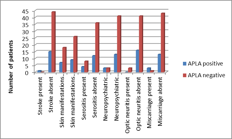
Figure 1 : Association Of Clinical Manifestations With Apla Positivity
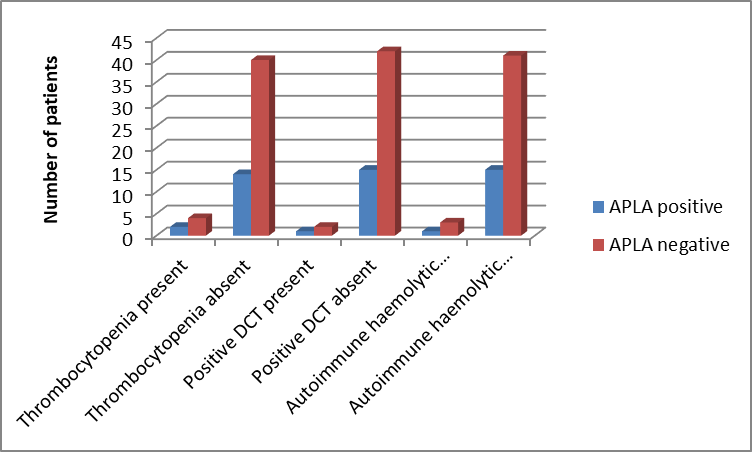
Figure 2 : Association Of Hematological Manifestations With Apla Positivity
The association of various clinical manifestations with APLA positivity among the study population. Stroke was present in 1 patient (APLA positive: 1, APLA negative: 0) and absent in 59 patients (APLA positive: 15, APLA negative: 44), with no statistically significant association (p=0.267).Skin manifestations were observed in 25 patients (APLA positive: 7, APLA negative: 18), while 35 patients had no skin manifestations (APLA positive: 9, APLA negative: 26). This association was not statistically significant (p=1.00).Serositis was present in 12 patients (APLA positive: 4, APLA negative: 8), whereas it was absent in 48 patients (APLA positive: 12, APLA negative: 36). The association was not significant (p=0.716).Neuropsychiatric manifestations were found in 6 patients (APLA positive: 3, APLA negative: 3), and absent in 54 patients (APLA positive: 13, APLA negative: 41), with no significant association (p=0.328).Optic neuritis was present in 3 patients (APLA positive: 0, APLA negative: 3), while absent in 57 patients (APLA positive: 16, APLA negative: 41). The association was not statistically significant (p=0.558).Miscarriage was reported in 4 patients (APLA positive: 3, APLA negative: 1), whereas 56 patients had no history of miscarriage (APLA positive: 13, APLA negative: 43). The association was found to be almost significant (p=0.054).
Deep vein thrombosis (DVT) was present in 3 patients (APLA positive: 3, APLA negative: 0), whereas 57 patients had no DVT (APLA positive: 13, APLA negative: 44). The association between DVT and APLA positivity was found to be statistically significant (p=0.016).Hypertension was present in 8 patients (APLA positive: 3, APLA negative: 5), while 52 patients did not have hypertension (APLA positive: 13, APLA negative: 39). This association was not statistically significant (p=0.429).Ischaemic heart disease (IHD) was not present in any patient (0 cases in both APLA positive and APLA negative groups); therefore, the p value could not be calculated (NA).
Thrombocytopenia was present in 6 patients (APLA positive: 2, APLA negative: 4), whereas 54 patients had no thrombocytopenia (APLA positive: 14, APLA negative: 40). The association between thrombocytopenia and APLA positivity was not statistically significant (p=0.653).Positive Direct Coomb’s Test (DCT) was observed in 3 patients (APLA positive: 1, APLA negative: 2), while 57 patients had negative DCT (APLA positive: 15, APLA negative: 42). This association was not statistically significant (p=1.00).Autoimmune haemolytic anemia (AIHA) was present in 4 patients (APLA positive: 1, APLA negative: 3), whereas 56 patients did not have AIHA (APLA positive: 15, APLA negative: 41). The association of AIHA with APLA positivity was not statistically significant (p=1.00).
Nephropathy was present in 33 patients (APLA positive: 7, APLA negative: 26), whereas 27 patients had no nephropathy (APLA positive: 9, APLA negative: 18). The association between nephropathy and APLA positivity was not statistically significant (p=0.382).Thyroid abnormalities were observed in 9 patients (APLA positive: 4, APLA negative: 5), while 51 patients had no thyroid abnormality (APLA positive: 12, APLA negative: 39). This association was also not statistically significant (p=0.230).
Among the 33 patients with nephropathy, 7 patients were APLA positive and 26 patients were APLA negative. In Class I lupus nephritis, no APLA positive cases were observed, whereas 7 APLA negative cases were present.In Class II lupus nephritis, 1 patient was APLA positive and 4 patients were APLA negative. In Class III lupus nephritis, no APLA positive cases were found, while 2 patients were APLA negative.The majority of cases belonged to Class IV lupus nephritis, with 16 patients in total (APLA positive: 6, APLA negative: 10). Class V lupus nephritis was observed in 3 patients, all of whom were APLA negative.
Autoimmune haemolytic anemia (AIHA) was observed in 4 patients (APLA positive: 1, APLA negative: 3). Bleeding manifestations were present in 1 patient (APLA positive: 0, APLA negative: 1), and hepatitis was also reported in 1 patient (APLA positive: 0, APLA negative: 1).Thyroid abnormalities were the most common organ involvement, affecting 9 patients (APLA positive: 4, APLA negative: 5). Myopathy was present in 2 patients (APLA positive: 1, APLA negative: 1), while menstrual abnormalities were noted in 4 patients (APLA positive: 1, APLA negative: 3).Peripheral neuropathy was found in 3 patients (APLA positive: 1, APLA negative: 2), and vasculitis was observed in 2 patients (APLA positive: 0, APLA negative: 2).Interstitial lung disease and megaloblastic anemia were each reported in 1 patient (APLA positive: 0, APLA negative: 1).
DISCUSSION:
In the present study, APLA positivity was associated with deep vein thrombosis (DVT) in a statistically significant manner (p=0.016). DVT was present in 3 patients, all of whom were APLA positive. Similar findings were reported by Cervera et al., [11]. who observed that antiphospholipid antibodies in SLE patients were strongly associated with thrombotic manifestations, particularly venous thrombosis. They emphasized that APLA positivity is an important risk factor for vascular complications in SLE patients Likewise, Petri et al. [2]. reported a higher frequency of thrombosis among lupus patients with antiphospholipid antibodies and identified APLA as an independent predictor of thrombotic events
In our study, miscarriage showed an almost significant association with APLA positivity (p=0.054), with 3 out of 4 patients having miscarriage being APLA positive. This finding is comparable with the study by Lockshin et al., [13]. who demonstrated that antiphospholipid antibodies are associated with recurrent pregnancy loss and adverse pregnancy outcomes in patients with autoimmune diseases Ruffatti et al. [14].also reported a significant relationship between APLA positivity and pregnancy morbidity in SLE patients, particularly recurrent miscarriage.
The present study showed that stroke was present in only 1 patient and was APLA positive; however, the association was not statistically significant (p=0.267). Similar observations were made by Brey et al., [15]. who reported that although APLA is associated with cerebrovascular events, the strength of association varies depending on patient characteristics and additional vascular risk factors.
Skin manifestations, serositis and neuropsychiatric manifestations were observed in our study but showed no significant association with APLA positivity. Skin manifestations were present in 25 patients, serositis in 12 patients and neuropsychiatric manifestations in 6 patients. These findings are consistent with the observations of Cervera et al., [11].who reported that APLA positivity does not consistently correlate with all clinical manifestations of SLE and that many non-thrombotic features occur independently of APLA status Regarding renal involvement, nephropathy was present in 33 patients, but the association with APLA positivity was not significant (p=0.382). Among lupus nephritis classes, Class IV lupus nephritis was the most common category, affecting 16 patients (APLA positive: 6, APLA negative: 10).
In the present study, hematological manifestations including thrombocytopenia, positive Direct Coomb’s Test and autoimmune haemolytic anemia were not significantly associated with APLA positivity. Thrombocytopenia was present in 6 patients, AIHA in 4 patients and positive DCT in 3 patients. Miyakis et al. [16 ].described thrombocytopenia as one of the recognized clinical associations of antiphospholipid syndrome; however, its relationship with antibody positivity in SLE patients may not always reach statistical significance. Other organ manifestations such as peripheral neuropathy, vasculitis, myopathy, menstrual abnormalities, interstitial lung disease and hepatitis were observed in smaller numbers. These findings reflect the heterogeneous nature of SLE, where multiple organ systems may be affected. Bertsias et al. [17]. highlighted that SLE clinical presentation varies widely and organ involvement depends on disease activity, duration and immunological profile.
CONCLUSION:
The present prospective observational study evaluated the association of antiphospholipid antibody (APLA) positivity with clinical manifestations and organ involvement in 60 patients with Systemic Lupus Erythematosus (SLE).APLA positivity was found to have an important association with thrombotic manifestations, particularly deep vein thrombosis (DVT), which showed a statistically significant relationship (p=0.016). This suggests that APLA-positive SLE patients are at increased risk of thrombotic events and require careful clinical monitoring. Among pregnancy-related manifestations, miscarriage showed an almost significant association with APLA positivity (p=0.054), indicating a possible relationship between antiphospholipid antibodies and adverse pregnancy outcomes.Other clinical manifestations including stroke, skin manifestations, serositis, neuropsychiatric manifestations and optic neuritis did not show statistically significant association with APLA positivity in this study.Renal involvement was common, with nephropathy observed in 33 patients, and Class IV lupus nephritis being the most frequent histopathological class. However, nephropathy and lupus nephritis class distribution did not show significant association with APLA positivity.Hematological abnormalities such as thrombocytopenia, positive Direct Coomb’s Test and autoimmune haemolytic anemia were observed among the patients but were not significantly related to APLA status.Among other organ involvements, thyroid abnormalities were the most commonly observed manifestation, followed by autoimmune haemolytic anemia, menstrual abnormalities and peripheral neuropathy; however, these were not significantly associated with APLA positivity.Overall, the study concludes that APLA positivity in SLE is mainly associated with thrombotic complications, especially DVT, and may contribute to pregnancy morbidity. Routine assessment of APLA status in SLE patients can help identify individuals at higher risk for thrombotic events and guide appropriate clinical management.
REFERENCES:
1. Ameer Ma, Chaudhry H, Mushtaq J, Khan Os, Babar M, Hashim T, Zeb S, Tariq Ma, Patlolla Sr, Ali J, Hashim Sn. An Overview Of Systemic Lupus Erythematosus (Sle) Pathogenesis, Classification, And Management. Cureus. 2022 Oct 15;14(10).
2. American College Of Rheumatology Ad Hoc Committee On Clinical Guidelines. Guidelines For The Management Of Rheumatoid Arthritis. Arthritis & Rheumatism. 1996 May;39(5):713-22.
3. Rampudda M, Marson P, Pasero G. The Main Stages In The History Of Systemic Lupus Erythematosus. Reumatismo. 2009 Jun 1;61(2):145.
4. Crissey Jt, Parish Lc, Holubar K. Historical Atlas Of Dermatology And Dermatologists. Crc Press; 2013 May 30.
5. Shankar S, Sharma P. Anti-Nucleosome Antibodies: In Quest Of Biomarkers Of Disease Activity In Lupus. Indian Journal Of Rheumatology. 2010 Dec;5(4):163-4.
6. Patriarcheas V, Tsamos G, Vasdeki D, Kotteas E, Kollias A, Nikas D, Kaiafa G, Dimakakos E. Antiphospholipid Syndrome: A Comprehensive Clinical Review. Journal Of Clinical Medicine. 2025 Jan 23;14(3):733.
7. Khamashta Ma, Hughes Gr. Phospholipid Autoantibodies—Cardiolipin. Inautoantibodies 1996 Jan 1 (Pp. 624-629). Elsevier Science Bv.
8. Pierangeli Ss. Anticardiolipin Testing. Inhughes Syndrome: Antiphospholipid Syndrome 2000 (Pp. 275-290). London: Springer London.
9. Hughes G. Hughes Syndrome: The Antiphospholipid Syndrome—A Clinical Overview. Clinical Reviews In Allergy & Immunology. 2007 Feb;32(1):3-11.
10. Katarzyna Pb, Wiktor S, Ewa D, Piotr L. Current Treatment Of Systemic Lupus Erythematosus: A Clinician's Perspective. Rheumatology International. 2023 Aug;43(8):1395-407.
11. Cervera R, Khamashta Ma, Font J, Sebastiani Gd, Gil A, Lavilla P, Mejía Jc, Aydintug Ao, Chwalinska-Sadowska H, De Ramón E, Fernández-Nebro A. Morbidity And Mortality In Systemic Lupus Erythematosus During A 10-Year Period: A Comparison Of Early And Late Manifestations In A Cohort Of 1,000 Patients. Medicine. 2003 Sep 1;82(5):299-308.
12. Petri M, Kiani An, Post W, Magder L. Lupus Atherosclerosis Prevention Study (Laps): A Randomized Double Blind Placebo Controlled Trial Of Atorvastatin Versus Placebo. Inarthritis And Rheumatism 2006 Sep 1 (Vol. 54, No. 9, Pp. S520-S520). Div John Wiley & Sons Inc, 111 River St, Hoboken, Nj 07030 Usa: Wiley-Liss.
13. Lockshin Md, Druzin Ml, Goei S, Qamar T, Magid Ms, Jovanovic L, Ferenc M. Antibody To Cardiolipin As A Predictor Of Fetal Distress Or Death In Pregnant Patients With Systemic Lupus Erythematosus. New England Journal Of Medicine. 1985 Jul 18;313(3):152-6.
14. Ruffatti A, Tonello M, Cavazzana A, Bagatella P, Pengo V. Laboratory Classification Categories And Pregnancy Outcome In Patients With Primary Antiphospholipid Syndrome Prescribed Antithrombotic Therapy. Thrombosis Research. 2009 Jan 1;123(3):482-7.
15. Brey Rl, Abbott Rd, Curb Jd, Sharp Ds, Ross Gw, Stallworth Cl, Kittner Sj. Β2-Glycoprotein 1–Dependent Anticardiolipin Antibodies And Risk Of Ischemic Stroke And Myocardial Infarction: The Honolulu Heart Program. Stroke. 2001 Aug 1;32(8):1701-6.
16. Miyakis S, Lockshin Md, Atsumi T, Branch Dw, Brey Rl, Cervera Rh, Derksen Rh, De Groot Pg, Koike T, Meroni Pl, Reber G. International Consensus Statement On An Update Of The Classification Criteria For Definite Antiphospholipid Syndrome (Aps). Journal Of Thrombosis And Haemostasis. 2006 Feb 1;4(2):295-306.
17. Bertsias G, Cervera R, Boumpas Dt. Systemic Lupus Erythematosus: Pathogenesis And Clinical Features. Eular Textbook On Rheumatic Diseases. 2012 Apr 21;5:476-505.