Role of 3-Tesla Magnetic Resonance Imaging in the Evaluation of Non-Traumatic Hip Joint Pain.
- Sachin Khanduri , Professor & HOD, Department of Radiodiagnosis, Era’s Lucknow Medical College & Hospital, Lucknow, Uttar Pradesh, India.
- Abhishek Singh , Junior Resident, Department of Radiodiagnosis, Era’s Lucknow Medical College & Hospital, Lucknow, Uttar Pradesh, India.
- Anmol Agrawal , Junior Resident, Department of Radiodiagnosis, Era’s Lucknow Medical College & Hospital, Lucknow, Uttar Pradesh, India.
- Deole Yash Milind , Junior Resident, Department of Radiodiagnosis, Era’s Lucknow Medical College & Hospital, Lucknow, Uttar Pradesh, India.
- Deshna Jain , Junior Resident, Department of Radiodiagnosis, Era’s Lucknow Medical College & Hospital, Lucknow, Uttar Pradesh, India.
- Nutan Patel , Junior Resident, Department of Radiodiagnosis, Era’s Lucknow Medical College & Hospital, Lucknow, Uttar Pradesh, India.
- Rubina Saudi , Junior Resident, Department of Radiodiagnosis, Era’s Lucknow Medical College & Hospital, Lucknow, Uttar Pradesh, India.
- Eraj Fatima Khan , Junior Resident, Department of Radiodiagnosis, Era’s Lucknow Medical College & Hospital, Lucknow, Uttar Pradesh, India.
Article Information:
Abstract:
Background: Non-traumatic hip joint pain is a common clinical problem with a broad spectrum of underlying pathologies. Conventional radiography often fails to detect early marrow, cartilage, and soft-tissue abnormalities, resulting in delayed diagnosis. Owing to its excellent soft-tissue contrast and multiplanar imaging capability, 3-Tesla MRI (Magnetic Resonance Imaging) plays a pivotal role in the early diagnosis and etiological characterization of hip disorders. The present study aimed to evaluate the diagnostic role of 3-Tesla MRI in patients presenting with non-traumatic hip joint pain and to determine the spectrum of underlying etiologies. Methods: A hospital-based cross-sectional observational study was conducted over a period of two years at Era's Lucknow Medical College and Hospital, Lucknow. Sixty-seven patients with non-traumatic hip joint pain who fulfilled the inclusion criteria underwent 3-Tesla MRI using a standard hip imaging protocol. MRI findings were interpreted by experienced radiologists, and the final etiological diagnosis was established. Data were analysed using SPSS version 26, with descriptive statistics and chi-square test applied. A p-value <0.05 was considered statistically significant. Results: The mean age of the study population was 45.0 ± 14.7 years, with a male predominance (61.19%). Most patients were older than 40 years and presented within three months of symptom onset. Avascular necrosis (35.82%) was the commonest etiology, followed by osteoarthritis (22.39%) and infective arthritis (14.93%). Bone marrow edema (68.66%) was the most frequent MRI finding, followed by joint effusion (58.21%) and articular cartilage thinning (46.27%). A significant association was observed between age and etiology of hip pain (χ²=10.32, p=0.016), whereas gender showed no significant association (p=0.110). Conclusion: Three-Tesla MRI is an excellent non-invasive imaging modality for evaluating non-traumatic hip joint pain. Its high sensitivity for detecting early marrow, cartilage, synovial, and soft-tissue abnormalities enables accurate etiological diagnosis and facilitates timely clinical management, thereby improving patient outcomes.
Keywords:
Article :
INTRODUCTION:
Magnetic resonance imaging has become an indispensable modality in the evaluation of hip disorders because it provides excellent visualisation of articular structures, peri-articular soft tissues, bone marrow, and osseous abnormalities. In patients with chronic hip pain, where clinical findings are often non-specific and radiographs may appear normal, MRI (Magnetic Resonance Imaging) plays a crucial role in identifying subtle pathological changes caused by trauma, infection, arthritis, AVN (Avascular Necrosis), tumours, and hip dysplasia.[1] Standard coronal and axial MRI planes provide symmetrical bilateral assessment, enabling accurate evaluation of the femoral head, neck, intertrochanteric region, articular space, supporting ligaments, and surrounding musculature, thereby improving diagnostic accuracy.[2,3]
MRI is regarded as the most sensitive imaging modality for the early detection of AVN, allowing diagnosis before radiographic abnormalities become evident. Early identification facilitates timely intervention, limits disease progression, and may preserve joint function, particularly in high-risk individuals.[4,5] In paediatric patients, MRI offers additional advantages because the immature hip is predominantly cartilaginous and cannot be adequately evaluated by radiography, CT (Computed Tomography), or ultrasonography after infancy. Its ability to directly visualise cartilage makes MRI especially valuable in assessing paediatric hip disorders.[6]
High-resolution MRI is currently the preferred technique for evaluating intra-articular hip pathology, whereas conventional radiography remains essential for detecting subtle bony abnormalities, particularly those associated with femoro-acetabular impingement. Therefore, both imaging modalities complement each other in the comprehensive assessment of hip pain.[7] Hip pain may result from a broad spectrum of conditions including osteonecrosis, osteoarthritis, septic arthritis, tubercular arthritis, femoro-acetabular impingement, transient osteoporosis, occult fractures, transient synovitis, sacroiliitis, and neoplasms.[8,9]
Although radiography and CT provide valuable information on bony architecture, their limitations in detecting early marrow, cartilage, labral, and soft-tissue abnormalities make MRI the preferred non-invasive imaging modality for comprehensive evaluation.[8,10] With superior tissue contrast and multiplanar capability, particularly at 3 Tesla field strength, MRI enables early and accurate diagnosis, guiding appropriate management and improving clinical outcomes in patients with non-traumatic hip pain.
Aims and Objectives
The study aimed to evaluate the role of 3-Tesla MRI in the diagnosis and etiological characterization of non-traumatic hip joint pain. The primary objective is to assess the diagnostic efficacy of 3-Tesla MRI in identifying the underlying causes of non-traumatic hip joint pain. The secondary objectives are to determine the spectrum and frequency of various etiologies responsible for non-traumatic hip joint pain as detected on 3-Tesla MRI and to describe the characteristic MRI findings associated with the major hip joint pathologies, thereby facilitating accurate diagnosis and appropriate clinical management.
MATERIALS AND METHODS:
Study Design
The present study was designed as a hospital-based cross-sectional study conducted over a period of two years at ERA'S Lucknow Medical College, Lucknow. The study included a total of 67 patients who fulfilled the predefined inclusion criteria and presented with non-traumatic hip joint pain to the Department of Orthopaedics, including both the OPD (Out-Patient Department) and Emergency Department, as well as the Department of Radiodiagnosis.
Inclusion and Exclusion Criteria
The study included patients of both sexes and all age groups presenting with acute or chronic unilateral or bilateral hip joint pain without a history of recent trauma (within the preceding one month), who were referred to the Department of Radiodiagnosis for MRI evaluation of the hip joint and provided written informed consent to participate in the study.
Patients were excluded if they had a history of recent trauma to the hip joint, a prior history of hip surgery, or congenital hip disorders. Additionally, patients who were unable to undergo MRI examination due to claustrophobia or the presence of contraindications such as a cardiac pacemaker, critically positioned metallic foreign bodies, or MRI-incompatible vascular implants were excluded from the study.
Sample Size Calculation
Sample size was calculated on the basis of the proportion of the most common etiology in MRI findings of non-traumatic hip joint pain cases using the formula:
Where p = 48%, proportion of most common etiology in MRI finding of non-traumatic Hip Joint pain.[7]
q = 100 – p,
Type I error α=5 %, for the significance level of 95%.
Allowable error L = 12% absolute for detecting the results with 90% power of study, the sample size comes out to be n = 67
Data Collection Procedure
Patients presenting with non-traumatic hip joint pain and fulfilling the inclusion criteria were referred from the Department of Orthopaedics to the Department of Radiodiagnosis for MRI evaluation. After obtaining written informed consent, all participants underwent MRI of the hip joint using a 3-Tesla MRI scanner following the standard imaging protocol. The acquired images were post-processed on a dedicated workstation and independently interpreted by experienced radiologists.
MRI findings involving the osseous structures, articular cartilage, synovium, bone marrow, and surrounding soft tissues were systematically recorded, and the final etiological diagnosis was established based on the imaging findings.
Statistical Analysis
The collected data were entered into Microsoft Excel and analysed using the Statistical Package for the Social Sciences (SPSS) software, version 26.0 (IBM Corp., Armonk, NY, USA). Descriptive statistics were used to summarize the study data. Continuous variables, such as age, were expressed as mean ± standard deviation (SD), whereas categorical variables were presented as frequencies and percentages. The association between categorical variables, including age group, gender, MRI findings, and final etiological diagnosis, was assessed using the Chi-square (χ²) test.
The diagnostic significance of key MRI findings, particularly bone marrow edema, was evaluated by analysing their association with the final etiological diagnosis. A p-value of <0.05 was considered statistically significant, while a p-value of <0.001 was considered highly statistically significant.
RESULTS:
Table 1. Demographic Profile of the Study Population (n = 67
|
Variable |
Category |
Number (n=67) |
Percentage (%) |
|
Age Group |
≤20 years |
4 |
5.97 |
|
21–30 years |
8 |
11.94 |
|
|
31–40 years |
11 |
16.42 |
|
|
41–50 years |
17 |
25.37 |
|
|
51–60 years |
15 |
22.39 |
|
|
>60 years |
12 |
17.91 |
|
|
Gender |
Male |
41 |
61.19 |
|
Female |
26 |
38.81 |
Table 1 illustrates the demographic characteristics of the study population. Most patients belonged to the 41–50 years age group (25.37%), followed by 51–60 years (22.39%). Males constituted the majority (61.19%) compared to females (38.81%).
Table 2. Clinical Characteristics of Patients (n = 67)
|
Variable |
Category |
Number |
Percentage (%) |
|
Hip Joint Involved |
Right |
28 |
41.79 |
|
Left |
24 |
35.82 |
|
|
Bilateral |
15 |
22.39 |
|
|
Duration of Symptoms |
<1 month |
14 |
20.90 |
|
1–3 months |
21 |
31.34 |
|
|
3–6 months |
18 |
26.87 |
|
|
>6 months |
14 |
20.90 |
Table 2 shows the clinical profile of patients. Right hip involvement (41.79%) was more common than left-sided disease (35.82%), while bilateral involvement was seen in 22.39% of patients. Most patients presented within 1–3 months of symptom onset (31.34%).
Table 3. Distribution of Final Etiological Diagnosis (n = 67)
|
Etiology |
Number |
Percentage (%) |
|
Avascular Necrosis (AVN) |
24 |
35.82 |
|
Osteoarthritis |
15 |
22.39 |
|
Infective Arthritis |
10 |
14.93 |
|
Inflammatory Arthritis |
7 |
10.45 |
|
Stress/Insufficiency Fracture |
6 |
8.96 |
|
Transient Synovitis |
5 |
7.46 |
Table 3 illustrates the final MRI diagnosis of patients. Avascular necrosis was the most common diagnosis (35.82%), followed by osteoarthritis (22.39%), whereas transient synovitis was the least frequent diagnosis (7.46%).
Table 4. Distribution of MRI Findings (n = 67)
|
MRI Finding |
Number |
Percentage (%) |
|
Bone Marrow Edema |
46 |
68.66 |
|
Joint Effusion |
39 |
58.21 |
|
Articular Cartilage Thinning |
31 |
46.27 |
|
Subchondral Cysts |
26 |
38.81 |
|
Double Line Sign |
24 |
35.82 |
|
Osteophyte Formation |
21 |
31.34 |
|
Synovial Thickening |
18 |
26.87 |
|
Femoral Head Contour Irregularity |
16 |
23.88 |
|
Soft Tissue Collection/Abscess |
9 |
13.43 |
Table 4 shows the overall MRI findings among painful hip joints. Bone marrow edema was the commonest imaging finding (68.66%), followed by joint effusion (58.21%), while soft tissue collection/abscess was the least common finding (13.43%).
Table 5. Association of Age and Gender with Etiology
|
Etiology |
≤40 years (n=23) |
>40 years (n=44) |
Male (n=41) |
Female (n=26) |
|
|
AVN |
6 (26.09%) |
18 (40.91%) |
18 (43.90%) |
6 (23.08%) |
|
|
Osteoarthritis |
2 (8.70%) |
13 (29.55%) |
9 (21.95%) |
6 (23.08%) |
|
|
Infective Arthritis |
7 (30.43%) |
3 (6.82%) |
7 (17.07%) |
3 (11.54%) |
|
|
Other Etiologies |
8 (34.78%) |
10 (22.73%) |
7 (17.07%) |
11 (42.31%) |
|
|
Statistical Test |
Value |
||||
|
Age vs Etiology |
χ²=10.32, p=0.0160* |
||||
|
Gender vs Etiology |
χ²=6.033, p=0.1100 |
||||
Table 5 illustrates the relationship between demographic variables and disease etiology. A statistically significant association was observed between age and etiology (p=0.0160), whereas gender did not show a statistically significant association with the underlying diagnosis (p=0.1100).
Table 6. MRI Findings According to Major Diagnoses
|
MRI Finding |
AVN (n=24) |
Osteoarthritis (n=15) |
Infective Arthritis (n=10) |
|
Double Line Sign |
24 (100%) |
— |
— |
|
Bone Marrow Edema |
20 (83.33%) |
9 (60.00%) |
9 (90.00%) |
|
Femoral Head Contour Irregularity |
18 (75.00%) |
— |
— |
|
Subchondral Collapse |
9 (37.50%) |
— |
— |
|
Joint Effusion |
11 (45.83%) |
7 (46.67%) |
8 (80.00%) |
|
Cartilage Thinning |
— |
15 (100%) |
— |
|
Osteophyte Formation |
— |
13 (86.67%) |
— |
|
Subchondral Cysts |
— |
11 (73.33%) |
— |
|
Synovial Thickening |
— |
— |
10 (100%) |
|
Soft Tissue Collection |
— |
— |
6 (60.00%) |
|
Cartilage Destruction |
— |
— |
5 (50.00%) |
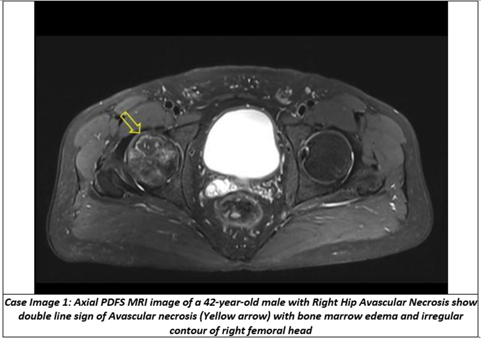
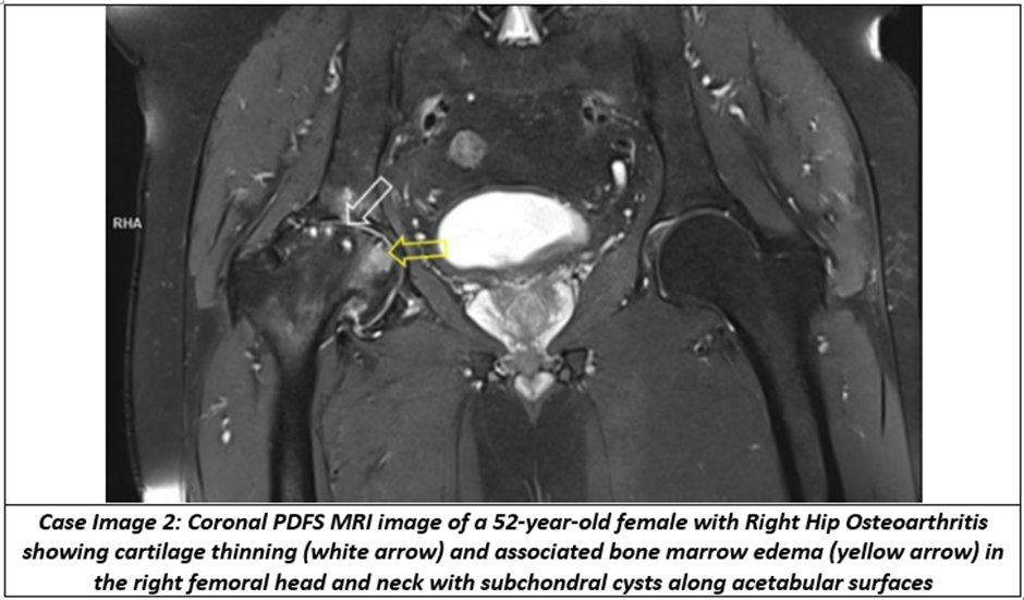
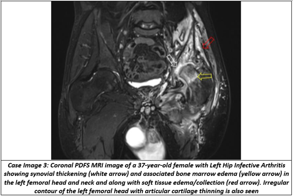
Table 6 shows disease-specific MRI findings. The double-line sign was present in all AVN cases, cartilage thinning was universal in osteoarthritis, and synovial thickening was present in all infective arthritis cases, demonstrating characteristic MRI features for each disease entity.
Table 7. Association Between Bone Marrow Edema and Final Diagnosis
|
Diagnosis |
BME Present (n=46) |
BME Absent (n=21) |
|
|
AVN |
20 (43.48%) |
4 (19.05%) |
|
|
Osteoarthritis |
9 (19.57%) |
6 (28.57%) |
|
|
Infective Arthritis |
9 (19.57%) |
1 (4.76%) |
|
|
Other Etiologies |
8 (17.39%) |
10 (47.62%) |
|
|
Statistical Test |
Value |
||
|
χ² |
9.945 |
||
|
p-value |
0.0190* |
||
Table 7 illustrates the association between bone marrow edema and the final diagnosis. Bone marrow edema showed a statistically significant association with disease etiology (p=0.0190), with AVN being the most common diagnosis among patients demonstrating bone marrow edema.
Table 8. Major Findings
|
Parameter |
Most Common Finding |
Percentage |
|
Predominant Age Group |
41–50 years |
25.37% |
|
Predominant Gender |
Male |
61.19% |
|
Most Common Hip Involved |
Right |
41.79% |
|
Most Common Duration |
1–3 months |
31.34% |
|
Most Common Etiology |
AVN |
35.82% |
|
Most Common MRI Finding |
Bone Marrow Edema |
68.66% |
|
Significant Association |
Age vs Etiology |
p=0.0160 |
|
Significant Association |
Bone Marrow Edema vs Diagnosis |
p=0.0190 |
Table 8 summarizes the principal findings of the study. Middle-aged males constituted the majority of the study population. Avascular necrosis was the most frequent cause of non-traumatic hip pain, bone marrow edema was the predominant MRI finding, and statistically significant associations were observed between age and etiology as well as between bone marrow edema and the final diagnosis.
DISCUSSION:
The present study evaluated the role of 3-Tesla MRI in assessing non-traumatic hip joint pain and demonstrated its usefulness in identifying a wide spectrum of hip pathologies at an early stage. MRI effectively detected bone marrow, cartilage, synovial, and soft tissue abnormalities, facilitating accurate etiological diagnosis.
Demographic Characteristics
The mean age of the study population was 45.0 ± 14.7 years, with the highest proportion of patients in the 41–50-year age group (25.37%). Nearly two-thirds (65.67%) of patients were older than 40 years, indicating that non-traumatic hip pain predominantly affects middle-aged and elderly individuals. Similar age distribution was reported by More et al. (2019),[11] who observed most patients between 31 and 50 years, and Shourbagy et al. (2022),[12] who also found maximum cases in the 40–50-year age group. Ghimire et al. (2025)[13] reported a higher mean age due to a greater prevalence of degenerative disorders, whereas Turan et al. (2022)[14] reported a younger population because their study involved paediatric hip disorders.
Male patients constituted 61.19%, while females accounted for 38.81%, demonstrating male predominance. Comparable findings were reported by More et al. (2019),[11] Shourbagy et al. (2022),[12] and Turan et al. (2022),[14] all of whom observed a higher prevalence among males. However, Cotoi et al. (2018)[15] emphasized that degenerative hip disease may also occur in females because of structural and biomechanical abnormalities.
Clinical Characteristics
Unilateral hip involvement (77.61%) was more common than bilateral disease (22.39%), with the right hip (41.79%) affected slightly more frequently than the left (35.82%). Similar observations were made by More et al. (2019)[11] and Shourbagy et al. (2022),[12] who reported unilateral involvement as the commonest presentation in non-traumatic hip disorders.
Etiological Spectrum
Avascular necrosis was the most common diagnosis (35.82%), followed by osteoarthritis (22.39%), infective arthritis (14.93%), inflammatory arthritis (10.45%), stress fractures (8.96%), and transient synovitis (7.46%). Similar predominance of AVN was reported by Tushar et al. (2018),[14] More et al. (2019),[11] and Osman et al. (2019),[16] whereas Ghimire et al (2025)[13] observed osteoarthritis as the leading pathology.
MRI Findings
Bone marrow edema (68.66%) was the most frequent MRI finding, followed by joint effusion (58.21%), cartilage thinning (46.27%), and subchondral cysts (38.81%). Verma et al. (2024)[17] similarly identified bone marrow edema as a common MRI finding in osteonecrosis, while Fang et al. (2024)[18] demonstrated a strong association between MRI-detected structural abnormalities and hip pain. Mak et al. (2020)[19] further emphasized MRI as the preferred imaging modality for painful hip disorders. Among AVN cases, the double-line sign was present in 100%, followed by bone marrow edema (83.33%), femoral head contour irregularity (75%), and subchondral collapse (37.5%). These findings closely resemble those of Verma et al. (2024),[17] confirming the diagnostic significance of these MRI features. In osteoarthritis, cartilage thinning (100%), osteophyte formation (86.67%), subchondral cysts (73.33%), and bone marrow edema (60%) were the predominant MRI findings, consistent with observations by Ghimire et al. (2025).[13] In infective arthritis, synovial thickening (100%), bone marrow edema (90%), joint effusion (80%), and soft tissue collections (60%) were the major findings. Similar MRI features were reported by Verma et al. (2024),[17] supporting the value of MRI in early diagnosis and treatment planning.
Role of MRI
The present study confirms that MRI is an indispensable imaging modality in evaluating non-traumatic hip pain because it detects bone marrow, cartilage, synovial, and periarticular soft tissue abnormalities before radiographic changes become evident. More et al. (2019)[11] reported an MRI diagnostic accuracy of 96.2%, significantly higher than plain radiography. Mak et al. (2020)[19] and Osman et al. (2019)[16] also emphasized MRI's comprehensive ability to identify both intra-articular and extra-articular causes of hip pain. Thus, MRI plays a pivotal role in early diagnosis, accurate etiological characterization, and appropriate clinical management of non-traumatic hip disorders.
Limitations
The present study has certain limitations that should be considered while interpreting the findings. First, the sample size was relatively small, comprising only 67 patients, which may limit the generalizability of the results to the broader population. Second, as the study was conducted at a single tertiary care centre, the findings may not fully represent the demographic and geographic diversity of patients presenting with hip pain in other healthcare settings. Finally, the absence of long-term follow-up prevented the assessment of disease progression, response to treatment, and long-term clinical outcomes. Therefore, larger multicentre studies with extended follow-up are recommended to validate the present findings and enhance their applicability to routine clinical practice.
CONCLUSION:
The present study concludes that 3-Tesla MRI is the imaging modality of choice for the evaluation of non-traumatic hip joint pain owing to its high sensitivity in detecting early bone marrow, cartilage, synovial, and soft-tissue abnormalities. Avascular necrosis was the most common etiology, followed by osteoarthritis and infective arthritis, with bone marrow edema and joint effusion being the predominant MRI findings. MRI enabled accurate characterization of a wide spectrum of hip pathologies, facilitating early diagnosis, appropriate treatment planning, and timely intervention, thereby helping to prevent disease progression and improve patient outcomes.
REFERENCES:
1. Manster BJ. Adult Chronic Hip Pain: Radiographic Evaluation. Radiographics 2000;20(2):S3-25.
2. Gabriel H, Fitzgerald SW, Myers MT, Donaldson JS, Poznanski AK. MR imaging of hip disorders. Radiographics 1994;14(1):763-81.
3. Berquist H. Thomas. MRI of the Musculoskeletal system: Hip. 6th edn. Philadelphia: Lippincott Williams and Wilkins 2012:204-318.
4. Steinberg ME, Hayken GD. A quantitative system for staging avascular necrosis. J Bone Joint Surg Br 1995;77(2):34-41.
5. Buckland-Wright JC, Verbruggen G, Haraoui PB. Session 3: Imaging. Radiological assessment of hand OA. Osteoarthritis and Cartilage 2000;8:S55-6.
6. Terjesen T, Gunderson RB. Radiographic evaluation of osteoarthritis of the hip: an inter-observer study of 61 hips treated for late-detected developmental hip dislocation. Acta Orthop 2012;83(4):185-9.
7. Kalekar T, Shriramwar P. MRI evaluation of non traumatic painful hip joint. International Journal of Contemporary Medicine Surgery and Radiology 2018;3(1):155-60.
8. Dutta P, Sonowal TN, Shahare SL, et al. Imaging of hip joint pathologies. J Evol Med Dent Sci 2016;5(63):4430-6.
9. Wilson JJ, Furukawa M. Evaluation of the patient with hip pain. Am Fam Physician 2014;89(1):27-34.
10. Mitchell DG, Rao VM, Dalinka M, et al. Hematopoietic and fatty bone marrow distribution in the normal and ischemic hip: new observations with 1.5-T MR imaging. Radiology 1986;161(1):199–202.
11. More D, Shaha P, Chougule SK, et al. The role of MRI in non-traumatic chronic hip joint pain. Executive Editor 2019;10(3):140.
12. Patel R, Gandhi N, Patel M. Magnetic Resonance Imaging (MRI) in Diagnosis of Non-Traumatic Hip Joint Pain. CME Journal Geriatric Medicine 2025;17:66-9.
13. Ghimire RK, Ghimire SK, Bhandari RM. Magnetic Resonance Imaging Parameters in the Evaluation of Non-traumatic Painful Hip Joint. Asian Journal of Basic Science & Research 2025;7(3):201-6.
14. Turan A, Akdag T, Yildirim F, et al. The evaluation of hip joint pathologies in pediatric age group with MRI. Medicine Science 2022;11(3):1166-71.
15. Cotoi AI, Dinu MS, Meetescu RE, et al. 3 Tesla Magnetic Resonance Imaging Role in Articular Degenerative Pathology. Curr Health Sci J 2018;44(3):299-303.
16. Osman NM, Hemeda TW, Thabit MT. Role of Magnetic Resonance Imaging in the evaluation of adult non-traumatic painful hip. Sohag Medical Journal 2019;23(2):150-7.
17. Sameer RV, Shrey M, Pankaj K, et al. Spectrum of MRI findings in non-traumatic hip pathologies. J Res Med Dent Sci 2024;12(8):26-30.
18. Fang H, Zhang X, Wang J, et al. The relationship between MRI-detected hip abnormalities and hip pain in hip osteoarthritis: a systematic review. Rheumatol Int 2024;44(10):1887-96.
19. Mak MS, Teh J. Magnetic resonance imaging of the hip: anatomy and pathology. Pol J Radiol 2020;85:e489-508.