Clinical, Electrophysiological and Magnetic Resonance Imaging Profile of Traumatic Brachial Plexopathy: A Descriptive Observational Study at a Tertiary Care Centre.
- Rashmi Devaraj , Assistant Professor, Department of Neurology, Vydehi Institute of Medical Sciences and Research Centre, Whitefield, Bengaluru, Karnataka, India.
- Shreyishi Ganguly , Senior Resident, Department of Neurology, Vydehi Institute of Medical Sciences and Research Centre, Whitefield, Bengaluru, Karnataka, India.
- Mohan P. Channapanavur , Professor, Department of Neurology, Vydehi Institute of Medical Sciences and Research Centre, Whitefield, Bengaluru, Karnataka,India.
Article Information:
Abstract:
Background: Traumatic brachial plexopathy produces major motor, sensory, and functional impairment. Accurate anatomical localization requires integration of clinical examination, electrodiagnostic testing, and magnetic resonance imaging (MRI), when preganglionic injury or root avulsion is suspected. Objectives: To describe the demographic, etiological, clinical, electrophysiological, and MRI profile of patients with traumatic brachial plexopathy presenting to a tertiary care neurology service. Methods: This descriptive observational study included 18 consecutive patients with traumatic brachial plexopathy evaluated between February 2024 and January 2026. Demographic and injury characteristics, neurological findings, nerve conduction/electromyographic interpretations, and brachial plexus MRI findings were recorded using a structured proforma. Continuous variables were summarized using mean, standard deviation, median, and range; categorical variables were expressed as frequencies and percentages. Results: The mean age was 30.5 ± 14.7 years, and 17 patients (94.4%) were male. Vehicle-related trauma accounted for 14 cases (77.8%); the right plexus was affected in 10 (55.6%). Weakness was universal, pain occurred in 10 (55.6%), and sensory symptoms in 8 (44.4%). Among patients with documented examination findings, proximal power was Medical Research Council grade 2/5 or lower in 11 of 17 (64.7%), reflexes were absent or depressed in 14 of 17 (82.4%), and sensation was impaired in 9 of 17 (52.9%). MRI was available for 12 patients and showed root avulsion with pseudomeningocele in 3 (25.0%). Electrodiagnostic data were available for 15 patients; pan-plexus involvement was identified in 7 (46.7%), and severe injury was explicitly reported in 6 (40.0%). Conclusion: Traumatic brachial plexopathy predominantly affected young men after vehicle-related trauma. The frequent proximal weakness, reflex loss, axonal abnormalities, and MRI evidence of root injury emphasize the value of coordinated clinical, electrophysiological, and imaging assessment for localization and management planning.
Keywords:
Article :
INTRODUCTION:
The brachial plexus is formed by the anterior rami of the C5-T1 spinal nerves and supplies motor, sensory, and sympathetic fibres to the upper limb. Its complex organization into roots, trunks, divisions, cords, and terminal branches creates substantial diagnostic difficulty when injury occurs at more than one anatomical level. Traumatic brachial plexus injury can result from traction, compression, rupture, penetrating trauma, or avulsion of the cervical roots. Lesions are commonly classified as preganglionic or postganglionic because this distinction directly influences prognosis and reconstructive options. Clinical patterns range from isolated upper-trunk weakness to complete flail limb with sensory loss, neuropathic pain, and autonomic disturbance. 1,2 Traumatic brachial plexopathy is particularly disabling because it affects hand use, self-care, employment, and social participation. Persistent pain and loss of function also contribute to psychological distress, depression, and post-traumatic stress symptoms. 3 Population-based and referral-centre studies consistently show that young men are disproportionately affected, usually following high-energy road traffic trauma. Motorcycle collisions carry a notable risk because forceful depression of the shoulder combined with contralateral neck movement produces severe traction across the supraclavicular plexus. Associated fractures of the clavicle, ribs, cervical spine, or upper limb can complicate assessment and delay definitive referral. 4,5. Initial localization depends on the mechanism of injury, posture of the limb, distribution of weakness, reflex changes, sensory loss, pain, Horner syndrome, and examination of muscles supplied by individual roots and peripheral nerves. However, examination alone cannot reliably define the full extent of a mixed lesion. Electrodiagnostic testing, including nerve conduction studies and needle electromyography, provides functional evidence regarding lesion level, axonal loss, severity, and early reinnervation. Sensory nerve action potentials and paraspinal muscle examination are useful in differentiating preganglionic from postganglionic injury, although timing after trauma and technical completeness influence interpretation. 6-8
Magnetic resonance imaging complements clinical and electrophysiological assessment by demonstrating root discontinuity, pseudomeningocele, plexus thickening, edema, neuroma, and associated soft-tissue or skeletal injury. 9,10 MRI is particularly important when root avulsion is suspected, but published diagnostic accuracy varies across techniques, field strengths, reporting criteria, and surgical cohorts. 11,12 Consequently, a multimodal approach provides more dependable anatomical localization than any isolated investigation. 13 Despite advances in diagnosis and reconstruction, detailed Indian data describing the combined clinical, electrodiagnostic, and MRI profile of traumatic brachial plexopathy remain limited outside major surgical referral units. The present study was undertaken to describe the demographic characteristics, mechanisms of injury, presenting symptoms, neurological examination findings, electrodiagnostic patterns, and MRI abnormalities among patients with traumatic brachial plexopathy evaluated at a tertiary care centre. The study also aimed to identify the predominant anatomical distribution and severity patterns documented through electrophysiological and imaging assessment.
MATERIALS AND METHODS:
Study design and setting: This prospective, hospital-based descriptive observational study was conducted in the Department of Neurology, Vydehi Institute of Medical Sciences and Research Centre, Whitefield, Bengaluru, India, over two years from February 2024 to January 2026. The study was designed to characterize the clinical, electrophysiological, and radiological spectrum of traumatic brachial plexopathy in patients attending neurology outpatient or inpatient services.
Study population and eligibility: Consecutive patients of any age and sex with a history of significant trauma and clinical findings compatible with brachial plexopathy were screened. Patients were included when traumatic causation was supported by the history, neurological examination, and available investigations, and informed consent was obtained. Patients with non-traumatic plexopathy or absent consent were excluded. Written informed consent was obtained from adults and from a parent or legal guardian for minors, with assent obtained when appropriate.
Data collection: Information was entered prospectively in a structured case-record proforma. Recorded variables included age, sex, affected side, mechanism and interval since injury, presenting weakness, pain, paresthesia or numbness, muscle wasting, associated injuries, and relevant comorbidities. A detailed neurological examination documented tone, proximal and distal muscle power using the Medical Research Council grading system, deep-tendon reflexes, sensory impairment, atrophy, and contractures. Clinical localization was based on the distribution of weakness and sensory loss across root, trunk, cord, and terminal nerve territories.
Electrophysiological assessment: Nerve conduction studies and needle electromyography were performed when clinically indicated according to the department's standard protocol. Interpretations were reviewed for sensory and motor axonal involvement, preganglionic or postganglionic localization, anatomical distribution, and reported severity. Electrodiagnostic evaluation was considered an adjunct to clinical localization because a comprehensive study can define lesion extent, quantify axonal loss, and detect denervation or reinnervation. 6-8
Imaging assessment: MRI of the brachial plexus and cervical region was obtained when clinically indicated. Reports were reviewed for nerve-root or plexus thickening, T2/STIR hyperintensity, edema, discontinuity, root avulsion, pseudomeningocele, cord-level involvement, mass effect, and associated muscular or osseous abnormalities. MRI findings were interpreted within a multimodal framework rather than as an isolated diagnostic standard. 9-12
Outcomes and statistical analysis: The principal outcomes were the demographic and etiological profile, neurological deficits, electrodiagnostic localization and severity, and MRI abnormalities. Treatment-response information was not analyzed because it was not consistently available in the finalized dataset. Data were entered in Microsoft Excel and analyzed using IBM SPSS Statistics version 23.0. Continuous variables were summarized as mean ± standard deviation and median with range. Categorical variables were presented as frequency and percentage. Missing observations were excluded variable-wise, and the relevant denominator was reported. Owing to the descriptive design and small sample, no inferential hypothesis testing was performed.
Ethical considerations: Institutional Ethics committee approval was obtained before starting the study. Participant confidentiality was maintained through de-identification of the analytical dataset.
RESULTS:
The finalized analysis included 18 patients with traumatic brachial plexopathy. The mean age was 30.5 ± 14.7 years, the median age was 29 years, and the range was 7-56 years. Four patients (22.2%) were younger than 18 years. Seventeen patients (94.4%) were male. Right-sided involvement occurred in 10 (55.6%) and left-sided involvement in 8 (44.4%). Vehicle-related trauma was the dominant mechanism, accounting for 14 cases (77.8%); 12 of these followed a fall from a motorized two-wheeler, one followed a bicycle fall, and one patient was hit by a car. The remaining causes were non-road falls in 2 (11.1%), direct occupational/hand trauma in 1 (5.6%), and birth injury in 1 (5.6%). Associated fractures or dislocation were documented in 3 patients (16.7%), including clavicular, cervical/rib, shoulder, and forearm injuries (Table 1).
Table 1. Demographic and injury-related characteristics of the study participants
|
Characteristic |
Value |
|
Total participants |
18 |
|
Age, years, mean ± SD |
30.5 ± 14.7 |
|
Age, years, median (range) |
29 (7-56) |
|
Age group <18 years |
4 (22.2%) |
|
Age group 18-30 years |
5 (27.8%) |
|
Age group 31-45 years |
5 (27.8%) |
|
Age group >45 years |
4 (22.2%) |
|
Male sex |
17 (94.4%) |
|
Female sex |
1 (5.6%) |
|
Right-sided involvement |
10 (55.6%) |
|
Left-sided involvement |
8 (44.4%) |
|
Vehicle-related trauma |
14 (77.8%) |
|
Non-road fall |
2 (11.1%) |
|
Direct occupational/hand trauma |
1 (5.6%) |
|
Birth injury |
1 (5.6%) |
|
Associated fracture or dislocation |
3 (16.7%) |
Data are presented as n (%) unless otherwise specified. SD: standard deviation.
Upper-limb weakness was present in every patient, and 16 (88.9%) specifically reported difficulty lifting the affected arm above shoulder level. Pain was documented in 10 (55.6%), while paresthesia, numbness, or impaired sensation was reported by 8 (44.4%). Clinical wasting or atrophy was present in 3 (16.7%), hypotonia in 5 (27.8%), and contracture in 1 (5.6%). Proximal muscle power was recorded in 17 patients; 11 (64.7%) had Medical Research Council grade 2/5 or lower and 6 (35.3%) had grade 3/5. Distal power was relatively preserved at grade 4/5 or higher in 10 patients (55.6%). Reflexes were available for 17 patients and were absent or depressed in 14 (82.4%). Sensory examination was also available for 17 patients; impairment was detected in 9 (52.9%) (Table 2).
Table 2. Presenting symptoms and neurological examination findings
|
Clinical characteristic |
Value |
|
Upper-limb weakness |
18 (100.0%) |
|
Difficulty lifting arm above shoulder |
16 (88.9%) |
|
Pain |
10 (55.6%) |
|
Paresthesia, numbness, or sensory complaint |
8 (44.4%) |
|
Clinical wasting or atrophy |
3 (16.7%) |
|
Hypotonia |
5 (27.8%) |
|
Contracture |
1 (5.6%) |
|
Proximal power MRC ≤2/5 (n=17) |
11 (64.7%) |
|
Proximal power MRC 3/5 (n=17) |
6 (35.3%) |
|
Distal power MRC ≥4/5 |
10 (55.6%) |
|
Absent or depressed reflexes (n=17) |
14 (82.4%) |
|
Normal reflexes (n=17) |
2 (11.8%) |
|
Brisk reflexes (n=17) |
1 (5.9%) |
|
Impaired or reduced sensation (n=17) |
9 (52.9%) |
|
Normal sensation (n=17) |
8 (47.1%) |
Denominators are shown where examination data were missing. MRC: Medical Research Council.
MRI of the brachial plexus was available for 12 patients (66.7%) and was abnormal in all evaluated cases. Nerve-root, trunk, division, or cord thickening with T2/STIR hyperintensity or edema was described in 11 (91.7%). Root avulsion was identified in 3 patients (25.0% of those imaged), and each of these reports also documented pseudomeningocele. One patient had near-complete loss of continuity at the C7 root/trunk level, and another had medial-cord signal abnormality with mass effect. Imaging additionally demonstrated adjacent muscular edema or skeletal trauma in selected patients. The MRI patterns are summarized in Table 3; categories overlap because several patients had more than one abnormality. Representative MRI abnormalities are illustrated in Figures 1–3.
Table 3. Magnetic resonance imaging findings
|
MRI characteristic |
Value |
|
MRI available among all participants |
12/18 (66.7%) |
|
Abnormal MRI among imaged patients |
12/12 (100.0%) |
|
Root/plexus thickening, hyperintensity, or edema |
11/12 (91.7%) |
|
Root avulsion |
3/12 (25.0%) |
|
Pseudomeningocele |
3/12 (25.0%) |
|
Root/trunk discontinuity |
1/12 (8.3%) |
|
Cord compression or mass effect |
1/12 (8.3%) |
MRI categories are not mutually exclusive. Percentages for imaging abnormalities use the 12 imaged patients as the denominator.
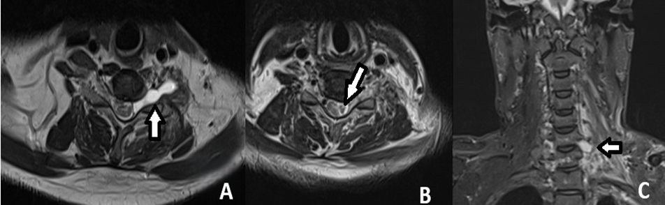
Figure 1. Magnetic resonance imaging demonstrating traumatic spinal nerve-root/trunk transection. Axial (A and B) and coronal (C) images show focal discontinuity of the cervical nerve-root/brachial plexus elements at the sites indicated by arrows.
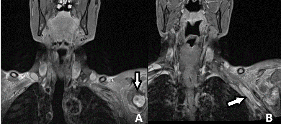 Figure 2. Coronal magnetic resonance images showing a pseudoaneurysm in the supraclavicular region producing compressive mass effect on the adjacent brachial plexus (arrows).
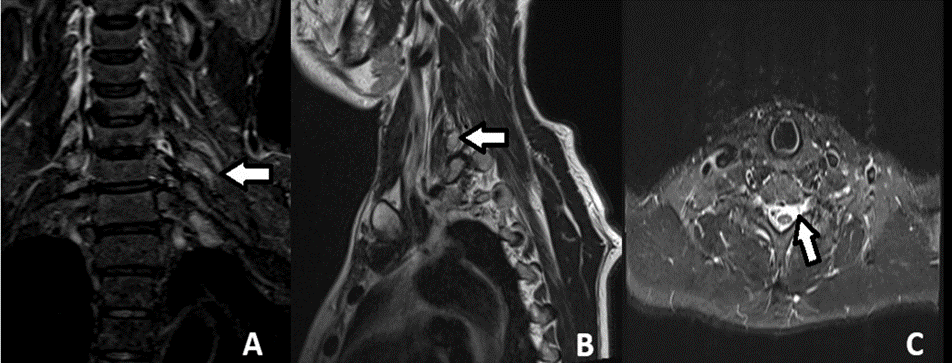
Figure 3. Magnetic resonance imaging demonstrating a traumatic pseudomeningocele. Coronal (A), sagittal (B), and axial (C) images show a cerebrospinal-fluid–intensity outpouching adjacent to the cervical neural foramen/nerve root (arrows).
Electrodiagnostic interpretations were available for 15 patients (83.3%) and were abnormal in all tested cases. Pan-plexus or global involvement was reported in 7 (46.7%). Upper-trunk, upper-cord, or other focal cord involvement was present in 5 (33.3%), lower-plexus/root involvement in 2 (13.3%), and diffuse postganglionic involvement without a more specific level in 1 (6.7%). A motor axonal lesion was explicitly described in 8 (53.3%), while severe injury was explicitly reported in 6 (40.0%). Preganglionic or mixed preganglionic-postganglionic localization was stated in 2 patients (13.3%), and definite postganglionic involvement in 4 (26.7%). These findings indicate that extensive axonal injury was common, although localization varied across the plexus (Table 4).
Table 4. Electrodiagnostic profile among tested patients
|
Electrodiagnostic characteristic |
Value |
|
Electrodiagnostic data available |
15/18 (83.3%) |
|
Abnormal study among tested patients |
15/15 (100.0%) |
|
Pan-plexus/global involvement |
7/15 (46.7%) |
|
Upper-trunk/upper-cord or focal cord involvement |
5/15 (33.3%) |
|
Lower-plexus/root involvement |
2/15 (13.3%) |
|
Diffuse postganglionic involvement, level unspecified |
1/15 (6.7%) |
|
Motor axonal lesion explicitly documented |
8/15 (53.3%) |
|
Severe injury explicitly documented |
6/15 (40.0%) |
|
Preganglionic or mixed pre-/postganglionic localization |
2/15 (13.3%) |
|
Definite postganglionic localization |
4/15 (26.7%) |
Percentages use the 15 patients with available nerve conduction/electromyographic interpretations as the denominator.
DISCUSSION:
The present study demonstrates that traumatic brachial plexopathy in a tertiary neurology setting predominantly affects young men after vehicle-related trauma. The mean age was 30.5 years, 94.4% were male, and 77.8% had vehicle-associated injury. This pattern closely resembles established epidemiological observations. Midha reported a strong predominance of young men and a disproportionate risk among motorcycle crash victims, while the Indian series by Jain et al. found road traffic accidents in 94% of patients and two-wheelers in 90% of those accidents.
4,5 The slightly lower proportion of road trauma in our cohort reflects the inclusion of falls, direct limb trauma, and one birth-related injury. Weakness was universal and usually showed proximal predominance. Nearly two-thirds of patients with documented proximal power had strength of grade 2/5 or lower, whereas distal strength remained grade 4/5 or better in more than half. Absent or depressed reflexes were frequent, and approximately half had objective sensory impairment. These findings are compatible with severe traction injuries affecting roots and trunks, although the near-equal distribution of right- and left-sided lesions indicates no clinically meaningful side preference. Clinical examination remains essential for defining functional loss, but mixed injury patterns and multilevel involvement reduce the accuracy of localization based on examination alone. 1,2
Electrodiagnostic testing was abnormal in all 15 evaluated patients. Pan-plexus involvement accounted for 46.7%, and severe injury was explicitly described in 40.0%. Puri et al. reported diffuse involvement in 30.8% of traumatic cases and found severe disability across the traumatic group. 7 Chuang et al. also demonstrated that mechanism of trauma influences the predominant anatomical level and that motorcycle- and fall-related injuries commonly produce severe incomplete lesions. 8 The higher proportion of global involvement in our cohort could reflect referral of more symptomatic patients to neurology and the small sample size.
Electrodiagnostic assessment remains valuable because it documents axonal loss, identifies denervation, supports preganglionic-postganglionic differentiation, and provides a baseline for serial recovery assessment. 6. MRI was abnormal in all 12 imaged patients, with signal alteration, thickening, or edema in most cases. Root avulsion and pseudomeningocele were identified in three patients. MRI is central to the structural evaluation of traumatic plexopathy, but its accuracy varies with acquisition technique, timing, and the reference standard. 9,10 Wade et al. reported high pooled sensitivity but only moderate specificity for root avulsion, whereas Acharya et al. found lower sensitivity and emphasized the association between pseudomeningocele and surgically confirmed avulsion.
11,12 The lower avulsion frequency in our cohort compared with surgical series is expected because our study included all clinically diagnosed cases rather than only patients selected for exploration. The findings support a coordinated diagnostic pathway integrating history, detailed examination, electrodiagnostic testing, and MRI. Combined assessment improves root-level localization and management planning compared with an isolated test. 13 Early rehabilitation, pain care, joint protection, and maintenance of muscle and sensory function should proceed alongside surgical evaluation, particularly in extensive injuries. 14
LIMITATIONS
This study has limitations. The sample was small and derived from a single tertiary neurology centre, restricting broader generalizability. MRI and electrodiagnostic testing were not available for every participant, and variable-wise denominators reduced direct cross-modality comparison. The dataset contained report-level interpretations rather than raw electrophysiological measurements or standardized MRI grading. Treatment, surgical findings, serial follow-up, pain scores, and functional outcome measures were not consistently recorded.
CONCLUSION:
Traumatic brachial plexopathy in this tertiary-care cohort mainly affected young men following vehicle-related trauma, with a slight predominance of right-sided lesions. Severe proximal weakness, depressed or absent reflexes, sensory impairment, and extensive axonal abnormalities were common. Nearly half of the patients undergoing electrodiagnostic testing had pan-plexus involvement, while MRI frequently demonstrated plexus edema or thickening and identified root avulsion with pseudomeningocele in selected cases. These observations underline the clinical value of combining neurological examination, nerve conduction studies, electromyography, and brachial plexus MRI. Early multimodal localization can guide referral, determine the need for reconstructive assessment, establish a baseline for recovery, and support timely rehabilitation, joint protection, pain control, and functional planning.
REFERENCES:
1. Moran SL, Steinmann SP, Shin AY. Adult brachial plexus injuries: mechanism, patterns of injury, and physical diagnosis. Hand Clin. 2005;21(1):13-24. doi:10.1016/j.hcl.2004.09.004.
2. Thatte MR, Babhulkar S, Hiremath A. Brachial plexus injury in adults: diagnosis and surgical treatment strategies. Ann Indian Acad Neurol. 2013;16(1):26-33. doi:10.4103/0972-2327.107686.
3. Landers ZA, Jethanandani R, Lee SK, Mancuso CA, Seehaus M, Wolfe SW. The psychological impact of adult traumatic brachial plexus injury. J Hand Surg Am. 2018;43(10):950.e1-950.e6. doi:10.1016/j.jhsa.2018.02.019.
4. Midha R. Epidemiology of brachial plexus injuries in a multitrauma population. Neurosurgery. 1997;40(6):1182-8; discussion 1188-9. doi:10.1097/00006123-199706000-00014.
5. Jain DKA, Bhardwaj P, Venkataramani H, Sabapathy SR. An epidemiological study of traumatic brachial plexus injury patients treated at an Indian centre. Indian J Plast Surg. 2012;45(3):498-503. doi:10.4103/0970-0358.105960.
6. Mansukhani KA. Electrodiagnosis in traumatic brachial plexus injury. Ann Indian Acad Neurol. 2013;16(1):19-25. doi:10.4103/0972-2327.107682.
7. Puri V, Chaudhry N, Jain KK, Chowdhury D, Nehru R. Brachial plexopathy: a clinical and electrophysiological study. Electromyogr Clin Neurophysiol. 2004;44(4):229-35.
8. Chuang TY, Chiu FY, Tsai YA, Chiang SC, Yen DJ, Cheng H. The comparison of electrophysiologic findings of traumatic brachial plexopathies in a tertiary care center. Injury. 2002;33(7):591-5. doi:10.1016/S0020-1383(02)00094-3.
9. Silbermann-Hoffman O, Teboul F. Post-traumatic brachial plexus MRI in practice. Diagn Interv Imaging. 2013;94(10):925-43. doi:10.1016/j.diii.2013.08.013.
10. Szaro P, Geijer M, Ciszek B, McGrath A. Magnetic resonance imaging of the brachial plexus. Part 2: traumatic injuries. Eur J Radiol Open. 2022;9:100397. doi:10.1016/j.ejro.2022.100397.
11. Wade RG, Takwoingi Y, Wormald JCR, Ridgway JP, Tanner S, Rankine JJ, et al. MRI for detecting root avulsions in traumatic adult brachial plexus injuries: a systematic review and meta-analysis of diagnostic accuracy. Radiology. 2019;293(1):125-33. doi:10.1148/radiol.2019190218.
12. Acharya AM, Cherian BS, Bhat AK. Diagnostic accuracy of MRI for traumatic adult brachial plexus injury: a comparison study with surgical findings. J Orthop. 2019;17:53-8. doi:10.1016/j.jor.2019.08.015.
13. Chanlalit C, Vipulakorn K, Jiraruttanapochai K, Mairiang E, Chowcheun P. Value of clinical findings, electrodiagnosis and magnetic resonance imaging in the diagnosis of root lesions in traumatic brachial plexus injuries. J Med Assoc Thai. 2005;88(1):66-70.
14. Li H, Chen J, Wang J, Zhang T, Chen Z. Review of rehabilitation protocols for brachial plexus injury. Front Neurol. 2023;14:1084223. doi:10.3389/fneur.2023.1084223.