Significance Of Crown Rump Length (CRL) and Foetal Heart Rate (FHR) In First Trimester Ultrasound In Predicting Spontaneous Abortion.
- Ramya T N , Assistant Professor, Department of OBG, Koppal Institue of Medical Sciences, Koppal
- Syeda Haafiza Begum , Consultant Obstetrician and Gynaecologist, Dept. of OBG, CHC Moka, Ballari District. India
- Sumitra Sangavi , Assistant Professor, Department of OBG, Mahavir Institue of Medical Sciences, Vikarabad
- Ramesh A C , Professor, Department of OBG, JJM Medical College, Davangere.
Article Information:
Abstract:
Background: There is inconsistent evidence for an association between early growth restriction, as defined by a deficit between the CRL and that predicted by the last menstrual period, and genetic abnormalities. A smaller than expected CRL has, however, been associated with poor pregnancy outcome. Objective: To study the Significance Of Crown Rump Length (CRL) and Foetal Heart Rate (FHR) In First Trimester Ultrasound In Predicting Spontaneous Abortion. Methods: This Prospective observational study was conducted among All pregnant women up to 12weeks of gestation visiting hospital for regular ANC check-up, visiting to OPD in hospitals attached to J.J.M. Medical College, Davangere ie Bapuji Hospital, Davangere. Chigateri General Hospital, Davangere and Women and Children Hospital, Davangere. Duration of study was Two years from September 2020 to September 2022. Result: The ultrasonographic parameters FHR less than 5th centile for the corresponding gestational age has been demonstrated as significant predictors of spontaneous abortion in the present study. The demographic characteristics such as high parity, maternal age and duration of married life had increased risk of poor pregnancy outcome in our study. The incidence of abortion was higher in early pregnancy. Conclusion: The present study adds to the growing body of evidence that the abnormal ultrasonographic biometry along with demographic characteristics can be used as predictors of pregnancy outcome.
Keywords:
Article :
INTRODUCTION:
Abortion is defined as spontaneous or induced termination of pregnancy before period of foetal viability. The National Centre for Health Statistics, the Centre for Disease Control and Prevention and the World Health Organization have all defined abortion as pregnancy termination or loss before 20 weeks of gestation or with a foetus delivered weighing less than 500 grams. Spontaneous abortion incidence varies according to the study population. In pregnancies of 5 to 20 weeks gestation, the incidence ranges from 11 to 22 % and is higher in earlier weeks of gestation.1
Pain abdomen in first trimester may be due to urinary tract infection, ligament pain, constipation, abortion, ectopic pregnancy and many more. The various reasons that might cause vaginal bleeding in first trimester include local causes such as cervical polyp, cervical erosions, carcinoma cervix, local inflammation, or placental pathology such as subchorionic haemorrhage, molar pregnancy, ectopic pregnancy and abortion.2 Backache in first trimester may be associated with calcium deficiency and stretching of ligaments due to enlarging uterus.
Depending on the history, period of gestation, clinical findings and ultrasonography abortion can be diagnosed.
Ultrasound is the primary imaging modality that has been rapidly replacing all other techniques used to study normal embryonic development in the first trimester.3
The use of ultrasound has clearly revolutionized the management of early pregnancy difficulties. The development of highly sensitive urinary hCG assays and larger awareness of early pregnancy ultrasound amongst health care providers and pregnant women has resulted in ever earlier presentation. This has resulted in an increase in the number of inconclusive scans and as a result an increase in the requirement for repeat ultrasound follow up to determine both pregnancy location and viability. Knowledge of the typical ultrasound appearances of normal early pregnancy development, a good understanding of its abnormalities and disadvantages are essential for the diagnosis and management of abortions. Use of appropriate terminology to describe clinical and ultrasound findings in early pregnancy failure is also required, and the use of obsolete descriptions like blighted ovum, anembryonic sac and trophoblastic bleeding should be abandoned. Such terms are of limited clinical usefulness and have no histopathological correlates and have therefore been replaced by more appropriate ultrasound-based terminology.
The significance of ultrasound markers, biochemical markers and demographic data in predicting abortion have been published in various literature.3,5 However, most of them are retrospective studies with contradictory results. Many of them have focused on one variable or a combination of only a few.
Wide range of research study has been published examining the measurement of foetal heart pulsation as the predictor of pregnancy outcome. Studies can be broadly divided into those examining foetal demise after confirmed foetal cardiac activity, and those examining foetal heart rate (FHR) in relation to pregnancy outcome.
Foetal cardiac activity is the earliest determinant of a viable pregnancy and it has been measured in utero by ultrasound as early as 36 days of menstrual age, approximately at the time when the heart tube initiates to beat. Theoretically, cardiac activity should always be present when the embryo measures more than 6 mm. However, in around 5–10% of embryos between 2 and 4 mm it cannot be measured, although the corresponding pregnancies will have a healthy outcome.
From 5 to 9 weeks of gestation there is a sudden increase in the mean foetal heart rate from 110 to 175 beats per minutes (bpm). The foetal heart rate then progressively decrease to around 160–170 bpm. Abnormal developmental pattern of FHR and/or bradycardia has been associated with poor pregnancy outcome. In specific, bradycardia at 6–8 weeks has shown to be associated with subsequent foetal death.
Therefore, the present study is a prospective study done to know the significance of Crown Rump Length (CRL) and Foetal Heart Rate (FHR) in first trimester ultrasound in predicting spontaneous abortion along with the impact of demographic factors. These also determine what constitutes abnormal early pregnancy and helps doctors counsel the patients under their care.
MATERIALS AND METHODS:
This Prospective observational study was conducted among All pregnant women up to 12weeks of gestation visiting hospital for regular ANC check-up, visiting to OPD in hospitals attached to J.J.M. Medical College, Davangere ie Bapuji Hospital, Davangere. Chigateri General Hospital, Davangere and Women and Children Hospital, Davangere. Duration of study was Two years from September 2020 to September 2022
SAMPLE SIZE ESTIMATION
Sample Size = Zα2 P (1-P)
d2 Zα2= Std normal variate 1.96
P = Expected proportion from population d = Absolute error
Sample size was estimated using the above formula
Prevalence of spontaneous abortion in First Trimester is 15%6 Considering 5% error sample needed to conduct this study is 196 cases.
It is follow-up study, dropout rate of 10% has been considered to calculate sample size.
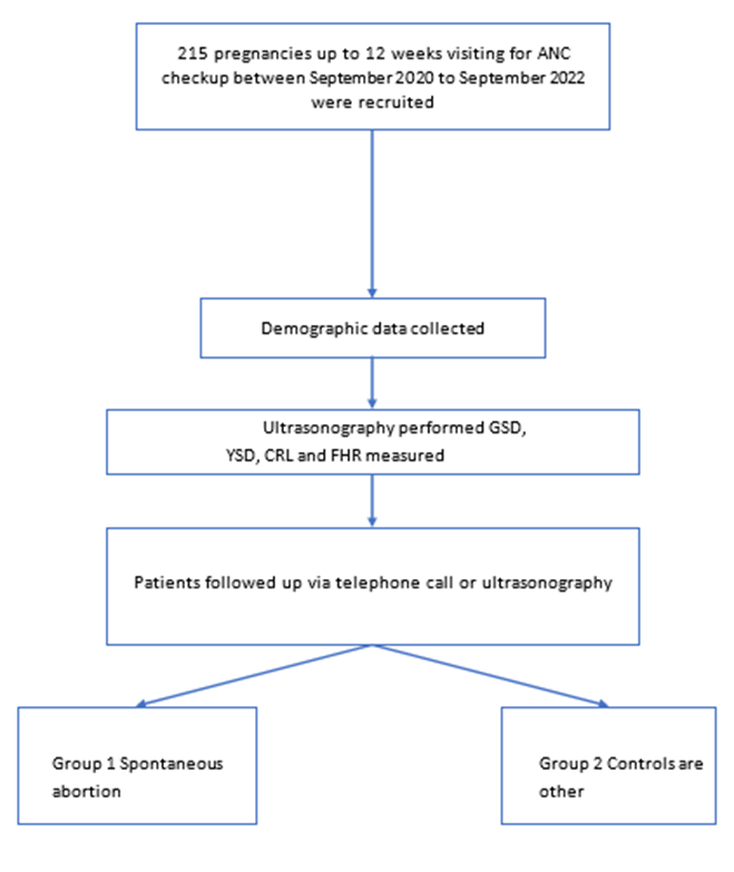
215 cases will be selected. Consecutive patients will be selected,
Pregnancy outcome at 20 completed weeks was divided into two groups.
Group-1 - spontaneous abortion group Group-2 - controls are the other cases.
Inclusion criteria:
• Pregnancies up to 12 weeks of gestation with documented embryonic cardiac activity
• Patient willing to give informed consent
• Singleton foetus
• Women with known LMP
• Regular menstrual cycles of 26 to 30 days.
Exclusion criteria:
• Multiple pregnancies
• Extrauterine pregnancy
• Chromosomal anomalies
• Elective termination of pregnancy
• Hormonal contraception use
• Pregnancy or breastfeeding in the 3 months preceding the pregnancy
• Women with unknown LMP
• Patient not willing to give informed consent
Procedure Of The Study:
After obtaining approval and clearance from the institutional ethics committee, the patients fulfilling the inclusion criteria was enrolled for the study after obtaining informed consent. All consecutive pregnant women up to 12 weeks of gestation consulting for regular ANC check-up were recruited.
Demographic data such as maternal age, parity, married life, method of conception, previous history of abortion and associated symptoms (bleeding per vagina, suprapubic pain, backache), if any were obtained. Transabdominal ultrasound was carried out to determine foetal CRL and FHR,
Measurement of foetal heart rate:
In the first trimester the measurement of heart rate should be performed using turnover M-mode. The heart rate increases rapidly from six to eight weeks and then remains relatively stable afterwards.
Measurement of crown rump length:
A properly performed measurement of CRL is the most accurate means of estimating the gestational age. An optimal CRL image, accurately measured, is more accurate in dating a pregnancy. It is measured by obtaining a true, unflexed, longitudinal section of the embryo or foetus, with the end-points of the crown and rump clearly defined, and then placing the callipers correctly on these defined end points. Three measurements are taken and the average of them is taken as the crown rump length.
All pregnancies were followed up to 20 weeks of gestation with subsequent ultrasound scan or telephone interview.
Group definitions:
Group-1 - spontaneous abortion group Group-2 - controls are the other cases
Statistical analysis: Statistical analysis was performed on the data obtained. Categorical data will be represented in the form of frequency and percentage. Association of variables will be assessed with Chi Square test, Quantitative data will be represented as Mean + Sd. Comparison will be done with unpaired t test.
The accuracy of the tests will be assessed using sensitivity, specificity, positive predictive value (PPV) and negative predictive value (NPV). Associations between measured variables and miscarriage will be estimated using receiver operating characteristic (ROC) curves. P value of <0.05 will be considered statistically significant. BM SPSS Version 25 for windows will be used for analysing the data.
RESULTS:
In our study mean age of study population is – 24.1±4.13 years with minimum 18 years and maximum of 42 years. Large number of patients were in between age group of 20 and 35 years that is 176. Among which 32 had spontaneous abortion and 144 had healthy pregnancy outcome. The mean age of patients in spontaneous abortion group is statistically higher than the ongoing pregnancy group with statistical significance of 0.0204
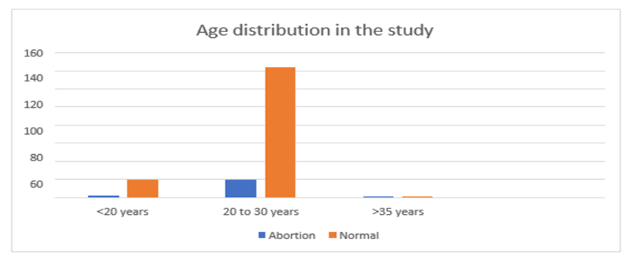
Graph 1: Age distribution in the study
Regarding parity, primiparous to multiparous women were included. In parity analysis primipara were 100 cases. Among 100 primiparous, 12 had spontaneous abortion and other 88 had normal pregnancy. Among 100 multiparous, 23 had spontaneous abortion and other 77 had normal pregnancy. In our study multiparous women had statistically higher abortion compared to nulliparous women. In spontaneous abortion group the mean years of married life is 4.36±2.78 years and in normal pregnancy group it is 3.24±2.72 years. Past history of abortion did not show any statistical significance in the present study.
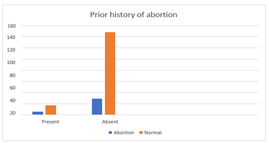
Graph 2: Prior history of abortion
The present study signifies that incidence of abortion is relatively high in early weeks of gestation. The result is statistically significant with a p value of <0.0001. In our study the most significant result is obtained about the fact that the incidence of abortion is higher in early weeks of gestation. The age of patient, years of married life and parity is statistically higher in women of spontaneous abortion group compared to ongoing pregnancy group. Prior miscarriage did not show statistical significance in the present study.
Bleeding per vagina and abdominal pain was found to be significantly associated with spontaneous abortion with odds ratio of 67.67 and 87.81 respectively. However, back pain although statistically significant in the present study its odds ratio was 3.4615 only.
Significance Of Ultrasound Markers In The Study
CROWN RUMP LENGTH
In the present study mean crown rump length in spontaneous abortion group is 10.39mm whereas in ongoing pregnancy group it is 22.94mm. The CRL is statistically higher in ongoing pregnancy group and it is statistically significant with p value- <0.0001
Table 1: CRL according to length of pregnancy in the study groups
|
Length
of pregnancy, d |
No. of cases |
CRL (in mm) |
|||
|
SA |
OP |
SA |
OP |
p-value |
|
|
42-48 |
17 |
17 |
4.28±1.79 |
5.88±2.18 |
<0.0001 |
|
49-55 |
8 |
25 |
7.77±2.34 |
12.1±1.48 |
<0.0001 |
|
56-62 |
3 |
41 |
12±2 |
18.56±2.59 |
<0.0001 |
|
63-69 |
1 |
28 |
25±0 |
25.5±3.33 |
<0.0001 |
|
70.76 |
6 |
54 |
28±6.6 |
35.3±5.12 |
<0.0001 |
|
Overall |
35 |
165 |
10.39±9.52 |
22.94±10.89 |
<0.0001 |
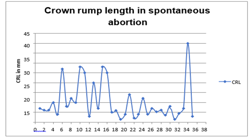
Graph 3: CRL in spontaneous abortion
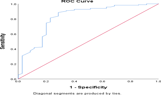
Graph 4: ROC Curve of CRL
|
Area Under the Curve |
||||
|
Test Result Variable(s): CRL |
||||
|
Area |
Std. Errora |
Asymptotic Sig.b |
Asymptotic 95% Confidence Interval |
|
|
Lower Bound |
Upper Bound |
|||
|
.823 |
.044 |
.000 |
.737 |
.909 |
Table 2: Significance of CRL in the study groups
|
CRL |
Total (n=200) |
SA (n=35) |
OP (n=165) |
p-value |
RR |
Sensitivity % |
Specificity % |
PPV |
NPV |
LR |
OR |
|
<5th
centile |
33 |
22 |
11 |
<0.001 |
8.46 |
59.25 |
96.77 |
94.11 |
73.17 |
0.42 |
23.6923 |
|
5th -95th
centile |
160 |
12 |
148 |
<0.001 |
0.13 |
37.04 |
6.45 |
25.64 |
10.53 |
0.40 |
0.0599 |
|
>95th centile |
7 |
1 |
6 |
0.229 |
0.81 |
3.70 |
96.77 |
50.00 |
53.57 |
1.15 |
0.7794 |
The above table depicts the significance of CRL less than 5th centile as a predictor of spontaneous abortion with statistical significance of p value- <0.001 and specificity of 96.877%. It also has positive predictive value of 94.11 and negative predictive value of 73.17. Hence, it can be considered as one of the good prognostic markers in prediction of spontaneous abortion.
FETAL HEART RATE
In the present study the mean foetal heart rate in the spontaneous abortion group is 111 bpm whereas in the ongoing pregnancy group it is 159 bpm. Hence the foetal heart rate in ongoing pregnancy is statistically higher than spontaneous abortion group with significance of <0.0001.
Table 3: FHR according to length of pregnancy in the study groups
|
Length
of pregnancy, d |
No. of cases |
FHR (in bpm) |
|||
|
SA |
OP |
SA |
OP |
p-value |
|
|
42-48 |
17 |
17 |
99.1±8.74 |
129.5±12 |
<0.0001 |
|
49-55 |
8 |
25 |
124.5±16.72 |
152.52±10.13 |
<0.0001 |
|
56-62 |
3 |
41 |
134.67±25.40 |
166.97±9.63 |
<0.0001 |
|
63-69 |
1 |
28 |
110±0 |
172±6.61 |
<0.0001 |
|
70.76 |
6 |
54 |
116±19.14 |
160.38±10.23 |
<0.0001 |
|
Overall |
35 |
165 |
111.17±18.78 |
159.67±15.32 |
<0.0001 |
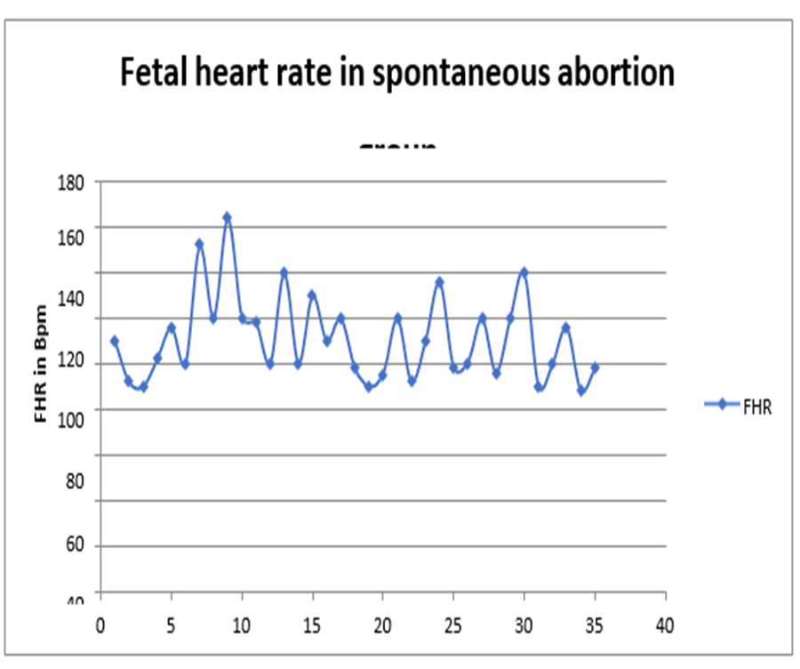
Graph 5: FHR in spontaneous abortion group
Table 4: FHR and Risk of spontaneous abortion
|
FHR, bpm |
Total (n=200) |
SA(n=35) |
OP(n=165) |
Abortion frequency, % |
|
≤ 80 |
0 |
0 |
0 |
0 |
|
81-90 |
4 |
4 |
0 |
100 |
|
91-100 |
12 |
12 |
0 |
100 |
|
101-110 |
4 |
4 |
0 |
100 |
|
111-120 |
14 |
9 |
5 |
64 |
|
121-130 |
12 |
1 |
11 |
8 |
|
131-140 |
11 |
3 |
8 |
27 |
|
141-150 |
16 |
1 |
15 |
6 |
|
151-160 |
38 |
1 |
37 |
3 |
|
161-170 |
55 |
0 |
55 |
0 |
|
171-180 |
32 |
0 |
32 |
0 |
|
≥ 181 |
2 |
0 |
2 |
0 |
There was a sharp increase in the frequency of spontaneous abortion when FHR was less than 130 bpm. The coefficient of determination for FHR below 130 bpm was 0.574. Therefore, for every 10-bpm decrease in FHR below 130, the risk of abortion increased by 57.4%. The optimum cut off value of FHR for the continuation of pregnancy was 132 bpm.
Table 5: Significance of FHR in the study groups
|
FHR |
Total (n=200) |
SA (n=35) |
OP (n=165) |
p-value |
RR |
Sensitivity % |
Specificity % |
PPV |
NPV |
LR |
OR |
|
<5th
centile |
27 |
23 |
4 |
<0.001 |
12.32 |
73.33 |
87.50 |
91.67 |
63.64 |
5.87 |
74.2708 |
|
5th -95th
centile |
167 |
12 |
155 |
<0.001 |
0.1 |
26.67 |
87.50 |
80.00 |
38.89 |
2.13 |
0.0337 |
|
>95th
centile |
6 |
0 |
6 |
0.652 |
0 |
0 |
100 |
- |
34.78 |
- |
0.3456 |
The above table depicts FHR less than 5th centile has strong association with spontaneous abortion with statistical significance of <0.001. It also has specificity of 87.5% and positive predictive value of 91.67. FHR more than 95th centile has 100% specificity for normal pregnancy outcome in the present study. Hence, FHR can be considered as a good prognostic marker for predicting outcome of pregnancy.
Table 6: Range and mean of the ultrasonographic markers
|
Variables |
Minimum |
Maximum |
Mean±SD |
|
CRL (mm) |
1.5 |
40 |
10.39±9.53 |
|
FHR (bpm) |
88 |
164 |
111.17±18.78 |
Table 7: Significance of ultrasonographic markers
|
Parameter |
AUC
(95% CI) |
SE |
Best
cut-off |
Sensitivity
(%) |
Specificity
(%) |
p-value |
|
CRL |
0.823(.737-
0.909) |
0.044 |
10.2 |
88.5 |
71.4 |
<0.0001 |
|
FHR |
0.963(0.92
9-0.996) |
0.017 |
132 |
90.3 |
85.7 |
<0.0001 |
The above table suggests area under the curve (AUC) was excellent for HR, which was 0.963. The best cut-off values were derived for all parameters in ROC and it was found that CRL of less than 10.2 mm, it was 88.5% and 71.4%; for FHR less than 132 bpm, it was 90.3% and 85.7% respectively. Hence, FHR can be considered as the best prognostic marker in the present study.
DISCUSSION:
Higher risk of spontaneous abortion is noted in earlier weeks of gestation compared to ongoing pregnancy group with statistical significance of p value <0.0001 Studies have demonstrated that in pregnancies aged 5 to 20 weeks gestation, the incidence of abortion ranges from 11 to 22 percent and is higher in earlier weeks.1 Many pregnancies abort before the pregnancy is confirmed by either ultrasound or a chemical pregnancy test. Approximately 50% of miscarriage is caused by chromosomal abnormalities.7
Early pathologic studies of Hertig and Rock et.al., also showed a high frequency of morphologic abnormalities in preimplantation embryos.8 Loss rates are increased with increased maternal age and prior history of early pregnancy failure. However, the past history of abortion did not show statistical significance in the present study.9 The association between maternal age and abortion reflects both biological mechanisms and forces of selection, meaning increased risk is attributable to chromosomal abnormalities and medical comorbidities at older age.10
There was no significant association of spontaneous abortion with previous abortion in our study, which was in accordance to the study by Makrydimas et al11 The duration of married life, age of the mother and parity was statistically higher in spontaneous abortion group compared to the ongoing pregnancy group with statistical significance in our study. This adds to the existing literature, supporting the association of above-mentioned factors with miscarriage.10
Makrydimas et al. noted that the incidence of miscarriage was 2.6 times higher among subjects with history of bleeding per vagina.11 Bleeding per vagina is an early feature of miscarriage. A study by Falco et al. found the rate of miscarriage in a woman with the bleeding episode in a viable pregnancy was 10%, whereas with nonviable pregnancy it was 40%.12 We found that abdominal pain is significantly associated with spontaneous abortion (OR-87.81). A study by Wie et al. found statistically significant higher incidence of miscarriage among woman with abdominal pain;5
Several previous studies have examined whether different sonographic variables measured in early pregnancy can predict pregnancy outcome. In a study of 556 pregnancies, Frates et al. observed that the spontaneous abortion rate was higher among women with abnormal sonography findings than among those with normal findings (15.2% versus 8.2%).13
The small CRL (<5th centile for gestational age) was found in 16.5% in the study, resulted in 66.66% abortion rate. It had a sensitivity of 59.25%, specificity of 96.77%, NPV of 73.17%, PPV of 94.11% and odds ratio of 23.6923. The CRL below 5th centile showed a significant association with spontaneous abortion with p value-<0.001. Small CRL indicates either delayed conception or growth restriction but not producing immediate mishaps, hence with less detection power (low sensitivity). The higher CRL >95th centile did not associate with higher pregnancy mishaps.
Abuelghar et al. reported that 56.6% of women who experienced abortion had CRL below the 5th percentile, and this variable predicted early abortion with a sensitivity of 56.6%, specificity of 81.9%, PPV of 36.6%, NPV value of 91.1%, and likelihood ratio positive of 3.1.14 D’Antonio et al. found that CRL below the 5th percentile was associated with abortion with an odds ratio of 2.21 (P=0.023).15 Similarly, Mukri et al. reported that 61% of women who experienced abortion had CRL below the 5th percentile.16
In the present study, FHR below the 5th percentile had a sensitivity of 73.33%, specificity of 87.5%, PPV of 91.67% and NPV of 63.64% for predicting early spontaneous abortion. FHR below 5th centile was significantly associated with spontaneous abortion with p value of <0.001. The present study also confirms that embryonic FHR has highest predictive value among different parameters. Hence all reported bradycardia needs to be evaluated after 7–10 days for the outcome.
Wie et al. also concluded that FHR below the 5th percentile had a significant correlation with spontaneous abortion (OR 6.43’ P=0.018).5 Papaioannou et al. showed that the sensitivity of predicting abortion was 23.9% with a false-positive rate of 5% for FHR values below the 5th percentile.4 Altay et al. concluded that FHR below the 5th percentile had a sensitivity of 50%, specificity of 94.9%, PPV of 50%, NPV of 98.9%, and P value 0.01 for prediction of abortion. 17 Similarly, Makrydimas et al. reported a sensitivity of 26.5% with a false-positive rate of 5% for FHR values below the 5th percentile.11
Dede et al. found that an FHR value below 130 bpm had 81.4% sensitivity and 85.1% specificity for predicting abortion.18 Chittacharoen and Herabutya reported that FHR values below 120 bpm predicted early abortion with a sensitivity of 54.2% and false-positive rate of 5%.19 In the present study, the optimum cut off for FHR in the prediction of abortion was 132 bpm.
CONCLUSION:
In the present time ultrasonography is routinely used by the obstetric care providers. It is widely used as it is a simple, non-invasive and cost effective. Hence it is also used in the prediction and diagnosis of abortion. It is also important to predict the risk of abortion as symptoms of pain abdomen, bleeding per vagina, advanced maternal age and patients conceived through infertility treatment are anxious about the outcome of their pregnancy. Patients with prediction of poor prognosis outcome could be counselled about the risk of abortion and need for intensive treatment. In contrast, the pregnant women having lower risk could be assured and further work up could be done at 11 to 13 weeks for examination of foetal aneuploidies. The predictive markers help the clinicians to provide counselling and further management to their patients.
In the present study we demonstrated that the demographic characteristics such as high parity, maternal age and duration of married life was significantly associated with abortion. However, history of prior abortion did show statistical significance. The incidence of abortion is higher in early pregnancy in our study which adds to the existing evidence which states that risk of abortion is higher in earlier weeks of gestation. The present study demonstrated that the symptoms of bleeding per vagina and lower abdominal pain has strong association with spontaneous abortion.
Our study demonstrates that CRL, FHR less than 5th centile has significant risk of abortion. Various studies have demonstrated the significance of these predictors individually or in combination few variables. Most of the studies were retrospective compared to prospective studies. Hence, we have done a prospective study to know the significance of ultrasonographic parameters (CRL and FHR) along with demographic characteristics in the prediction of spontaneous abortion. Hence, the present study adds to the growing body of evidence that the abnormal ultrasonographic biometry along with demographic characteristics can be used as predictors of pregnancy outcome.
REFERENCES:
1. Williams obstetrics. 25th ed. Chapter 18 on page 346, 347
2. Predictors of vaginal bleeding during pregnancy and its relation to preterm birth. The University of North Carolina at Chapel Hill; 2003.
3. Jauniaux E, Johns J, Burton GJ. The role of ultrasound imaging in diagnosing and investigating early pregnancy failure. Ultrasound in Obstetrics and Gynecology: The Official Journal of the International Society of Ultrasound in Obstetrics and Gynecology. 2005 Jun;25(6):613-24.
4. Papaioannou GI, Syngelaki A, Maiz N, Ross JA, Nicolaides KH. Ultrasonographic prediction of early miscarriage. Human reproduction. 2011 Jul 1;26(7):1685-92.
5. Wie JH, Choe S, Kim SJ, Shin JC, Kwon JY, Park IY. Sonographic parameters for prediction of miscarriage: role of 3-dimensional volume measurement. Journal of Ultrasound in Medicine. 2015 Oct;34(10):1777-84.
6. Shah KH, Sultana R, Nambiar R, Bhat P, Bhat S. Prediction of early pregnancy loss by ultrasound markers. Journal of South Asian Federation of Obstetrics and Gynaecology. 2019;11(3).
7. Stephenson MD, Awartani KA, Robinson WP. Cytogenetic analysis of miscarriages from couples with recurrent miscarriage: a case–control study. Human reproduction. 2002 Feb 1;17(2):446-51.
8. Hertig AT, Rock J. A series of potentially abortive ova recovered from fertile women prior to the first missed menstrual period. American journal of obstetrics and gynecology. 1949 Nov 1;58(5):968-93.
9. Practice Committee of the American Society for Reproductive Medicine.
10. Evaluation and treatment of recurrent pregnancy loss: a committee opinion. Fertility and sterility. 2012 Nov 1;98(5):1103-11.
11. Andersen AM, Wohlfahrt J, Christens P, Olsen J, Melbye M. Maternal age and fetal loss: population based register linkage study. Bmj. 2000 Jun 24;320(7251):1708-12.
12. Makrydimas G, Sebire NJ, Lolis D, Vlassis N, Nicolaides KH. Fetal loss following ultrasound diagnosis of a live fetus at 6–10 weeks of gestation. Ultrasound in Obstetrics and Gynecology: The Official Journal of the International Society of Ultrasound in Obstetrics and Gynecology. 2003 Oct;22(4):368-72.
13. Falco P, Zagonari S, Gabrielli S, Bevini M, Pilu G, Bovicelli L. Sonography of pregnancies with first-trimester bleeding and a small intrauterine gestational sac without a demonstrable embryo. Ultrasound in Obstetrics and Gynecology: The Official Journal of the International Society of Ultrasound in Obstetrics and Gynecology. 2003 Jan;21(1):62-5.
14. Frates MC, Benson CB, Doubilet PM. Pregnancy outcome after a first trimester sonogram demonstrating fetal cardiac activity. Journal of ultrasound in medicine. 1993 Jul;12(7):383-6.
15. Abuelghar WM, Fathi HM, Ellaithy MI, Anwar MA. Can a smaller than expected crown–rump length reliably predict the occurrence of subsequent miscarriage in a viable first trimester pregnancy?. Journal of Obstetrics and Gynaecology Research. 2013 Oct;39(10):1449-55.
16. D'antonio F, Khalil A, Mantovani E, Thilaganathan B, Southwest Thames Obstetric Research Collaborative (STORK), Hamid R, Gandhi H, Ellis C, Deans A, Peregrine L, Breeze A. Embryonic growth discordance and early fetal loss: the STORK multiple pregnancy cohort and systematic review. Human Reproduction. 2013 Oct 1;28(10):2621-7.
17. Mukri F, Bourne T, Bottomley C, Schoeb C, Kirk E, Papageorghiou AT. Evidence of early first-trimester growth restriction in pregnancies that subsequently end in miscarriage. BJOG: An International Journal of Obstetrics & Gynaecology. 2008 Sep;115(10):1273-8.
18. Altay MM, Yaz H, Haberal A. The assessment of the gestational sac diameter, crown–rump length, progesterone and fetal heart rate measurements at the 10th gestational week to predict the spontaneous abortion risk. Journal of Obstetrics and Gynaecology Research. 2009 Apr;35(2):287-92.
19. Dede FS, Ulucay U, Kose MF, Dede H, Dilbaz S. Fetal loss in threatened abortion after demonstration of fetal cardiac activity in a low socioeconomic population. Journal of Obstetrics and Gynaecology. 2010 Aug 1;30(6):622-5.
20. Chittacharoen A, Herabutya Y. Slow fetal heart rate may predict pregnancy outcome in first-trimester threatened abortion. Fertility and sterility. 2004 Jul 1;82(1):227-9.