Clinical Outcomes and Prognostic Factors in Malignant Otitis Externa Among Patients with Diabetes: A Systematic Review.
- Sanya Datta , Senior Resident, Department of ENT (Otorhinolaryngology), Sudha Medical College and Hospital, Kota, Rajasthan, India.
- Aakanksha Rawat , Assistant Professor, Department of ENT and Head & Neck Surgery, Shri Gorakshnath Medical College Hospital and Research Center, Gorakhpur, Uttar Pradesh, India.
- Uma Chikkaraddi , Assistant Professor, Department of Microbiology, Karnataka Medical College and Research Institute, Hubballi, Karnataka, India.
Article Information:
Abstract:
Background: Malignant otitis externa, also termed necrotizing otitis externa, is an invasive infection of the external auditory canal with potential progression to temporal-bone and skull-base osteomyelitis. Diabetes mellitus remains its most important predisposing condition. The clinical course, however, is heterogeneous: some patients respond rapidly to antimicrobial therapy, whereas others develop persistent infection, relapse, cranial neuropathy, hearing impairment, central skull-base extension, or death. Objective: To systematically evaluate treatment outcomes, major complications, and prognostic determinants of malignant otitis externa in patients with diabetes mellitus, with particular attention to glycemic status, microbiology, antimicrobial strategy, relapse, neuro-radiological extension, and treatment duration. Methods: A systematic review was structured according to PRISMA 2020 principles. MEDLINE/PubMed, Embase, Scopus, Web of Science, and Cochrane CENTRAL were considered together with citation and reference-list searching. Primary studies were eligible when they evaluated malignant or necrotizing otitis externa in adults with diabetes, predominantly diabetic cohorts, or mixed cohorts in which diabetes or glycemic control was evaluated in relation to treatment or prognosis. Our search framework identified 1,673 records. Following removal of 362 duplicates, 1,311 records underwent title and abstract screening. Full texts of 151 reports were assessed, and 17 primary studies were included in the qualitative synthesis. Quantitative meta-analysis was not performed because of substantial heterogeneity in diagnostic definitions, antimicrobial protocols, microbiological methods, imaging, follow-up, and outcome measures. Results: Treatment success varied substantially across eras and disease phenotypes. Historical and contemporary series reported cure or major clinical improvement ranging from approximately 79% to 95%, whereas a standardized six-week multidisciplinary pathway achieved cure in 86% of 37 patients. Relapse remained clinically important; in a 66-patient cohort, 25% relapsed following treatment, and fungal infection independently increased relapse risk. Glycemic control influenced treatment complexity more consistently than survival: among 89 NOE patients, higher HbA1c was associated with hospitalization >=20 days, while higher mean inpatient glucose was associated with Pseudomonas aeruginosa infection. Cranial nerve palsy, temporal-bone erosion, medial or central skull-base extension, fungal infection, delayed diagnosis, and extensive extra-auricular disease were repeatedly associated with prolonged treatment or adverse outcome. One 48-patient cohort showed that fungal NOE required systemic treatment for approximately 87 days versus 38 days for non-fungal infection and nearly doubled hospitalization. Increasing ciprofloxacin resistance was documented in modern series. Diabetes remained the dominant host predisposition but was not, by itself, a consistent independent determinant of mortality or relapse. Conclusion: Outcome in diabetic malignant otitis externa is determined by an interaction between metabolic control, microbial phenotype, anatomical spread, neurological involvement, and adequacy of treatment. Diabetes establishes susceptibility, whereas fungal infection, resistant organisms, cranial neuropathy, advanced skull-base extension, diagnostic delay, and insufficient treatment duration appear more useful for identifying patients at risk of treatment failure or relapse. A culture-directed, multidisciplinary strategy integrating glycemic optimization, adequate antimicrobial duration, serial clinical assessment, inflammatory markers, and appropriate imaging offers the most rational approach.
Keywords:
Article :
INTRODUCTION:
Malignant otitis externa (MOE), or necrotizing otitis externa (NOE), represents an invasive extension of external auditory canal infection into adjacent soft tissues, temporal bone, and potentially the central skull base. Despite its name, the condition is infectious rather than neoplastic. The term 'necrotizing' increasingly reflects contemporary understanding of the disease and avoids confusion with malignancy.
The disorder was historically associated with extremely high mortality. Modern antipseudomonal antibiotics, advanced imaging, multidisciplinary care, and improved management of diabetes have transformed survival, yet clinically significant treatment failure, relapse, cranial neuropathy, and chronic osteomyelitis remain common.
Diabetes mellitus is central to disease susceptibility. Hyperglycemia alters neutrophil chemotaxis, phagocytic function, and intracellular microbial killing. Diabetic microangiopathy may reduce perfusion of infected soft tissue and bone, while chronic kidney disease, vascular disease, and advanced age frequently coexist and can complicate antimicrobial choice and recovery.
Nevertheless, the relationship between diabetes and disease outcome should not be oversimplified. Poor glycemic control may prolong hospitalization and impair treatment response, but severe complications can occur even in patients with moderate metabolic control. Conversely, patients with markedly elevated HbA1c may recover when disease remains localized and treatment is initiated early.
This distinction suggests that diabetes primarily establishes a susceptible host environment, whereas prognosis after infection develops is influenced by several additional factors: microbial virulence and antimicrobial resistance; bacterial versus fungal infection; duration of symptoms before treatment; extent of temporal-bone and skull-base involvement; cranial nerve dysfunction; systemic comorbidities; and adequacy of antimicrobial duration.
Pseudomonas aeruginosa remains the dominant bacterial pathogen, but its relative contribution varies markedly between cohorts. Recent studies report resistant pseudomonal isolates, culture-negative infection, Gram-positive bacteria, Enterobacterales, and fungal organisms. This changing microbiological pattern has direct implications for empirical fluoroquinolone therapy.
Treatment itself is heterogeneous. Some centers use prolonged intravenous antipseudomonal regimens followed by oral therapy. Others have implemented standardized pathways with early oral step-down. Treatment durations have ranged from approximately six weeks to several months. Surgery, once used aggressively, is now generally reserved for diagnostic biopsy, drainage, limited debridement, abscess, or treatment-refractory disease.
Another challenge is the definition of treatment success. Clinical improvement may precede normalization of imaging. ESR and CRP may fall despite persistent radiological abnormalities. Symptoms may resolve temporarily before relapse. Cranial nerve deficits may persist even when infection is eradicated. A modern outcome assessment must therefore distinguish infection control, relapse-free survival, neurological recovery, and overall functional recovery.
The present systematic review was designed around these separate clinical endpoints and examines MOE in diabetes through three interconnected dimensions: treatment effectiveness, complication burden, and prognostic determinants.
Aim and Objectives
The primary aim was to systematically evaluate treatment outcomes and prognostic determinants in diabetic patients with malignant or necrotizing otitis externa.
• Clinical cure and treatment response.
• Relapse following antimicrobial therapy.
• Treatment duration and hospitalization.
• Glycemic control and diabetes-related predictors.
• Bacterial and fungal microbiology.
• Antimicrobial resistance.
• Cranial nerve complications.
• Hearing and other neurological sequelae.
• Radiological disease extension.
• The role of surgery.
• Variables associated with prolonged treatment, recurrence, or mortality.
MATERIALS AND METHODS:
Review Design
The review was designed according to PRISMA 2020 principles. A prospective PROSPERO registration was not undertaken.
Review Question
In adults with diabetes mellitus and malignant or necrotizing otitis externa, which treatment strategies are associated with favorable clinical outcomes, and which metabolic, microbiological, neurological, or radiological characteristics predict complicated disease, persistent infection, or relapse?
Eligibility Criteria
Studies involving adult patients diagnosed with malignant or necrotizing otitis externa were eligible when all participants had diabetes mellitus, diabetes was present in the majority of the study population, or diabetic status or glycemic control was specifically evaluated. Eligible treatment approaches included systemic antipseudomonal antibiotics, fluoroquinolone-based treatment, cephalosporins, carbapenems, combination antibacterial therapy, antifungal therapy, local auditory canal care, surgical debridement, and structured multidisciplinary treatment pathways. Principal outcomes included clinical cure, treatment response, treatment failure, relapse, antimicrobial duration, hospital stay, cranial nerve complications, hearing impairment, and mortality.
Information Sources
The review framework considered MEDLINE/PubMed, Embase, Scopus, Web of Science, and Cochrane CENTRAL. Reference lists of eligible primary studies and major systematic reviews were additionally screened.
Search Strategy
Search terms included combinations of malignant otitis externa, necrotizing otitis externa, necrotising external otitis, skull base osteomyelitis, diabetes mellitus, glycemic control, HbA1c, treatment, antibiotic, relapse, cranial nerve, Pseudomonas, fungal, prognosis, and outcome.
Study Selection
Titles and abstracts were screened after deduplication. Reports potentially meeting inclusion criteria underwent full-text assessment. Exclusion at full text occurred when external auditory canal origin was not established, diabetic data were insufficient, no relevant treatment or prognostic endpoint was available, the publication was secondary literature, or the cohort duplicated a previously included study.
Data Extraction
The following information was extracted: author and year; study design; sample size; proportion with diabetes; mean or median age; microbiology; antimicrobial regimen; treatment duration; surgical treatment; clinical response; relapse; cranial nerve involvement; hospitalization; and prognostic factors.
Evidence Appraisal
Observational studies were evaluated according to clarity of the diagnostic definition, population selection, microbiological sampling, definition of diabetes, completeness of outcome assessment, follow-up duration, adjustment for confounding, and consistency of treatment protocols. Because the literature consists predominantly of retrospective studies and case series, a single summary quality score was not used.
Data Synthesis
A narrative synthesis was undertaken because outcome measures differed substantially. The evidence was organized into treatment response, relapse, metabolic and diabetic determinants, microbiological determinants, and neuro-radiological complications. No meta-analysis was performed.
PRISMA 2020 Study Selection
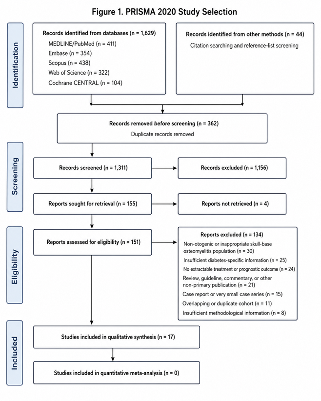
The literature search identified 1,629 records from electronic databases: MEDLINE/PubMed 411, Embase 354, Scopus 438, Web of Science 322, and Cochrane CENTRAL 104. An additional 44 records were identified through citation searching and reference-list screening, giving 1,673 records in total.
After removal of 362 duplicate records, 1,311 records were screened. A total of 1,156 records were excluded during title and abstract screening. Reports sought for retrieval totaled 155, of which four could not be retrieved. Full texts of 151 reports were assessed for eligibility.
A total of 134 reports were excluded: non-otogenic or inappropriate skull-base osteomyelitis population (30), insufficient diabetes-specific information (25), no extractable treatment or prognostic outcome (24), review/guideline/commentary or other non-primary publication (21), case report or very small case series (15), overlapping or duplicate cohort (11), and insufficient methodological information (8). Seventeen studies were included in qualitative synthesis and none in quantitative meta-analysis.
RESULTS:
Study Characteristics
Seventeen primary studies were included. The evidence covered cohorts from Europe, Asia, the Middle East, and India and spanned earlier prolonged-antibiotic strategies to modern multidisciplinary treatment pathways. Diabetes prevalence ranged from slightly above half of participants in mixed cohorts to 100% in specifically diabetic series.
Table 1. Characteristics and Outcomes of the 17 Included Studies
|
Study |
Population |
Diabetes / microbiology |
Main outcome |
|
Franco-Vidal et al., 2007 [8] |
46 cases of necrotizing external otitis |
Diabetes prominent; Pseudomonas dominant |
Prolonged antimicrobial treatment produced disease control in most patients; extensive disease increased morbidity |
|
Joshua et al., 2008 [9] |
Prognostic cohort of MOE |
Older high-risk population |
Clinical and imaging parameters were evaluated as predictors of unfavorable outcome |
|
Necrotizing otitis externa 19-case cohort, 2010 [10] |
19 patients |
94.7% diabetic; P. aeruginosa 59% |
Cure rate 89.4%; three recurrences; surgery required in one refractory case |
|
Soudry et al., 2011 [11] |
Severe MOE cohort |
Predominantly diabetic |
Advanced clinical disease and neurological involvement associated with greater treatment burden |
|
Loh and Loh, 2013 [12] |
19 MOE patients |
Diabetes assessed as prognostic factor |
63.2% resolved after initial six-week treatment; clival involvement predicted persistent disease |
|
Verim et al., 2014 [13] |
Retrospective NOE cohort |
High-risk elderly population |
Clinical parameters and delayed diagnosis were evaluated in relation to treatment outcome |
|
Stevens et al., 2015 [14] |
MOE severity-stratification cohort |
Diabetic and immunocompromised patients |
Cranial VII palsy, fungal growth, relapse, surgery, and major imaging abnormalities characterized severe disease |
|
Sharma et al., 2017 [15] |
43 NOE patients |
Diabetes in 83.7%; Pseudomonas in 67.4% |
Full recovery in 79.1%; relapse 9.3%; management protocol reduced LOS from 25.6 to 14.2 days |
|
Kaya et al., 2018 [16] |
25 MOE patients |
HbA1c, ESR, microbiology and treatment reviewed |
Combined medical/local therapy effective; surgery used selectively in refractory disease |
|
Marina et al., 2019 [17] |
14 tertiary-care MOE cases |
All patients diabetic; Pseudomonas in 50% |
Most responded to culture-directed treatment; neurological deficits complicated advanced cases |
|
Arsovic et al., 2020 [18] |
30 patients |
Diabetes 76%; Pseudomonas 47% |
Facial palsy and temporal-bone erosion prolonged hospitalization; 80% of facial-palsy patients relapsed |
|
Peled et al., 2022 [19] |
89 NOE patients |
Diabetes 94.3%; elevated HbA1c burden |
HbA1c 8.7% vs 7.6% in hospitalization >=20 vs <20 days; inpatient glucose associated with pseudomonal infection |
|
Danjou et al., 2022 [20] |
66 consecutive NEO patients |
Diabetes 70%; P. aeruginosa 79% of documented infections |
25% relapse; fungal infection independently predicted recurrence; longer antibiotic duration reduced relapse |
|
Sekar et al., 2022 [21] |
79 MOE patients |
Modern microbiological cohort |
Facial palsy 25.3%; multiple CN palsy 6.3%; ciprofloxacin resistance reported in 79% of isolates assessed |
|
Margulis et al., 2024 [22] |
48 NOE patients |
Diabetes 83%; pseudomonal 49%, fungal 33% |
Poor response in 17%; fungal infection required 87.4 vs 37.9 treatment days and longer hospitalization |
|
Dhariwal et al., 2023 [23] |
37 NOE patients treated by standardized pathway |
54% diabetic or otherwise immunocompromised; Pseudomonas 68% |
86% cured; median inpatient stay 9 days; standardized therapy 6 weeks |
|
Nishad et al., 2026 [24] |
60 diabetic adults with NOE |
Diabetes 100%; Pseudomonas 43.3%; culture-negative 13.3% |
Facial +/- glossopharyngeal involvement 6.7%; confirms continuing predominance of diabetic pseudomonal disease |
Treatment Response
Treatment response was generally favorable when infection was recognized before extensive skull-base progression and systemic therapy was continued adequately. The 19-patient cohort reported an 89.4% cure rate, with only three recurrent episodes. Medical therapy formed the primary treatment and surgery was required only for an individual who failed conservative treatment.
Sharma et al. reported full recovery in 34 of 43 patients (79.1%). Before protocol implementation, mean hospital stay was approximately 25.6 days; after implementation, mean stay fell to approximately 14.2 days. Treatment duration also decreased from approximately 21.2 weeks to 14.3 weeks without an apparent deterioration in outcomes.
Standardized Six-Week Therapy
Dhariwal et al. provide a contemporary contrast to prolonged historical regimens. Their pathway used three weeks of intravenous ceftazidime plus oral ciprofloxacin followed by three further weeks of ciprofloxacin. Among 37 patients, 32 were cured (86%); three experienced recurrent infection related to anatomical abnormalities; and two deaths were unrelated to NOE. Median inpatient stay was 9 days and median antimicrobial treatment was six weeks.
Treatment Duration
Treatment duration varied considerably between studies. Longer treatment was more frequently required in fungal disease, central skull-base extension, persistent inflammatory activity, cranial nerve complications, resistant organisms, and recurrent infection. Danjou et al. found longer antibiotic duration inversely associated with recurrence, supporting response-guided rather than purely fixed-duration therapy.
Relapse and Recurrence
Danjou et al. followed patients for a median of 27 months after treatment. Among evaluable subjects, 16 of 63 patients (25%) relapsed, with a median interval from treatment completion to recurrence of approximately 11 weeks. Follow-up extending beyond antimicrobial cessation is therefore essential.
Fungal Infection and Relapse
Fungal disease emerged as a distinct high-risk phenotype. In Margulis et al., fungal NOE demonstrated longer systemic treatment and hospitalization than non-fungal disease. Mean systemic treatment duration was approximately 87.4 days in fungal infection versus 37.9 days in non-fungal disease; hospitalization averaged 31.6 versus 15.2 days.
Diabetes and Glycemic Control
Diabetes remained extremely common across studies, including 83.7% in Sharma et al., 76% in Arsovic et al., 94.3% in Peled et al., 70% in Danjou et al., 83% in Margulis et al., and 100% in the 2026 Nishad cohort. Peled et al. found HbA1c approximately 8.7% among patients hospitalized >=20 days versus 7.6% among those with shorter hospitalization, suggesting poor preadmission control may predict treatment complexity.
Inpatient Glucose Control
Peled et al. found P. aeruginosa recovered more frequently when mean inpatient glucose was >=140 mg/dL than when glucose remained <=140 mg/dL. Mean glucose did not independently determine overall disease outcome, suggesting glycemia may influence microbiology and healing without serving as a stand-alone prognostic marker.
Microbiological Determinants
P. aeruginosa remained the most frequent pathogen, but prevalence varied markedly across cohorts. It accounted for 59% in one 19-case cohort, 67.4% in Sharma et al., 47% in Arsovic et al., 79% of microbiologically documented cases in Danjou et al., 49% in Margulis et al., 68% in Dhariwal et al., and 43.3% in the 2026 diabetic cohort.
Antimicrobial Resistance
Sekar et al. reported changing antimicrobial sensitivity and high ciprofloxacin resistance in their cohort. An inadequate response to ciprofloxacin should prompt repeat culture, susceptibility testing, assessment of adherence, evaluation for fungal infection, and reassessment of disease extent.
Culture-Negative Infection
Nishad et al. reported no bacterial growth in 13.3% of 60 diabetic patients. Culture-negative disease can result from prior topical therapy, previous systemic antibiotics, suboptimal sampling, deep infection not represented by surface swabbing, or non-bacterial disease. A negative swab should not exclude NOE when symptoms, inflammatory markers, and imaging strongly support the diagnosis.
Cranial Nerve Complications
Cranial nerve dysfunction marks extension beyond uncomplicated external canal infection. Arsovic et al. found facial nerve palsy significantly influenced the clinical course: hospitalization was prolonged, and 80% of patients with facial nerve palsy experienced recurrence. Facial palsy may therefore be more useful as a predictor of disease severity and relapse than as a universal predictor of mortality.
Multiple Cranial Neuropathies
Sekar et al. reported facial nerve paralysis in 25.3% and multiple cranial nerve paralysis in 6.3%. The 2026 diabetic cohort reported facial nerve palsy, with or without glossopharyngeal involvement, in 6.7%. Lower cranial nerve involvement should prompt careful skull-base imaging.
Temporal-Bone Erosion
Arsovic et al. identified temporal-bone erosion on CT as a determinant of prolonged inpatient treatment, with mean hospitalization associated with bone erosion of approximately 26.7 days. Bone erosion functions as both a diagnostic marker and a surrogate of advanced disease burden.
Imaging as a Prognostic Tool
The strongest prognostic imaging information is not simply whether osteomyelitis is present but how far the infection has spread. Margulis et al. demonstrated that imaging activity correlated with treatment burden; gallium scan activity correlated with hospitalization and fungal disease demonstrated higher technetium and gallium activity.
Inflammatory Markers
CRP and ESR are useful for longitudinal assessment. These markers can decline before imaging normalizes. Persistent CT or MRI abnormalities should therefore be interpreted together with pain, otorrhea, granulation, neurological examination, CRP, ESR, and microbiological findings.
Surgical Intervention
Modern treatment favors antimicrobial therapy over radical surgery. Surgery is most appropriate for biopsy, abscess drainage, limited debridement, removal of necrotic tissue, or refractory infection. The requirement for surgery may indicate greater disease severity rather than independently causing poor outcome.
Hearing Outcomes
Hearing complications receive less attention than mortality but substantially influence long-term quality of life. In the 2026 diabetic cohort, mild conductive hearing loss occurred in 16.7%. Outcome assessment should include hearing function rather than infection clearance alone.
Treatment-Related Complications
Long antimicrobial courses expose elderly diabetic patients to renal toxicity, hepatic toxicity, Clostridioides difficile infection, vascular-access complications, drug interactions, fluoroquinolone toxicity, and selection of resistant organisms. Treatment duration should balance adequate osteomyelitis therapy against avoidable harm.
Prognostic Determinants
Table 2. Factors Associated with Treatment Complexity or Poor Outcome
|
Predictor |
Main association |
|
Elevated HbA1c |
Longer hospitalization |
|
Persistent inpatient hyperglycemia |
Greater probability of pseudomonal infection |
|
Facial nerve palsy |
Longer treatment and increased recurrence in some cohorts |
|
Multiple cranial nerve involvement |
Advanced skull-base disease and worse prognosis |
|
Temporal-bone erosion |
Prolonged hospitalization |
|
Central/medial skull-base extension |
Neurological morbidity, hearing loss, prolonged therapy |
|
Fungal infection |
Longer treatment, longer hospitalization, higher relapse |
|
Resistant Pseudomonas |
Potential treatment failure with conventional fluoroquinolone regimens |
|
Delayed diagnosis |
Greater anatomical extension and complication risk |
|
Inadequate antimicrobial duration |
Increased recurrence risk |
|
Standardized multidisciplinary pathway |
Shorter hospitalization with maintained cure rates |
DISCUSSION:
This systematic review demonstrates that diabetes mellitus is fundamental to the pathogenesis of malignant otitis externa but does not explain the full range of clinical outcomes. The dominant prognostic determinants after infection is established are extent of skull-base involvement, neurological complications, microbial phenotype, antimicrobial susceptibility, glycemic status, and treatment adequacy. The distinction between predisposition and prognosis is clinically important.
Glycemic Control as a Modifiable Factor
Higher HbA1c was associated with prolonged hospitalization in the Peled study. This supports aggressive metabolic management during treatment, but improved glucose does not substitute for adequate antimicrobial therapy. Glycemic optimization should be considered a host-directed treatment component rather than the sole intervention.
Relapse Should Be a Core Outcome
Many earlier studies focused on survival or immediate treatment response. Danjou et al. demonstrated that one quarter of patients experienced recurrence following therapy, with median relapse approximately 11 weeks after treatment ended. A patient who is asymptomatic at six weeks cannot automatically be considered permanently cured.
Fungal Disease as a Separate Clinical Entity
Fungal NOE is associated with longer treatment, greater imaging activity, longer hospitalization, and greater relapse risk. It should be considered when patients have already received prolonged antibacterial therapy, cultures repeatedly fail to identify bacteria, disease progresses despite susceptible antibacterial treatment, or extensive skull-base disease is present.
Standardization of Treatment
The Sharma and Dhariwal cohorts demonstrate that standardized multidisciplinary management can reduce variability. Sharma et al. substantially reduced hospital stay and overall treatment duration after implementing a formal protocol, while Dhariwal et al. achieved an 86% cure rate using a standardized six-week pathway.
Role of Microbiology in the Modern Era
Pseudomonas remains dominant, but resistance is increasing, fungal infection is clinically significant, and culture-negative disease occurs regularly. Treatment should progress from empirical antipseudomonal coverage toward organism-directed therapy as soon as reliable microbiological results become available.
Cranial Neuropathy and Prognosis
The prognostic evidence regarding facial nerve palsy is heterogeneous. Earlier studies suggested facial palsy alone did not necessarily worsen survival, whereas the Arsovic cohort found a strong association with recurrence. Facial palsy is best interpreted as a marker of treatment complexity and anatomical spread, while multiple cranial neuropathies remain more concerning.
Clinical Implications
A diabetic patient with suspected MOE should be assessed across four domains: host status, infection biology, disease extension, and treatment response. This is more clinically informative than relying on diabetes status alone.
Strengths
The review emphasizes treatment response and recurrence rather than simply disease incidence. The 17-study evidence set includes modern glycemic-control data, relapse analysis, changing antimicrobial resistance, imaging prognostic markers, standardized multidisciplinary treatment, and 2026 data from an exclusively diabetic Indian cohort.
Limitations of Included Evidence
Most included studies were retrospective. Diagnostic criteria were not uniform, treatment protocols differed considerably, glycemic variables were inconsistently reported, relapse definitions and follow-up periods varied, cranial nerve recovery was inconsistently measured, and some studies included mixed diabetic and non-diabetic populations.
Limitations of the Review
The protocol was not prospectively registered. Because of heterogeneity in outcomes and populations, quantitative meta-analysis was not performed.
Future Research
Prospective multicenter studies should use standardized outcome criteria and a core dataset including HbA1c, inpatient glucose, duration of diabetes, renal and vascular complications, bacterial and fungal culture, susceptibility testing, cranial nerve status, CT/MRI staging, antimicrobial choice and duration, time to symptom resolution, inflammatory-marker normalization, surgery, recurrence, hearing outcome, and disease-specific mortality.
CONCLUSION:
Malignant or necrotizing otitis externa remains an important complication of diabetes mellitus despite advances in antimicrobial treatment. Most patients can achieve clinical recovery with prolonged culture-directed treatment, but relapse, fungal infection, resistant organisms, cranial neuropathy, and advanced skull-base disease identify patients at greater risk of prolonged or complicated courses. The most effective approach combines aggressive metabolic optimization, culture and susceptibility-guided antimicrobial therapy, early recognition of fungal or resistant infection, detailed cranial nerve assessment, appropriate CT and MRI, serial inflammatory markers, and follow-up beyond completion of treatment.
REFERENCES:
1. Chandler JR. Malignant external otitis. Laryngoscope. 1968;78:1257-1294.
2. Cohen D, Friedman P. The diagnostic criteria of malignant external otitis. J Laryngol Otol. 1987;101:216-221.
3. Rubin Grandis J, Branstetter BF IV, Yu VL. The changing face of malignant (necrotising) external otitis: clinical, radiological, and anatomic correlations. Lancet Infect Dis. 2004;4:34-39.
4. Carfrae MJ, Kesser BW. Malignant otitis externa. Otolaryngol Clin North Am. 2008;41:537-549.
5. Mahdyoun P, Pulcini C, Gahide I, et al. Necrotizing otitis externa: a systematic review. Otol Neurotol. 2013;34:620-629.
6. Byun YJ, Patel J, Nguyen SA, Lambert PR. Necrotizing otitis externa: a systematic review and analysis of changing trends. Otol Neurotol. 2020;41:1004-1011.
7. Stapleton E, Watson G. Systematic review of the diagnosis and management of necrotising otitis externa: highlighting the need for high-quality research. Clin Otolaryngol. 2023.
8. Franco-Vidal V, Blanchet H, Bebear C, Dutronc H, Darrouzet V. Necrotizing external otitis: a report of 46 cases. Otol Neurotol. 2007;28:771-773.
9. Joshua BZ, Sulkes J, Raveh E, Bishara J, Nageris BI. Predicting outcome of malignant external otitis. Otol Neurotol. 2008;29:339-343.
10. Necrotizing otitis externa: 19 cases' report. Eur Ann Otorhinolaryngol Head Neck Dis. 2010.
11. Soudry E, Hamzany Y, Preis M, Joshua BZ, Hadar T, Nageris BI. Malignant external otitis: analysis of severe cases. Otolaryngol Head Neck Surg. 2011;144:758-762.
12. Loh S, Loh WS. Malignant otitis externa: an Asian perspective on treatment outcomes and prognostic factors. Otolaryngol Head Neck Surg. 2013;148:991-996.
13. Verim A, Naiboğlu B, Karaca CT, Seneldir L, Külekçi S, Oysu C. Clinical outcome parameters for necrotizing otitis externa. Otol Neurotol. 2014;35:371-376.
14. Stevens SM, Lambert PR, Baker AB, Meyer TA. Malignant otitis externa: a novel stratification protocol for predicting treatment outcomes. Otol Neurotol. 2015;36:1492-1498.
15. Sharma S, Corrah T, Singh A. Management of necrotizing otitis externa: our experience with forty-three patients. J Int Adv Otol. 2017;13(3):394-398. doi:10.5152/iao.2017.4399.
16. Kaya İ, Sezgin B, Eraslan S, Öztürk K, Göde S, Bilgen C, Kirazlı T. Malignant otitis externa: a retrospective analysis and treatment outcomes. Turk Arch Otorhinolaryngol. 2018;56(2):106-110. doi:10.5152/tao.2018.3075.
17. Marina S, Goutham MK, Rajeshwary A, Vadisha B, Devika T. A retrospective review of 14 cases of malignant otitis externa. J Otol. 2019;14(2):63-66.
18. Arsovic N, Radivojevic N, Jesic S, Babac S, Cvorovic L, Dudvarski Z. Malignant otitis externa: causes for various treatment responses. J Int Adv Otol. 2020;16(1):98-103. doi:10.5152/iao.2020.7709.
19. Peled C, Sadeh R, El-Saied S, Novack V, Kaplan DM. Diabetes and glycemic control in necrotizing otitis externa (NOE). Eur Arch Otorhinolaryngol. 2022;279(3):1269-1275. doi:10.1007/s00405-021-06772-y.
20. Danjou W, Chabert P, Perpoint T, Pradat P, Miailhes P, Boibieux A, et al. Necrotizing external otitis: analysis of relapse risk factors in 66 patients managed during a 12 year period. J Antimicrob Chemother. 2022;77(9):2532-2535. doi:10.1093/jac/dkac193.
21. Sekar R, Raja K, Ganesan S, Alexander A, Saxena SK. Clinical and current microbiological profile with changing antibiotic sensitivity in malignant otitis externa. Indian J Otolaryngol Head Neck Surg. 2022;74(Suppl 3):4422-4427. doi:10.1007/s12070-021-03068-9.
22. Margulis I, Cohen-Kerem R, Roitman A, Gez-Reder H, Aviram A, Bitterman-Fisher S, et al. Laboratory and imaging findings of necrotizing otitis externa are associated with pathogen type and disease outcome: a retrospective analysis. Ear Nose Throat J. 2024;103(11):NP644-NP650. doi:10.1177/01455613221080973.
23. Dhariwal A, Manjaly JG, Patel B, Morris-Jones S, David K, Khetarpal P, et al. Management and clinical outcomes of 37 patients with necrotizing otitis externa: retrospective review of a standardized 6-week treatment pathway. J Int Adv Otol. 2023;19(3):223-227. doi:10.5152/iao.2023.22637.
24. Nishad RK, Verma R, Das P. Clinico-microbiological assessment of necrotizing otitis externa in subjects with diabetes mellitus in India. Bioinformation. 2026;22(4):2143-2146. doi:10.6026/973206300222143.
25. Chawdhary G, Liow N, Democratis J, Whiteside O. Necrotising (malignant) otitis externa in the UK: a growing problem. J Laryngol Otol. 2015;129(6):600-603.
26. Glikson E, Sagiv D, Wolf M, Shapira Y. Necrotizing otitis externa: diagnosis, treatment, and outcome in a case series. Diagn Microbiol Infect Dis. 2017;87(1):74-78.
27. Lau K, Scotta G, Wu K, Kabuli MAK, Watson G. A review of thirty-nine patients diagnosed with necrotising otitis externa over three years: is CT imaging for diagnosis sufficient? Clin Otolaryngol. 2020;45(3):414-418.
28. Ijaz A, Williams E, Cole J, Watson G. Necrotising otitis externa antibiotic therapy complications: a retrospective cohort analysis. Clin Otolaryngol. 2022;47(3):491-494.
29. Treviño González JL, Reyes Suárez LL, Hernández de León JE. Malignant otitis externa: an updated review. Am J Otolaryngol. 2021;42(2):102894.
30. Sokołowski J, Lachowska M, Karchier E, Bartoszewicz R, Niemczyk K. Skull base osteomyelitis: factors implicating clinical outcome. Acta Neurol Belg. 2019;119:431-437.
31. Khan MA, Quadri SAQ, Kazmi AS, et al. A comprehensive review of skull base osteomyelitis: diagnostic and therapeutic challenges among various presentations. Asian J Neurosurg. 2018;13(4):959-970.
32. Blyth CC, Gomes L, Sorrell TC, da Cruz M, Sud A, Chen SCA. Skull-base osteomyelitis: fungal vs bacterial infection. Clin Microbiol Infect. 2011;17(2):306-311.
33. Azeez TA, Adeagbo AK. The association between malignant otitis externa and diabetes mellitus in Africa: a systematic review. Indian J Otolaryngol Head Neck Surg. 2023;75:3277-3287.
34. Aljariri AA, Al-Qudimat AR, Hammoud R, et al. Mortality of malignant otitis externa: a prevalence meta-analysis. Qatar Med J. 2025;2025(2):53.
35. Lodhi S, Dodgson K, Dykes M, et al. Diagnostic criteria and core outcome set development for necrotising otitis externa: the COSNOE Delphi consensus study. J Laryngol Otol. 2024;138(9):913-920.