Clinicopathological and Immunohistochemical Spectrum of Thyroid Carcinoma: From Differentiated to Anaplastic Forms with Emphasis on Diagnostic Mimickers.
- Mrunmayee Patra , Assistant Professor, Department of Pathology, Fakir Mohan Medical College & Hospital (Fmmch), Balasore, Odisha, India.
- Prakash Kumar Nath , Assistant Professor, Department of Obstetrics & Gynaecology, Government Medical College & Hospital (GMCH), Sundargarh, Odisha, India.
- Alok Ranjan Panda , Assistant Professor, Department of Community Medicine, Government Medical College & Hospital (GMCH), Sundargarh, Odisha, India.
- Laxmidhara Padhy , Associate Professor, Department of General Surgery, Government Medical College & Hospital (GMCH), Sundargarh, Odisha, India.
Article Information:
Abstract:
Background: Thyroid carcinoma represents a heterogeneous group of endocrine malignancies, ranging from indolent differentiated tumors to highly aggressive undifferentiated forms. Poorly Differentiated Thyroid Carcinoma (PDTC) serves as an intermediate entity in the dedifferentiation spectrum, whereas Anaplastic Thyroid Carcinoma (ATC) is associated with rapid progression and poor prognosis. Accurate diagnosis is often challenging because of overlapping morphology with several mimickers, necessitating the use of immunohistochemistry. Aim: To evaluate the clinicopathological and immunohistochemical spectrum of thyroid carcinomas, ranging from differentiated to anaplastic forms, and to analyze their diagnostic mimickers. Methods: This was a (retrospective observational study was conducted in the Department of Pathology at a tertiary care center over a period of 3 years. A total of 100 thyroid carcinoma cases were included in the study. Clinical details, histomorphological features, and immunohistochemical findings were analyzed. Tumors were classified as differentiated thyroid carcinoma (DTC), PDTC, or ATC based on established histopathological criteria. Immunohistochemical markers, including TTF-1, thyroglobulin, PAX8, cytokeratin, calcitonin, and Ki-67, were applied where required. Data were analyzed using appropriate statistical methods. Results: DTC constituted the majority of cases, predominantly affecting females in the third to fifth decades. PDTC cases demonstrate intermediate morphological features with increased mitotic activity and necrosis. ATC was observed mainly in elderly patients presenting with rapidly enlarging neck masses and aggressive clinical behaviour. A progressive increase in the Ki-67 proliferation index was observed from DTC to ATC. Immunohistochemistry proved essential in differentiating thyroid carcinomas from mimickers such as Medullary Thyroid Carcinoma, lymphoma, and metastatic malignancies. Conclusion: Thyroid carcinomas exhibit a broad clinicopathological spectrum reflecting tumour dedifferentiation. Integration of histomorphology with immunohistochemistry is crucial for accurate diagnosis, especially in high-grade tumours and their mimickers. Early recognition of aggressive variants such as PDTC and ATC is essential for appropriate management and prognostication.
Keywords:
Article :
INTRODUCTION:
Thyroid carcinomas is the most common malignant neoplasm of the endocrine system and account for the majority of endocrine cancer-related diagnoses worldwide. It comprises a heterogeneous group of tumours with diverse histogenesis, morphology, biological behaviour, and prognosis. Differentiated thyroid carcinomas (DTC), including papillary and follicular thyroid carcinomas, account for the majority of cases and generally have favourable long-term survival when diagnosed early and managed appropriately.[1,2]
At the opposite end of the spectrum lies Anaplastic Thyroid Carcinoma (ATC), a rare but highly aggressive undifferentiated malignancy associated with rapid local invasion, distant metastasis, and poor overall survival. Although ATC accounts for a small proportion of thyroid malignancies, it contributes disproportionately to thyroid cancer-related mortality.[3,4]
Poorly Differentiated Thyroid Carcinoma (PDTC) occupies an intermediate position between differentiated and anaplastic carcinomas in terms of morphology, molecular alterations, and clinical aggressiveness. The recognition of PDTC as a distinct entity has important prognostic and therapeutic implications, particularly because these tumours often behave more aggressively than DTC but less aggressively than ATC.[5,6]
The concept of tumour dedifferentiation suggests progression from well-differentiated thyroid carcinoma to poorly differentiated and, ultimately, anaplastic carcinoma through the accumulation of additional molecular abnormalities, including alterations involving TP53, the TERT promoter, BRAF, and RAS pathways.[7,8]
Oncogene activation is common in follicular carcinomas, with approximately 80% harbouring RAS mutations or PAX8/PPARγ rearrangements.[9] Additional alterations such as TP53 mutation, TERT promoter activation and PI3K/AKT pathway dysregulation are associated with tumour progression and aggressive behaviour.[10,11]
Fine Needle Aspiration Cytology (FNAC) is the primary diagnostic modality for thyroid nodules and is standardised by the Bethesda System for Reporting Thyroid Cytopathology.[12] However, significant overlap exists between cytological features of malignant thyroid tumours and various benign and malignant mimickers, leading to diagnostic dilemmas and potential misinterpretation
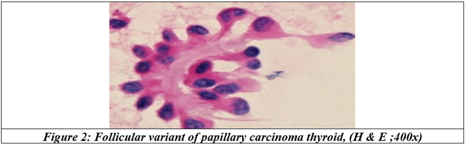
Similarly, Histopathological diagnosis of high-grade thyroid malignancies can be challenging due to overlap with several diagnostic mimickers. ATC may mimic lymphoma, metastatic carcinoma and sarcomas, while PDTC can resemble follicular neoplasms and medullary carcinoma. Similarly, benign conditions such as nodular goitre and Hashimoto thyroiditis may exhibit nuclear features overlapping with papillary carcinoma. These diagnostic pitfalls can result in both false-positive and false-negative interpretations. In such situations, immunohistochemistry serves as an indispensable adjunct in establishing lineage differentiation and excluding non-thyroid lesions.[13,14]
Immunohistochemical markers such as Ki-67 and p53 play an important role in distinguishing aggressive tumours and assessing tumour biology.[3,15,16]
Despite several studies describing cytomorphological features of thyroid carcinomas, limited data is available focusing comprehensively on their diagnostic mimickers and associated cytological pitfalls in a single study. The present study aimed to evaluate the clinicopathological and immunohistochemical spectrum of thyroid carcinomas, ranging from differentiated to anaplastic forms, with an emphasis on their diagnostic mimickers.
MATERIALS AND METHODS:
This is a hospital-based cross-sectional study, carried out in a tertiary care centre hospital, from January 2022 to January 2026.
A total of 100 cases of solitary thyroid nodules were examined. FNAC was performed, and smears were stained with Giemsa and Hematoxylin & Eosin. Cases were categorised according to the Bethesda System (3rd edition, 2023). Histopathological examination was performed in surgically resected specimens. Immunohistochemistry (IHC) using Pan Cytokeratin, Ki-67 and p53 was performed in selected cases (Category IV and V lesions). Cases showing overlapping cytological features with non-neoplastic or other neoplastic conditions were analysed for diagnostic pitfalls. Data analysed using SPSS version 19.0. Chi-square test applied. p<0.05 is considered significant.
RESULTS:
Out of 100 cases of solitary thyroid nodule, a total of 22 cases of Anaplastic Thyroid Carcinoma (ATC), 15 cases of Poorly Differentiated Thyroid Carcinoma (PDTC) and 63 cases of DTC were identified. The clinico-pathological characteristics, including age distribution, tumour size, lymph node status and metastasis, are summarised.
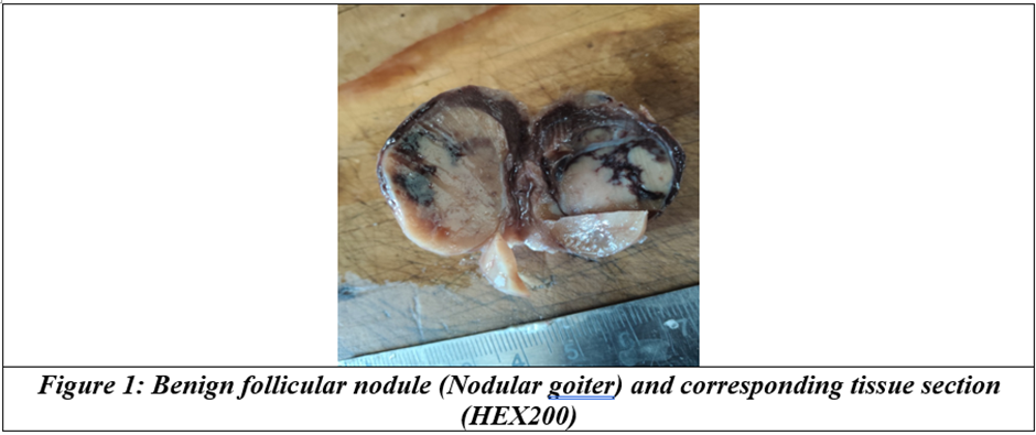
Table 1: Clinicopathologic findings of the patients
|
Parameters |
ATC (N=22) |
PDTC (N=15) |
DTC (N=63) |
|
|
Sample type =N (%) |
Pure tumour |
9 (43%) |
5(33.33%) |
63(100%) |
|
Tumour with DTC |
12(57%) |
10(66.66%) |
||
|
Age(years) |
Mean (range) |
64.5(43-82) |
52(27-79) |
45(35-55) |
|
Sex |
Female |
11(50%) |
9(60%) |
48(76.2%) |
|
Male |
11(50%) |
6(40%) |
15(23.8%) |
|
|
Tumour diameter(cm) |
Mean (range) |
5.8(1.8-10) |
4.6(1.6-9) |
2.5(1-3) |
|
≤2cm [n(%)] |
1(4.8%) |
1(6.7%) |
41(65%) |
|
|
2-4 cm[n(%)] |
5(31%) |
6(40%) |
18(28.6%) |
|
|
≥4cm[n(%)] |
14(72.7%) |
8(53.3%) |
4(6.3%) |
|
|
Lymph node metastasis [n(%)] |
N0 |
10(45.2%) |
4(26.6%) |
33(52.4%) |
|
N1a |
2(9.6%) |
3(20%) |
8 (12.7%) |
|
|
N1b |
10(45.2%) |
8(53.3%) |
2 (3.17%) |
|
|
Distant metastasis [n(%)] |
Present |
8(36.4%) |
8(53.3%) |
0 |
|
Absent |
14(63.6%) |
7(46.7%) |
63(100%) |
|
ATC predominantly affected elderly patients (median age: 64.5 years), whereas PDTC occurred in relatively younger individuals (median age: 52 years) and DTC occurs at young population (mean age 45). Female predominance was noted in ATC.
Cytological evaluation of ATC cases showed highly pleomorphic tumour cells, including spindle cells, giant cells and squamoid cells, with marked nuclear atypia, frequent mitotic figures and necrosis. In contrast, PDTC demonstrated insular, trabecular and solid growth patterns.
Table 2: Cytomorphological table comparing ATC, PDTC & DTC
|
Cytomorphological features |
ATC |
PDTC |
DTC |
|
Cellular pleomorphism |
Marked |
Mild to moderate |
Minimal to nil |
|
Spindle cells |
Common |
Rare |
rare |
|
Giant cells |
Common |
Rare |
rare |
|
Squamoid cells |
Present |
Usually, absent |
absent |
|
Mitotic activity |
Frequent |
moderate |
low |
|
tumour necrosis |
Common |
occasional |
rare |
|
Growth pattern |
Disorganizing sheet/dispersed cells |
Insular, trabecular, solid pattern |
Papillary/follicular |
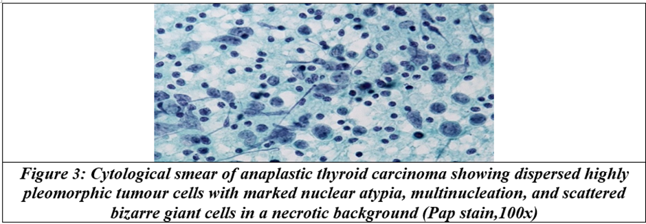
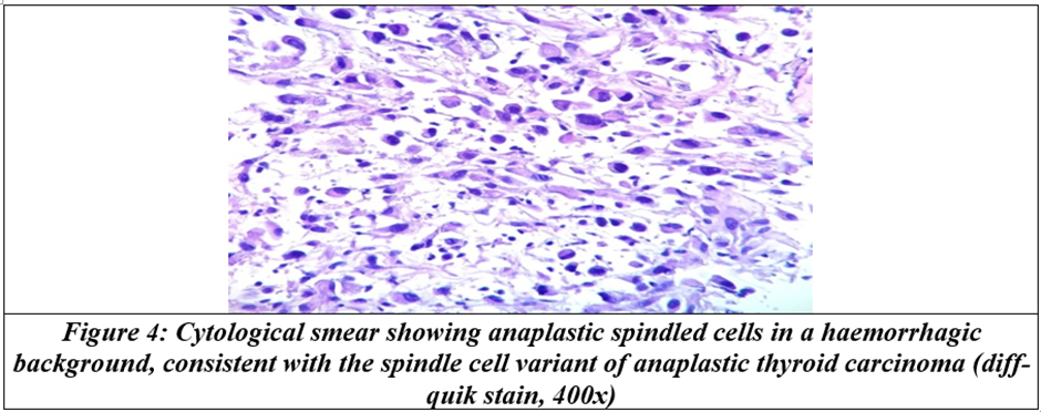
Immunohistochemical analysis showed a significantly higher Ki-67 proliferation index in ATC cases (75%) compared to differentiated thyroid carcinoma components (37.5%). Mutant p53 expression was predominantly observed in ATC (70.8%), whereas DTC components showed wild-type expression. Pan-cytokeratin positivity was observed in the majority of epithelial tumours; the findings are summarised as below.
Table 3: Immunohistochemical Expression of DTC, PDTC & ATC
|
Marker |
DTC |
PDTC |
ATC |
|
Ki-67 index |
low |
moderate |
high |
|
P53 |
Wild type |
Variable |
mutant |
|
Thyroglobulin |
positive |
reduced |
Negative |
|
TTF-1 |
positive |
Variable |
Reduced |
|
PAX-8 |
Positive |
Positive |
variable |
|
Pan-CK |
Positive |
Positive |
Positive |
|
calcitonin |
Negative |
Negative |
Negative |
Table 4: Differential Diagnosis with IHC
|
Lesion |
Key Morphology |
IHC Profile |
|
Medullary Thyroid Carcinoma |
Amyloid, plasmacytoid cells |
Calcitonin+, CEA+ |
|
Thyroid lymphoma |
Diffuse lymphoid cells |
CD45+, LCA+ |
|
Metastatic carcinoma |
Variable |
Variable |
|
Sarcoma |
Spindle cells |
Vimentin+, CK− |
ATC (other cytological mimickers were identified during evaluation. These included thyroid lymphoma, Metastatic carcinoma, Medullary carcinoma, Nodular goitre, and Hashimoto thyroiditis. Diagnostic confusion arose due to overlapping features such as nuclear grooves, intranuclear inclusions and necrotic background. The spectrum of mimickers and their distinguishing features is summarised in Error! Reference source not found.
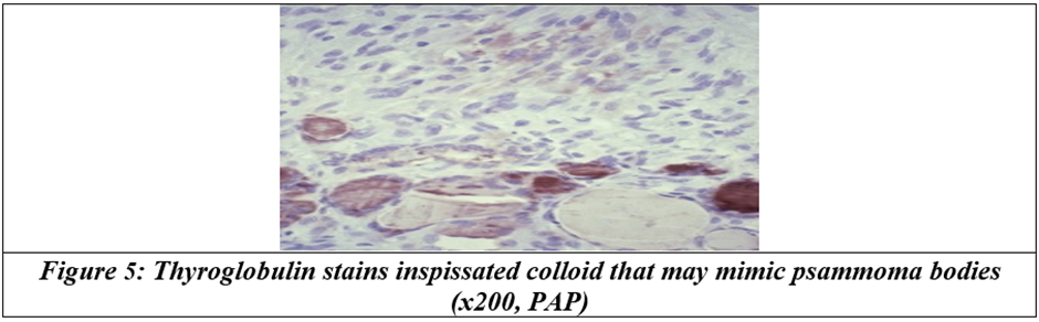
Calcified debris and inspissated colloid mimicking psammoma bodies were observed in a benign thyroid lesion, which may lead to overdiagnosis of papillary carcinoma.
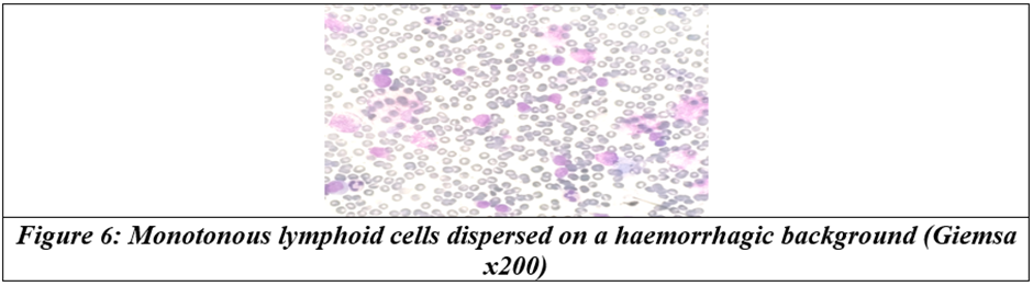
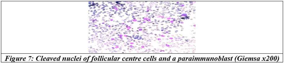
Necrosis mimics high-grade malignancy. In certain cases, a monotonous population of lymphoid cells was noted, raising suspicion of lymphoma; however, further evaluation helped in distinguishing reactive from neoplastic conditions.
DISCUSSION:
The present study highlights the wide cytomorphological spectrum of ATC and PDTC and emphasises the diagnostic challenges posed by their mimickers. The coexistence of differentiated thyroid carcinoma (DTC) components observed in a significant proportion of cases supports the concept of tumour progression through differentiation.[10,11,17]
ATC frequently mimics lymphoma and metastatic tumours due to marked cellular pleomorphism, necrosis and inflammatory background. Similarly, PDTC may resemble follicular neoplasms, leading to diagnostic ambiguity. These findings are consistent with previous studies that have documented significant overlap in cytological features among thyroid neoplasms.[12]
Benign lesions such as nodular goitre and Hashimoto thyroiditis may also show nuclear grooves and atypia, mimic papillary thyroid carcinoma and contributing to false-positive diagnoses. Careful evaluation of cytological details along with clinicoradiological correlation is therefore essential.
The significantly higher Ki-67 proliferation index observed in ATC reflects increased tumour aggressiveness and rapid growth, in agreement with previous studies.[15] Likewise, mutant p53 expression predominantly seen in ATC supports its role as a marker of tumour progression and genetic instability.[18,19]
The role of immunohistochemistry is crucial in differentiating ATC from its mimickers, particularly lymphoma and metastatic carcinoma. Expression of epithelial markers such as Pan Cytokeratin helps confirm epithelial origin, while additional markers may be required in challenging cases.
Recent advances in molecular and immune profiling, including PD-L1 expression and targeted therapies, have further improved the understanding and management of advanced thyroid cancers.[13,16]
The present study demonstrates the wide morphological and biological spectrum of thyroid carcinomas, extending from differentiated neoplasms with indolent clinical behaviour to highly aggressive undifferentiated tumours. This progression model has been widely supported in literature, where PDTC is considered a transitional entity between DTC and ATC.[5,6]
In our series, DTC occurred predominantly in younger to middle-aged patients with female preponderance, consistent with previously published epidemiological studies showing higher incidence of papillary thyroid carcinoma in women.[1,2] In contrast, ATC was more frequently encountered in elderly individuals presenting with rapidly enlarging neck masses, compressive symptoms, and locally advanced disease, findings similar to those reported by Ain et al. and ATA guidelines.[4,15]
Histologically, PDTC showed solid, trabecular, and insular growth patterns with increased mitotic activity and focal necrosis. These features align with the Turin diagnostic criteria proposed by Volante et al., which remain widely accepted for classification of PDTC.[20]
A progressive increase in proliferative activity, as reflected by Ki-67 labelling index, was observed from DTC to PDTC and ATC. Similar findings have been reported in previous studies, supporting the role of Ki-67 as a marker of tumour aggressiveness and rapid cell turnover.[13] Aberrant p53 expression was more frequent in ATC, reflecting underlying TP53 mutation and genomic instability associated with dedifferentiation.[7,8]
One of the most important practical aspects of this study was the distinction of ATC and PDTC from their mimickers. Spindle cell morphology may simulate sarcoma or metastatic squamous carcinoma, while small cell or diffuse infiltrative patterns may mimic lymphoma. Use of markers such as cytokeratin, PAX8, thyroglobulin, TTF-1, calcitonin, and leukocyte common antigen can significantly aid in resolving these differentials.[12,13]
Recognition of dedifferentiated thyroid carcinoma is clinically significant because treatment strategies and prognosis differ markedly across the spectrum. While DTC often responds to surgery and radioiodine therapy, PDTC and ATC frequently require multimodal management including surgery, radiotherapy, chemotherapy, and targeted therapy.[4]
Thus, integration of clinical presentation, histomorphology, and immunohistochemistry remains essential for accurate diagnosis and appropriate management of thyroid carcinomas and their mimickers.
CONCLUSION:
ATC and PDTC exhibit diverse cytomorphological features with significant overlap with various benign and malignant mimickers. Overall, awareness of cytological mimickers, adequate sampling, and the use of adjunct techniques such as immunohistochemistry are essential to minimise diagnostic errors and improve patient outcomes.
Limitations
• Single-centre study.
• Limited number of PDTC cases.
• Lack of long-term follow-up.
REFERENCES:
1. Delellis RA, Lloyd R V, Heitz PU, et al. WHO classification of tumours of endocrine organs. World Health Organisation 2017.
2. Cibas ES, Ali SZ. The Bethesda system for reporting thyroid cytopathology. Thyroid 2009;19(11):1159-65.
3. Ahn S, Kim TH, Kim SW, et al. Comprehensive screening for PD-L1 expression in thyroid cancer. Endocr Relat Cancer 2017;24(2):97-106.
4. Bible KC, Kebebew E, Brierley J, et al. 2021 American thyroid association guidelines for management of patients with anaplastic thyroid cancer: American thyroid association anaplastic thyroid cancer guidelines task force. Thyroid 2021;31(3):337-86.
5. Asa SL, Mete O. Oncocytic change in thyroid pathology. Front Endocrinol (Lausanne) 2021;12:678119.
6. Volante M, Collini P, Nikiforov YE, et al. Poorly differentiated thyroid carcinoma: the Turin proposal for the use of uniform diagnostic criteria and an algorithmic diagnostic approach. Am J Surg Pathol 2007;31(8):1256-64.
7. Xu B, Ghossein R. Thyroid Gland. In: Endocrine Pathology. Springer 2022:784-8.
8. Nikiforova MN, Tseng GC, Steward D, et al. MicroRNA expression profiling of thyroid tumors: biological significance and diagnostic utility. J Clin Endocrinol Metab 2008;93(5):1600-8.
9. Rosai J, Carcangiu ML, DeLellis RA. Tumors of the thyroid gland. American Registry of Pathology 1992.
10. Mills SE, Greenson JK, Hornick JL, et al. Sternberg’s diagnostic surgical pathology. Lippincott Williams & Wilkins 2015.
11. Siegel RL, Miller KD, Fuchs HE, et al. Cancer statistics, 2022. CA Cancer J Clin 2022;72(1):7-33.
12. Leandro-García LJ, Landa I. Mechanistic insights of thyroid cancer progression. Endocrinology 2023;164(9):bqad118.
13. Tang AL, Kloos RT, Aunins B, et al. Pathologic features associated with molecular subtypes of well-differentiated thyroid cancer. Endocrine Practice 2021;27(3):206-11.
14. Baloch ZW, Asa SL, Barletta JA, et al. Overview of the 2022 WHO classification of thyroid neoplasms. Endocr Pathol 2022;33(1):27-63.
15. Agrawal N, Akbani R, Aksoy BA, et al. Integrated genomic characterization of papillary thyroid carcinoma. Cell 2014;159(3):676–90.
16. Capdevila J, Mayor R, Mancuso FM, et al. Early evolutionary divergence between papillary and anaplastic thyroid cancers. Annals of Oncology 2018;29(6):1454-60.
17. Ahn J, Jin M, Song E, et al. Immune profiling of advanced thyroid cancers using fluorescent multiplex immunohistochemistry. Thyroid 2021;31(1):61-7.
18. Capdevila J, Wirth LJ, Ernst T, et al. PD-1 blockade in anaplastic thyroid carcinoma. Journal of Clinical Oncology 2020;38(23):2620-7.
19. Ragazzi M, Torricelli F, Donati B, et al. Coexisting well-differentiated and anaplastic thyroid carcinoma in the same primary resection specimen: immunophenotypic and genetic comparison of the two components in a consecutive series of 13 cases and a review of the literature. Virchows Archiv 2021;478(2):265-81.
20. Pita JM, Figueiredo IF, Moura MM, et al. Cell cycle deregulation and TP53 and RAS mutations are major events in poorly differentiated and undifferentiated thyroid carcinomas. J Clin Endocrinol Metab 2014;99(3):E497-507.