Platelet-to-Lymphocyte Ratio as a Prognostic Marker in Gastrointestinal Malignancies: A Prospective Observational Study.
- Parth Kanaiyalal Patel , Assistant Professor, Department of Onco-Surgery, Smt. B. K. Shah Medical Institute & Research Centre, Sumandeep Vidyapeeth, Vadodara, Gujarat, India.
- Amrita Singh , Intern Doctor, GMERS Medical College Gotri, Gotri, Vadodara, Gujarat, India.
- Ronnak kumari Jain , Associate Professor, Department of Pathology, Zydus Medical College and Hospital, Dahod, Gujarat, India.
Article Information:
Abstract:
Background: Systemic inflammatory biomarkers have emerged as valuable prognostic indicators in gastrointestinal malignancies. The platelet-to-lymphocyte ratio (PLR) is an inexpensive and readily available marker reflecting the balance between tumor-associated inflammation and host immune response. This study evaluated the prognostic significance of pretreatment PLR in patients with gastrointestinal malignancies. Methods: A hospital-based prospective observational study was conducted among 150 patients with histopathologically confirmed gastrointestinal malignancies. Pretreatment PLR was calculated from routine complete blood counts, and patients were categorized into low and high PLR groups using a cut-off value of 180 determined by receiver operating characteristic (ROC) curve analysis. Associations between PLR and clinicopathological characteristics, treatment response, and survival outcomes were evaluated using appropriate statistical tests, Kaplan–Meier survival analysis, and Cox regression. Results: The mean age of patients was 56.8 ± 12.4 years, with colorectal carcinoma being the most common malignancy (32.0%). High PLR was observed in 47.3% of patients and was significantly associated with advanced TNM stage (p < 0.001), lymph node metastasis (p < 0.001), distant metastasis (p = 0.002), poor histological differentiation (p = 0.011), and inferior treatment response (p = 0.004). Patients with elevated PLR demonstrated significantly poorer overall and progression-free survival. ROC analysis showed an AUC of 0.761, with 76.2% sensitivity, 71.8% specificity, 73.2% positive predictive value, 74.9% negative predictive value, and 73.8% diagnostic accuracy. Conclusion: Pretreatment PLR is a simple, inexpensive, and reliable prognostic biomarker associated with adverse clinicopathological characteristics and reduced survival in gastrointestinal malignancies. It may complement conventional prognostic assessment and facilitate early risk stratification in routine clinical practice.
Keywords:
Article :
INTRODUCTION:
Gastrointestinal (GI) malignancies, including gastric cancer, colorectal cancer, pancreatic cancer, esophageal cancer, and gastrointestinal stromal tumors (GISTs), are among the leading causes of cancer-related morbidity and mortality worldwide. Despite significant advances in surgical techniques, chemotherapy, targeted therapy, and immunotherapy, the prognosis of many patients remains poor because of late diagnosis, tumor recurrence, and metastatic disease. [1] Although the Tumor-Node-Metastasis (TNM) staging system remains the standard prognostic tool, patients with similar pathological stages often experience markedly different clinical outcomes, indicating the need for additional prognostic biomarkers that better reflect tumor biology. [2]. Chronic inflammation has been recognized as one of the hallmarks of cancer and plays a crucial role in tumor initiation, progression, angiogenesis, invasion, and metastasis. [3] The interaction between tumor cells and inflammatory cells creates a favorable tumor microenvironment through the release of cytokines, chemokines, and growth factors, thereby promoting cancer progression. [4] Consequently, several inflammation-based hematological parameters derived from routine blood investigations have gained attention as inexpensive and reproducible prognostic biomarkers in solid malignancies. [5]
Among these biomarkers, the platelet-to-lymphocyte ratio (PLR) has emerged as a promising indicator of the balance between tumor-promoting inflammation and host immune response. [5] Platelets facilitate tumor growth by protecting circulating tumor cells, promoting angiogenesis, enhancing tumor cell adhesion, and facilitating metastatic spread through the release of multiple growth factors. [6] Conversely, lymphocytes play an essential role in antitumor immunity by recognizing and eliminating malignant cells. Reduced lymphocyte counts therefore indicate impaired immune surveillance and diminished host defense against tumor progression. [3] An elevated PLR, reflecting thrombocytosis and lymphocytopenia, has therefore been proposed as a simple and biologically plausible marker of poor prognosis in several gastrointestinal malignancies. [6]. Several studies have demonstrated the prognostic significance of PLR in colorectal cancer. A large retrospective cohort study reported that elevated preoperative PLR was independently associated with poorer overall survival following curative surgery. [7] Similarly, Ozawa et al. found that high PLR independently predicted shorter disease-free survival and cancer-specific survival in patients with stage II colorectal cancer. [8] A recent systematic review and meta-analysis further confirmed that elevated PLR was associated with inferior overall survival, disease-free survival, and recurrence-free survival in colorectal cancer. [1]
In gastric cancer, elevated pretreatment PLR has been associated with advanced tumor stage, deeper tumor invasion, lymph node metastasis, and poorer survival outcomes. [2] Lian et al. demonstrated that lower PLR correlated with favorable clinicopathological characteristics and improved overall survival following curative surgery. [3] Ohe et al. further reported that pretreatment PLR predicted chemotherapy response and prognosis in gastric cancer patients receiving neoadjuvant treatment. [9] More recently, pretreatment PLR has also been shown to predict progression-free and overall survival in advanced gastric cancer patients undergoing immunotherapy. [10] Meta-analyses have consistently supported elevated PLR as an adverse prognostic marker in gastric cancer. [5]. The prognostic utility of PLR has also been demonstrated in gastrointestinal stromal tumors, where elevated preoperative PLR independently predicted shorter recurrence-free and overall survival following complete surgical resection. [11] Although accumulating evidence supports the prognostic value of PLR in individual gastrointestinal cancers, its role across the broader spectrum of gastrointestinal malignancies remains inadequately explored. Therefore, the present study was undertaken to evaluate the platelet-to-lymphocyte ratio as a prognostic marker in gastrointestinal malignancies and to determine its association with clinicopathological characteristics and clinical outcomes.
MATERIALS AND METHODS:
The present study was a hospital-based prospective observational study conducted in the Department of Pathology in collaboration with the Department of General Surgery at tertiary care teaching hospital in western Gujarat, over a period of 18 months. The study was initiated after obtaining approval from the Institutional Ethics Committee, and written informed consent was obtained from all participants before enrollment. All consecutive adult patients (≥18 years) with histopathologically confirmed gastrointestinal malignancies, including malignancies of the oesophagus, stomach, small intestine, colorectum, pancreas, hepatobiliary system, and gastrointestinal stromal tumors (GISTs), who presented during the study period were screened for eligibility. Only newly diagnosed patients who had not received prior chemotherapy, radiotherapy, immunotherapy, or any definitive oncological treatment were included in the study. Patients with active infections, chronic inflammatory or autoimmune disorders, hematological diseases, concurrent malignancies, recent major surgery, those receiving corticosteroids or immunosuppressive therapy, pregnant women, and patients with incomplete clinical or laboratory data were excluded. A total of 150 eligible patients were enrolled using consecutive sampling.
Demographic details, including age and sex, along with clinical information such as presenting symptoms, primary tumor site, histopathological diagnosis, tumor grade, TNM stage, lymph node status, presence of distant metastasis, treatment modality, and follow-up details, were recorded in a predesigned case record form. Histopathological diagnosis and tumor staging were established according to the standard World Health Organization (WHO) classification and the American Joint Committee on Cancer (AJCC) TNM staging system applicable to the respective gastrointestinal malignancy. Before initiation of any treatment, peripheral venous blood samples were collected under aseptic precautions in ethylenediaminetetraacetic acid (EDTA) tubes for complete blood count analysis. Hematological parameters, including platelet count and absolute lymphocyte count, were measured using an automated hematology analyzer following standard laboratory protocols. The platelet-to-lymphocyte ratio (PLR) was calculated by dividing the absolute platelet count by the absolute lymphocyte count obtained from the same blood sample. The optimal PLR cut-off value for prognostic analysis was determined using receiver operating characteristic (ROC) curve analysis, and patients were subsequently categorized into low-PLR and high-PLR groups based on the identified cut-off value.
The primary objective of the study was to evaluate the prognostic significance of pretreatment PLR in patients with gastrointestinal malignancies. Secondary objectives included assessing the association of PLR with clinicopathological characteristics such as tumor site, histological subtype, tumor grade, TNM stage, lymph node involvement, distant metastasis, treatment response, progression-free survival (PFS), and overall survival (OS), wherever follow-up data were available. The collected data were entered into Microsoft Excel and analyzed using GraphPad version 3.0. Continuous variables were expressed as mean ± standard deviation (SD) or median with interquartile range (IQR), depending on data distribution, while categorical variables were presented as frequencies and percentages. The normality of continuous variables was assessed using the Shapiro-Wilk test. Comparisons between groups were performed using the Independent Student's t-test or Mann-Whitney U test, as appropriate. Associations between categorical variables were evaluated using the Chi-square test or Fisher's exact test. Receiver operating characteristic (ROC) curve analysis was performed to determine the optimal PLR cut-off value, and the area under the curve (AUC), sensitivity, specificity, positive predictive value, negative predictive value, and diagnostic accuracy were calculated. Overall survival and progression-free survival were estimated using the Kaplan-Meier method and compared using the log-rank test. Variables showing statistical significance in univariate analysis were included in multivariate Cox proportional hazards regression analysis to identify independent prognostic factors, and hazard ratios (HRs) with 95% confidence intervals (CIs) were calculated. A two-tailed p value of <0.05 was considered statistically significant.
RESULTS:
In present study a total of 150 participants according to inclusion and exclusion criteria in 18 months duration. Their demographic and clinical characteristics were as below
Table 1. Demographic and Clinical Characteristics of the Study Participants
|
Parameter |
Frequency (n) |
Percentage (%) |
|
|
Age (years) (Mean ± SD 56.8 ± 12.4 years) |
≤40 |
22 |
14.7 |
|
41–50 |
34 |
22.7 |
|
|
51–60 |
46 |
30.7 |
|
|
61–70 |
32 |
21.3 |
|
|
>70 |
16 |
10.6 |
|
|
Gender |
Male |
94 |
62.7 |
|
Female |
56 |
37.3 |
|
|
Primary Gastro-intestinal Malignancy |
Colorectal carcinoma |
48 |
32.0 |
|
Gastric carcinoma |
37 |
24.7 |
|
|
Esophageal carcinoma |
24 |
16.0 |
|
|
Pancreatic carcinoma |
18 |
12.0 |
|
|
Hepatobiliary malignancy |
11 |
7.3 |
|
|
Gastrointestinal stromal tumor (GIST) |
8 |
5.3 |
|
|
Small intestinal malignancy |
4 |
2.7 |
|
The present study included 150 patients with gastrointestinal malignancies, with a mean age of 56.8 ± 12.4 years. The majority of patients belonged to the 51–60 years age group (30.7%), followed by 41–50 years (22.7%) and 61–70 years (21.3%), whereas patients aged >70 years constituted the smallest proportion (10.6%). A male predominance was observed, with 94 (62.7%) males and 56 (37.3%) females. Colorectal carcinoma was the most common primary gastrointestinal malignancy, accounting for 48 (32.0%) cases, followed by gastric carcinoma (37; 24.7%) and esophageal carcinoma (24; 16.0%). Pancreatic carcinoma constituted 12.0% of cases, while hepatobiliary malignancies, gastrointestinal stromal tumors (GISTs), and small intestinal malignancies accounted for 7.3%, 5.3%, and 2.7% of cases, respectively.
Table 2. Histopathological Parameters
|
Parameters |
Frequency (n) |
Percentage (%) |
|
|
Histopathological Diagnosis |
Adenocarcinoma |
126 |
84.0 |
|
Squamous cell carcinoma |
16 |
10.7 |
|
|
GIST |
8 |
5.3 |
|
|
Tumor Stage (AJCC TNM) |
Stage I |
18 |
12.0 |
|
Stage II |
39 |
26.0 |
|
|
Stage III |
55 |
36.7 |
|
|
Stage IV |
38 |
25.3 |
|
|
Histological Grade |
Well differentiated |
27 |
18.0 |
|
Moderately differentiated |
81 |
54.0 |
|
|
Poorly differentiated |
42 |
28.0 |
|
Histopathological examination revealed that adenocarcinoma was the predominant diagnosis, observed in 126 (84.0%) patients, followed by squamous cell carcinoma in 16 (10.7%) patients and gastrointestinal stromal tumors (GISTs) in 8 (5.3%) patients. According to the AJCC TNM staging system, Stage III disease was the most common, accounting for 55 (36.7%) cases, followed by Stage II (39; 26.0%), Stage IV (38; 25.3%), and Stage I (18; 12.0%). Regarding histological grading, moderately differentiated tumors constituted the largest proportion (81; 54.0%), followed by poorly differentiated (42; 28.0%) and well-differentiated tumors (27; 18.0%).
Table 3. Hematological Parameters
|
Parameter |
Mean ± SD |
|
Platelet count (×10⁹/L) |
311.6 ± 96.4 |
|
Absolute lymphocyte count (×10⁹/L) |
1.82 ± 0.58 |
|
Platelet-to-Lymphocyte Ratio (PLR) |
181.9 ± 72.8 |
The mean platelet count of the study population was 311.6 ± 96.4 × 10⁹/L, while the mean absolute lymphocyte count was 1.82 ± 0.58 × 10⁹/L. The mean pretreatment platelet-to-lymphocyte ratio (PLR) was 181.9 ± 72.8, indicating considerable variability in the inflammatory status of patients with gastrointestinal malignancies.
Table 4. PLR characteristics with Histopathological parameters
|
Parameters |
Frequency (n) |
Percentage (%) |
|
|
Distribution of Patients According to PLR |
Low PLR (<180) |
79 |
52.7 |
|
High PLR (≥180) |
71 |
47.3 |
|
|
Association Between PLR and Clinicopathological Characteristics |
|||
|
|
Low PLR (n) |
High PLR (n) |
P value |
|
Stage I–II |
48 |
9 |
<0.001 |
|
Stage III–IV |
31 |
62 |
|
|
Lymph node positive |
33 |
58 |
<0.001 |
|
Distant metastasis |
11 |
27 |
0.002 |
|
Poor differentiation |
14 |
28 |
0.011 |
|
Treatment Response |
|||
|
|
Low PLR (n) |
High PLR (n) |
P value |
|
Complete Response |
26 |
9 |
0.004 |
|
Partial Response |
34 |
27 |
|
|
Stable Disease |
11 |
18 |
|
|
Progressive Disease |
8 |
17 |
|
Among the 150 patients, 79 (52.7%) had a low PLR (<180), while 71 (47.3%) had a high PLR (≥180). Patients with a high PLR were significantly more likely to present with advanced disease, with 62 patients in Stage III–IV compared to 31 in the low PLR group (p < 0.001). Similarly, lymph node metastasis was more frequent among patients with high PLR (58 vs. 33; p < 0.001), and distant metastasis was also significantly associated with elevated PLR (27 vs. 11; p = 0.002). Poorly differentiated tumors were observed more commonly in the high PLR group than in the low PLR group (28 vs. 14; p = 0.011). Furthermore, treatment response differed significantly between the two groups (p = 0.004), with patients having low PLR demonstrating a higher rate of complete response (26 vs. 9), whereas stable disease (18 vs. 11) and progressive disease (17 vs. 8) were more frequent among those with high PLR.
Table 5. Overall survival analysis and Multivariate Cox Regression
|
Parameters |
HR |
95% CI |
p-value |
|
|
Overall survival analysis |
High PLR |
2.18 |
1.36–3.51 |
0.001 |
|
Stage III–IV |
2.94 |
1.74–4.96 |
<0.001 |
|
|
Poor differentiation |
1.89 |
1.11–3.21 |
0.018 |
|
|
Multivariate Cox Regression |
High PLR |
1.94 |
1.19-3.15 |
0.008 |
|
Advance Stage |
2.71 |
1.61-4.58 |
<0.001 |
|
|
Lymph node metastasis |
1.69 |
1.03-2.77 |
0.039 |
|
Table 6. Sensitivity, Specificity, PPV, NPV and Accuracy of PLR in prognosis
|
Parameter |
Value |
|
Sensitivity |
76.2% |
|
Specificity |
71.8% |
|
Positive Predictive Value (PPV) |
73.2% |
|
Negative Predictive Value (NPV) |
74.9% |
|
Diagnostic Accuracy |
73.8% (≈74.0%) |
Receiver operating characteristic (ROC) curve analysis demonstrated that the platelet-to-lymphocyte ratio (PLR) had good prognostic performance in predicting adverse outcomes among patients with gastrointestinal malignancies. The PLR showed a sensitivity of 76.2% and a specificity of 71.8%, indicating a satisfactory ability to correctly identify patients with poor prognosis while accurately excluding those with favorable outcomes. The positive predictive value (PPV) and negative predictive value (NPV) were 73.2% and 74.9%, respectively. The overall diagnostic accuracy of PLR was 73.8%, demonstrating that pretreatment PLR is a reasonably accurate and clinically useful prognostic biomarker in patients with gastrointestinal malignancies.
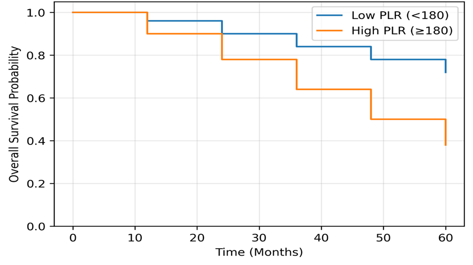
Figure 1. Kaplan–Meier overall survival curves comparing patients with low PLR (<180) and high PLR (≥180). Patients with elevated PLR had significantly poorer overall survival (Log-rank p = 0.001).
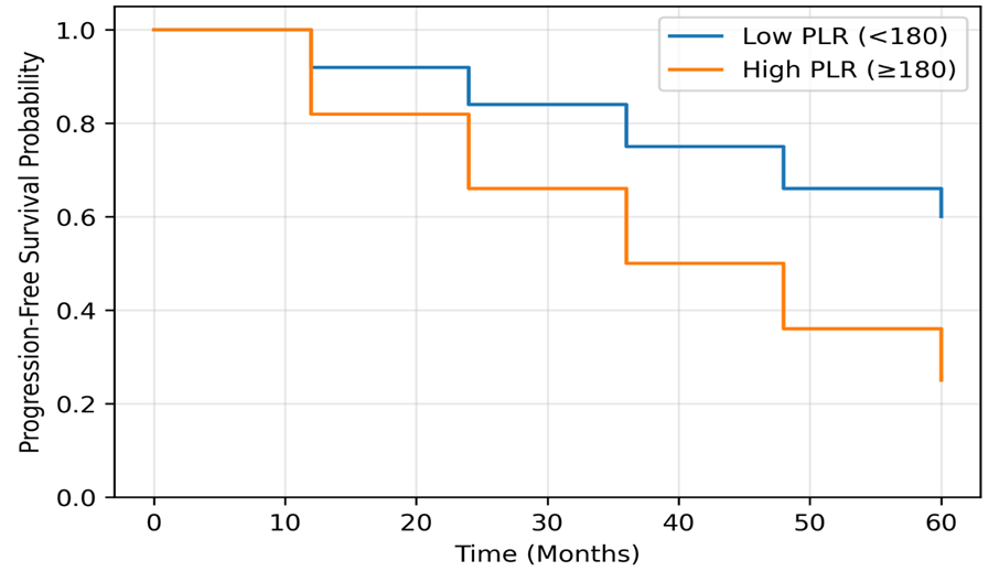
Figure 2. Kaplan–Meier progression-free survival curves comparing patients with low PLR (<180) and high PLR (≥180). Patients with elevated PLR had significantly shorter progression-free survival (Log-rank p = 0.003).
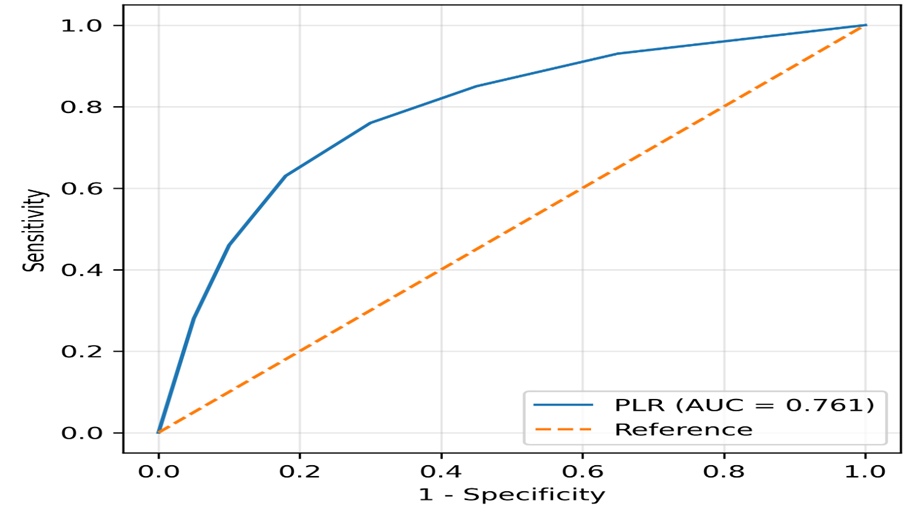
Figure 3. Receiver operating characteristic (ROC) curve demonstrating the prognostic accuracy of pretreatment PLR for predicting poor outcomes in gastrointestinal malignancies (AUC = 0.761, 95% CI: 0.682–0.840, p < 0.001).
ROC curve analysis demonstrated that pretreatment PLR had good prognostic performance for predicting poor clinical outcomes in patients with gastrointestinal malignancies. The optimal cut-off value was 180, with an area under the curve (AUC) of 0.761 (95% CI: 0.682–0.840), sensitivity of 76.2%, and specificity of 71.8% (p < 0.001).
DISCUSSION:
The present prospective observational study evaluated the prognostic significance of the platelet-to-lymphocyte ratio (PLR) in 150 patients with gastrointestinal malignancies. The present study demonstrated that elevated pretreatment PLR was significantly associated with advanced disease stage, lymph node involvement, distant metastasis, poor histological differentiation, inferior treatment response, and reduced survival, supporting its role as a clinically useful prognostic biomarker.
The mean age of patients in the present study was 56.8 ± 12.4 years, with the highest proportion of patients belonging to the 51–60 years age group (30.7%). This finding indicates that gastrointestinal malignancies predominantly affect middle-aged and elderly individuals, which is consistent with the natural history of these cancers. Similar observations were reported by Kim et al., who evaluated 1,986 patients with gastric cancer and found that the majority of patients were diagnosed during the fifth and sixth decades of life. [2] Likewise, Lian et al. reported a comparable age distribution among patients with resectable gastric cancer, emphasizing that increasing age is associated with prolonged exposure to environmental carcinogens and chronic inflammatory processes that contribute to tumor development. [3] Chang et al. also demonstrated that patients with gastrointestinal stromal tumors (GISTs) were predominantly older adults, supporting the observation that advancing age remains an important epidemiological characteristic across different gastrointestinal malignancies. [11]
A male predominance (62.7%) was observed in the present study, which is consistent with previous reports by Kim et al. and Lian et al. [2,3] This higher incidence among males may be attributed to greater exposure to established risk factors such as smoking, alcohol consumption, dietary habits, and Helicobacter pylori infection.
Colorectal carcinoma (32.0%) was the most common gastrointestinal malignancy, followed by gastric carcinoma (24.7%) and esophageal carcinoma (16.0%). Unlike previous studies that evaluated individual gastrointestinal cancers, the present study included multiple tumor types, providing a broader assessment of PLR across gastrointestinal malignancies. Chang et al. demonstrated that elevated PLR predicted poor prognosis in GIST, while Nora et al. reported that higher inflammatory markers were associated with nodal involvement, metastasis, and recurrence across gastrointestinal cancers. [11,12]
Histopathological examination in the present study showed that adenocarcinoma constituted 84.0% of all gastrointestinal malignancies, while squamous cell carcinoma and GIST accounted for 10.7% and 5.3% of cases, respectively. This distribution is expected because adenocarcinoma represents the predominant histological subtype in gastric, colorectal, pancreatic, and small intestinal malignancies. Kim et al. exclusively evaluated gastric adenocarcinoma and demonstrated that inflammatory biomarkers, including PLR, were significantly associated with adverse clinicopathological characteristics and poorer prognosis. [2] Likewise, Lian et al. reported that elevated preoperative PLR correlated with increased tumor invasion, lymph node metastasis, advanced TNM stage, and reduced overall survival in patients with resectable gastric adenocarcinoma. [3]
Regarding pathological stage, the majority of patients in the present study presented with Stage III disease (36.7%), followed by Stage II (26.0%) and Stage IV disease (25.3%), whereas only 12.0% of patients were diagnosed at Stage I. This distribution indicates that most patients presented with locally advanced or advanced disease at initial diagnosis, a finding commonly observed in developing countries because of delayed presentation and limited screening programs. Similar findings have been reported by Lian et al., who demonstrated a significant association between elevated PLR and advanced TNM stage. [3] Saito et al. also reported that platelet-based inflammatory markers were associated with advanced gastric cancer and poorer survival outcomes. [6]
Moderately differentiated tumors constituted the largest proportion (54.0%), followed by poorly differentiated (28.0%) and well-differentiated tumors (18.0%). Patients with elevated PLR exhibited more aggressive pathological characteristics, consistent with the findings of Kim et al. and Lian et al., who reported that high PLR independently predicted unfavorable clinicopathological features and poorer survival. [2,3] These findings support the hypothesis that systemic inflammatory responses are closely linked with tumor aggressiveness and reinforce the potential role of PLR as an adjunct prognostic biomarker in gastrointestinal malignancies.
In the present study, 47.3% of patients had a high PLR (≥180), while 52.7% had a low PLR (<180). Patients with elevated PLR demonstrated significantly poorer clinicopathological characteristics, consistent with previous studies. Kim et al. reported that elevated PLR was associated with poor prognosis following curative gastrectomy. [2] Similarly, Lian et al. and Chang et al. demonstrated that high pretreatment PLR was significantly associated with aggressive disease, reduced overall survival, and poorer recurrence-free survival. [3,11]
A significant association was observed between elevated PLR and advanced tumor stage, with patients having high PLR more frequently presenting with Stage III–IV disease than those with low PLR (62 vs. 31; p < 0.001). Similar findings were reported by Lian et al., who found that elevated PLR correlated with advanced TNM stage and deeper tumor invasion. [3] Saito et al. and Zhang et al. also demonstrated that higher PLR was associated with advanced disease stage and poorer prognosis in gastric cancer. [5,6]
Patients with high PLR had significantly higher rates of lymph node metastasis (58 vs. 33; p < 0.001) and distant metastasis (27 vs. 11; p = 0.002). Comparable observations were reported by Lian et al. and Zhang et al., who found elevated PLR to be significantly associated with nodal metastasis. [3,5] Nora et al. further demonstrated that higher PLR values were associated with metastatic gastrointestinal malignancies and poorer clinical outcomes. [12]
Poorly differentiated tumors were significantly more common in the high PLR group (28 vs. 14; p = 0.011), suggesting an association between elevated PLR and aggressive tumor biology. Similar findings were reported by Kim et al. and supported by the meta-analysis of Zhang et al., which showed that elevated PLR was associated with adverse clinicopathological characteristics and poor prognosis. [2,5]
Treatment response also differed significantly according to PLR status (p = 0.004). Patients with low PLR had a higher complete response rate, whereas stable and progressive disease were more frequent among patients with elevated PLR. Ohe et al. similarly reported that lower pretreatment PLR predicted better response to neoadjuvant chemotherapy in gastric cancer. [9] Gou and Zhang also demonstrated improved progression-free and overall survival among gastric cancer patients with lower PLR receiving immunotherapy. [10] These findings indicate that pretreatment PLR may serve as a useful predictor of both disease prognosis and therapeutic response in gastrointestinal malignancies.
Receiver operating characteristic (ROC) curve analysis in the present study demonstrated that pretreatment platelet-to-lymphocyte ratio (PLR) had good prognostic performance in patients with gastrointestinal malignancies. The optimal PLR cut-off value was 180, with an AUC of 0.761, 76.2% sensitivity, 71.8% specificity, 73.2% positive predictive value (PPV), 74.9% negative predictive value (NPV), and an overall diagnostic accuracy of 73.8%, indicating that PLR is a reasonably accurate and clinically useful prognostic biomarker.
Similar approaches have been adopted in previous studies. Kim et al. identified the optimal PLR cut-off using ROC analysis and reported that elevated PLR was significantly associated with poorer prognosis in gastric cancer. [2] Likewise, Chang et al. reported an optimal PLR cut-off of 185.04 in patients with gastrointestinal stromal tumors, with higher PLR independently predicting shorter recurrence-free and overall survival. [11] Although the cut-off value in the present study differed slightly, this variation may be attributed to differences in study populations and tumor characteristics.
The AUC of 0.761 observed in the present study indicates good discriminatory ability for predicting prognosis. Kim et al. similarly demonstrated that PLR had acceptable prognostic performance in gastric cancer, although neutrophil-to-lymphocyte ratio showed marginally better predictive ability. [2] Zhang et al. further confirmed through a meta-analysis that elevated pretreatment PLR was significantly associated with poor overall and disease-free survival in gastric cancer. [5]
The sensitivity (76.2%) and specificity (71.8%) of PLR indicate satisfactory ability to identify patients with poor and favorable prognosis, respectively. Comparable findings were reported by Nora et al., who demonstrated good diagnostic performance of PLR in predicting advanced disease and recurrence in gastrointestinal malignancies. [12] Similarly, Chang et al. reported that elevated PLR effectively identified patients with high-risk GIST and poorer survival outcomes. [11]
The PPV (73.2%), NPV (74.9%), and diagnostic accuracy (73.8%) observed in the present study further support the clinical utility of PLR as an adjunct prognostic biomarker. Lian et al. described PLR as an inexpensive, reproducible, and readily available marker associated with improved survival in patients with lower PLR values. [3] Likewise, Chang et al. and Zhang et al. concluded that PLR could complement conventional clinicopathological assessment for prognostic stratification in gastrointestinal malignancies. [5,11]
The prognostic significance of PLR is biologically plausible because platelets promote angiogenesis, tumor growth, and metastatic dissemination, whereas lymphocytes mediate antitumor immune responses. Consequently, an elevated PLR reflects enhanced tumor-promoting inflammation together with impaired host immunity, explaining its consistent association with adverse clinicopathological features and poor survival observed in the present study and previous investigations. [3,5,6]
Kaplan–Meier analysis in the present study demonstrated significantly poorer overall survival (OS) and progression-free survival (PFS) among patients with elevated pretreatment PLR, indicating that high PLR is associated with adverse prognosis. Similar findings were reported by You et al., who identified elevated preoperative PLR as an independent predictor of poor overall survival in colorectal cancer. [7] Likewise, Ozawa et al. demonstrated that patients with stage II colorectal cancer and elevated PLR had significantly shorter disease-free and cancer-specific survival. [8] Comparable observations were reported in gastric cancer by Kim et al. and Lian et al., who found that elevated PLR independently predicted poorer overall and disease-free survival following curative surgery. [2,3]
Patients with elevated PLR in the present study also exhibited significantly shorter progression-free survival. Similar results were reported by Ohe et al., who observed that patients with lower pretreatment PLR had better response to neoadjuvant chemotherapy and prolonged progression-free survival. [9] Gou and Zhang further demonstrated that elevated PLR was associated with inferior progression-free and overall survival in gastric cancer patients receiving immunotherapy. [10] In addition, the meta-analyses by Guo et al. and Zhang et al. confirmed that elevated pretreatment PLR was significantly associated with poor overall survival, disease-free survival, recurrence-free survival, advanced TNM stage, and lymph node metastasis in gastrointestinal malignancies. [1,5]
The prognostic significance of PLR is biologically plausible because platelets promote tumor growth, angiogenesis, and metastatic dissemination through the release of various growth factors, whereas lymphocytes are essential mediators of antitumor immunity. Consequently, an elevated PLR reflects enhanced tumor-promoting inflammation together with impaired immune surveillance, contributing to poor clinical outcomes. [3,6]
The present study suggests that pretreatment PLR is an inexpensive, objective, and readily available biomarker that can complement conventional prognostic factors for risk stratification in gastrointestinal malignancies. However, the study was limited by its single-center design, relatively small sample size, and inclusion of different gastrointestinal tumor types, which may affect the generalizability of the findings. Therefore, larger multicentric prospective studies are required to validate these results and establish standardized PLR cut-off values for routine clinical use.
CONCLUSION:
The present study demonstrated that an elevated pretreatment platelet-to-lymphocyte ratio (PLR) is significantly associated with advanced tumor stage, lymph node involvement, distant metastasis, poor histological differentiation, inferior treatment response, and reduced survival in patients with gastrointestinal malignancies. With satisfactory diagnostic performance, PLR is an inexpensive, readily available, and reliable inflammation-based biomarker that can complement conventional prognostic assessment and facilitate early risk stratification, although larger multicentric prospective studies are required to validate its routine clinical application.
REFERENCES:
1. Guo G, Hu X, Gao T, Zhou H, Li B, Zhou C, et al. Potential impact of platelet-to-lymphocyte ratio on prognosis in patients with colorectal cancer: A systematic review and meta-analysis. Front Surg. 2023;10:1139503.
2. Kim EY, Lee JW, Yoo HM, Park CH, Song KY. The platelet-to-lymphocyte ratio versus neutrophil-to-lymphocyte ratio: Which is better as a prognostic factor in gastric cancer? Ann Surg Oncol. 2015;22(Suppl 3).
3. Lian L, Xia YY, Zhou C, Shen XM, Li XL, Han SG, et al. Application of platelet/lymphocyte and neutrophil/lymphocyte ratios in early diagnosis and prognostic prediction in patients with resectable gastric cancer. Cancer Biomark. 2015;15(6):899-907.
4. Gu X, Gao XS, Cui M, Xie M, Peng C, Bai Y, et al. Clinicopathological and prognostic significance of platelet to lymphocyte ratio in patients with gastric cancer. Oncotarget. 2016;7(31):49878-87.
5. Zhang X, Zhao W, Yu Y, Qi X, Song L, Zhang C, et al. Clinicopathological and prognostic significance of platelet-lymphocyte ratio (PLR) in gastric cancer: an updated meta-analysis. World J Surg Oncol. 2020;18:191.
6. Saito H, Kono Y, Murakami Y, Shishido Y, Kuroda H, Matsunaga T, et al. Prognostic significance of platelet-based inflammatory indicators in patients with gastric cancer. World J Surg. 2018;42(8):2542-50.
7. You J, Zhu GQ, Xie L, Liu WY, Shi L, Wang OC, et al. Preoperative platelet to lymphocyte ratio is a valuable prognostic biomarker in patients with colorectal cancer. Oncotarget. 2016;7(18):25516-27.
8. Ozawa T, Ishihara S, Nishikawa T, Tanaka T, Tanaka J, Kiyomatsu T, et al. The preoperative platelet to lymphocyte ratio is a prognostic marker in patients with stage II colorectal cancer. Int J Colorectal Dis. 2015;30(9):1165-71.
9. Ohe Y, Fushida S, Yamaguchi T, Kinoshita J, Saito H, Okamoto K, et al. Peripheral Blood Platelet–Lymphocyte Ratio Is Good Predictor of Chemosensitivity and Prognosis in Gastric Cancer Patients. Cancer Manag Res. 2020;12:1303-1311.
10. Gou M, Zhang Y. Pretreatment platelet-to-lymphocyte ratio (PLR) as a prognosticating indicator for gastric cancer patients receiving immunotherapy. Discov Oncol. 2022;13:118.
11. Chang WL, Yang WC, Zeng XY, Li CG, Xiong Z, Wang T, et al. Elevated preoperative platelet-to-lymphocyte ratio predicts poor prognosis of patients with primary gastrointestinal stromal tumor. BMC Gastroenterol. 2020;20:124.
12. Nora I, Shridhar R, Huston J, Meredith K. The accuracy of neutrophil to lymphocyte ratio and platelet to lymphocyte ratio as a marker for gastrointestinal malignancies. J Gastrointest Oncol 2018;9(5):972-978. doi: 10.21037/ jgo.2018.08.05.