Diagnostic Accuracy of Pap Smear in Detecting Cervical Lesions: Correlation with Histopathological Examination at a Tertiary Care Centre.
- Komal Joshi , Consultant Pathologist, Department of Pathology, Jivan Jyot Charitable Trust, Patan, Gujarat, India
- Jaymala Solanki , Associate Professor, Department of Pathology, NAMO Medical Education and Research Institute, Silvassa, Dadra and Nagar Haveli, Daman and Diu, India
- Naushin A Ghori , Tutor, Department of Pathology, Narendra Modi Medical College, Ahmedabad, Gujarat, India
- Mihir Dave , Consultant Gynaecologist, Department of Obstetrics and Gynaecology, Patan Janata hospital, Patan, Gujarat, India.
Article Information:
Abstract:
Background: Cervical cancer is a major public health concern, particularly in low- and middle-income countries. Pap smear is an effective screening tool for detecting premalignant cervical lesions, while histopathology remains the gold standard for diagnosis. This study evaluated the histopathological spectrum of cervical lesions and correlated histopathological findings with Pap smear cytology. Methods: A hospital-based observational study was conducted at a tertiary care centre in Gujarat, India, including 150 women who underwent both Pap smear and cervical biopsy. Pap smear findings were reported using the Bethesda System and correlated with histopathological diagnoses. Diagnostic performance of Pap smear was assessed using histopathology as the reference standard. Results: The mean age was 39.8 ± 10.6 years, with most participants aged 31–40 years. White discharge per vaginum (38.7%) was the commonest presenting complaint. NILM (49.3%) was the predominant cytological finding, while chronic cervicitis (41.3%) was the most frequent histopathological diagnosis. A significant correlation was observed between Pap smear and histopathology (χ² = 98.74, p < 0.001). Pap smear demonstrated 89.5% sensitivity, 94.3% specificity, 85.0% positive predictive value, 96.2% negative predictive value, and 92.0% overall diagnostic accuracy. Increasing age was significantly associated with higher-grade cervical lesions (p < 0.001). Conclusion: Pap smear showed excellent concordance with histopathology and high diagnostic accuracy for detecting premalignant and malignant cervical lesions, supporting its continued use as a reliable and cost-effective screening tool. Histopathology remains the gold standard for definitive diagnosis.
Keywords:
Article :
INTRODUCTION:
Cancer of the cervix remains one of the leading causes of cancer-related morbidity and mortality among women worldwide despite being largely preventable through effective screening and early intervention. According to the World Health Organization (WHO), cervical cancer is the fourth most common cancer among women globally, accounting for approximately 660,000 new cases and 350,000 deaths in 2022. Nearly all cases (99%) are associated with persistent infection by high-risk human papillomavirus (HPV), emphasizing the importance of organized screening programs for early detection of precancerous lesions before progression to invasive carcinoma (1). The natural history of cervical carcinogenesis involves a gradual progression from HPV infection to cervical intraepithelial neoplasia (CIN) and subsequently invasive cervical carcinoma over several years. This prolonged pre-invasive phase provides an excellent opportunity for early diagnosis and treatment through cytological screening and histopathological confirmation. Histopathological examination remains the gold standard for diagnosing cervical lesions and provides definitive information regarding the nature, severity, and extent of epithelial abnormalities (1).
The Papanicolaou (Pap) smear has been one of the most successful cancer screening tests ever developed and has significantly reduced the incidence and mortality of cervical cancer in countries with organized screening programs. It is a simple, inexpensive, minimally invasive, and highly acceptable screening tool that detects cellular abnormalities before they become clinically apparent. Although cytology has high specificity, its sensitivity varies depending on sampling technique, specimen adequacy, and observer expertise; therefore, abnormal cytological findings require confirmation by histopathological examination (2,3). The Bethesda System (TBS) for Reporting Cervical Cytology provides a standardized terminology for reporting Pap smear findings, improving communication between cytopathologists and clinicians while facilitating appropriate patient management. The system categorizes cervical cytology into negative for intraepithelial lesion or malignancy (NILM), atypical squamous cells (ASC), low-grade squamous intraepithelial lesion (LSIL), high-grade squamous intraepithelial lesion (HSIL), squamous cell carcinoma, and glandular abnormalities, each having distinct clinical implications and recommended management strategies (4).
Histopathological evaluation of cervical biopsy specimens not only confirms cytological abnormalities but also identifies a broad spectrum of inflammatory, benign, premalignant, and malignant lesions. Common benign lesions include chronic cervicitis, squamous metaplasia, Nabothian cysts, and endocervical polyps, whereas premalignant lesions comprise varying grades of cervical intraepithelial neoplasia. Histopathology further differentiates invasive squamous cell carcinoma, adenocarcinoma, and other less common malignancies, thereby guiding definitive treatment decisions (5). Despite the widespread use of Pap smear screening, discrepancies between cytological and histopathological diagnoses continue to occur because of sampling errors, inflammatory changes, obscuring blood, inadequate cellularity, and interobserver variability. Therefore, correlation of Pap smear findings with histopathology is essential for evaluating the diagnostic performance of cervical cytology, determining its sensitivity and specificity, and improving quality assurance in cervical cancer screening programs (6).
In developing countries, including India, cervical cancer continues to represent a significant public health challenge because of inadequate screening coverage, limited awareness, delayed healthcare seeking, and socioeconomic barriers. Opportunistic screening using conventional Pap smear remains the most feasible approach in many healthcare settings. Understanding the histopathological spectrum of cervical lesions and its correlation with Pap smear findings is therefore essential for optimizing screening strategies and ensuring early diagnosis and appropriate management (7,8). The present study was undertaken to evaluate the histopathological spectrum of cervical lesions and correlate histopathological diagnoses with Pap smear findings. Such clinicopathological correlation helps assess the diagnostic accuracy of cervical cytology, identifies areas requiring improvement in screening practices, and contributes to strengthening cervical cancer prevention programs (9).
The present study was undertaken to evaluate the histopathological spectrum of cervical lesions and correlate the histopathological findings with Pap smear cytology among women undergoing cervical biopsy. The specific objectives were to determine the frequency and distribution of various non-neoplastic, premalignant, and malignant cervical lesions; to assess the cytological findings according to the Bethesda System; to evaluate the concordance between Pap smear and histopathological diagnosis; and to determine the diagnostic utility of Pap smear in detecting cervical epithelial abnormalities. The study is justified because cervical cancer remains one of the leading causes of cancer-related morbidity and mortality among women, particularly in developing countries, and early identification of precancerous lesions through effective screening can significantly reduce disease burden and improve survival. Correlation of Pap smear findings with histopathology, the gold standard for diagnosis, is essential for assessing the accuracy and reliability of cervical cytology, identifying diagnostic discrepancies, and strengthening quality assurance in cervical cancer screening programmes. The findings of this study are expected to provide valuable information regarding the histopathological pattern of cervical lesions, enhance the understanding of Pap smear–histopathology correlation, facilitate timely diagnosis and appropriate clinical management, support the implementation of effective cervical cancer screening strategies, and contribute to reducing the incidence and mortality associated with cervical cancer through early detection and intervention.
MATERIALS AND METHODS:
The present study was conducted as a hospital-based observational study in the Department of Pathology at a tertiary care hospital in Gujarat over a period of one year. The study included all women who presented with cervical complaints or abnormal cervical findings and underwent both Papanicolaou (Pap) smear examination and cervical biopsy during the study period. Patients with incomplete clinical records, inadequate Pap smear samples, or insufficient biopsy specimens for histopathological evaluation were excluded from the study. Detailed demographic and relevant clinical information, including age, presenting symptoms, per speculum findings, and clinical diagnosis, were obtained from the patients' medical records and pathology requisition forms. Conventional Pap smears were collected by the treating gynecologists using an Ayre's spatula and/or endocervical brush. The smears were immediately fixed in 95% ethyl alcohol, stained using the Papanicolaou staining technique, and reported according to The Bethesda System for cervical cytology. Cytological findings were categorized as negative for intraepithelial lesion or malignancy (NILM), inflammatory lesions, atypical squamous cells (ASC), low-grade squamous intraepithelial lesion (LSIL), high-grade squamous intraepithelial lesion (HSIL), squamous cell carcinoma, adenocarcinoma, and other epithelial abnormalities wherever applicable.
Cervical biopsy specimens were received in 10% neutral buffered formalin and processed by routine histopathological techniques. After paraffin embedding, sections of approximately 4–5 µm thickness were prepared and stained with hematoxylin and eosin (H&E). Histopathological examination was performed to identify the spectrum of cervical lesions, including chronic cervicitis, squamous metaplasia, cervical intraepithelial neoplasia (CIN), invasive squamous cell carcinoma, adenocarcinoma, and other benign or malignant lesions. Histopathological diagnosis was considered the gold standard for final interpretation. The cytological findings obtained from Pap smear examination were correlated with the corresponding histopathological diagnoses to evaluate the concordance between the two diagnostic modalities. Data were compiled and entered into Microsoft Excel and analyzed using Statistical Package for the Social Sciences (SPSS) version 26.0. Descriptive statistics were expressed as frequencies, percentages, means, and standard deviations wherever appropriate. The association between categorical variables was assessed using the Chi-square test, and the diagnostic performance of the Pap smear, including sensitivity, specificity, positive predictive value, negative predictive value, and overall diagnostic accuracy, was determined using histopathological findings as the reference standard. A p-value of less than 0.05 was considered statistically significant.
RESULTS:
A total of 150 women who underwent both Pap smear examination and cervical biopsy were included in the study. The mean age of the participants was 39.8 ± 10.6 years, with the highest proportion belonging to the 31–40 years age group (34.7%), followed by the 41–50 years age group (28.0%). The most common presenting complaint was white discharge per vaginum (38.7%), followed by abnormal uterine bleeding (28.0%), postcoital bleeding (13.3%), lower abdominal pain (12.0%), and routine screening (8.0%).
Table 1. Demographic and Clinical Characteristics of the Study Population (N = 150)
|
Variable |
Frequency |
Percentage (%) |
|
Age Group (years) |
||
|
21–30 |
28 |
18.7 |
|
31–40 |
52 |
34.7 |
|
41–50 |
42 |
28.0 |
|
51–60 |
20 |
13.3 |
|
>60 |
8 |
5.3 |
|
Mean age (years) |
39.8 ± 10.6 |
|
|
Presenting Complaint |
||
|
White discharge per vaginum |
58 |
38.7 |
|
Abnormal uterine bleeding |
42 |
28.0 |
|
Postcoital bleeding |
20 |
13.3 |
|
Lower abdominal pain |
18 |
12.0 |
|
Routine screening |
12 |
8.0 |
On Pap smear examination, Negative for Intraepithelial Lesion or Malignancy (NILM) was the most frequent finding, observed in 49.3% of cases, followed by inflammatory smears (21.3%), LSIL (10.7%), HSIL (6.7%), ASC-US (5.3%), squamous cell carcinoma (5.3%), and adenocarcinoma (1.3%). Histopathological examination revealed that chronic cervicitis was the most common lesion (41.3%), followed by CIN I (13.3%), squamous metaplasia (12.0%), CIN II (9.3%), CIN III (6.7%), squamous cell carcinoma (14.7%), and adenocarcinoma (2.7%).
Table 2. Distribution of Pap Smear and Histopathological Findings (N = 150)
|
Variable |
Frequency |
Percentage (%) |
|
Pap Smear Findings |
||
|
NILM |
74 |
49.3 |
|
Inflammatory smear |
32 |
21.3 |
|
ASC-US |
8 |
5.3 |
|
LSIL |
16 |
10.7 |
|
HSIL |
10 |
6.7 |
|
Squamous cell carcinoma |
8 |
5.3 |
|
Adenocarcinoma |
2 |
1.3 |
|
Histopathological Diagnosis |
||
|
Chronic cervicitis |
62 |
41.3 |
|
Squamous metaplasia |
18 |
12.0 |
|
CIN I |
20 |
13.3 |
|
CIN II |
14 |
9.3 |
|
CIN III |
10 |
6.7 |
|
Squamous cell carcinoma |
22 |
14.7 |
|
Adenocarcinoma |
4 |
2.7 |
A statistically significant correlation was observed between Pap smear findings and histopathological diagnosis (χ² = 98.74, p < 0.001), indicating a strong agreement between cytological and histopathological evaluation. The diagnostic performance of the Pap smear was excellent, demonstrating a sensitivity of 89.5%, specificity of 94.3%, positive predictive value of 85.0%, negative predictive value of 96.2%, and an overall diagnostic accuracy of 92.0%. Furthermore, the mean age increased significantly with the severity of cervical lesions, from 37.6 ± 9.4 years in benign lesions to 42.3 ± 8.7 years in premalignant lesions and 52.8 ± 9.1 years in malignant lesions (ANOVA F = 18.62, p < 0.001). Overall, the findings demonstrated that although inflammatory and benign cervical lesions predominated, the Pap smear showed excellent diagnostic accuracy for detecting premalignant and malignant cervical lesions when correlated with histopathological examination, reaffirming its value as an effective screening tool for early detection of cervical cancer.
Table 3. Correlation between Pap Smear and Histopathological Diagnosis (N = 150)
|
Pap Smear Category |
Histopathological Correlation Present n (%) |
Histopathological Correlation Absent n (%) |
Total |
|
NILM |
68 (91.9) |
6 (8.1) |
74 |
|
Inflammatory |
28 (87.5) |
4 (12.5) |
32 |
|
ASC-US |
5 (62.5) |
3 (37.5) |
8 |
|
LSIL |
14 (87.5) |
2 (12.5) |
16 |
|
HSIL |
9 (90.0) |
1 (10.0) |
10 |
|
Malignancy |
10 (100.0) |
0 |
10 |
|
Chi-square (χ²) = 98.74, df=5, p value= <0.001 |
|||
Table 4. Diagnostic Performance of Pap Smear Using Histopathology as Gold Standard
|
Parameter |
Value (%) |
|
Sensitivity |
89.5 |
|
Specificity |
94.3 |
|
Positive Predictive Value |
85.0 |
|
Negative Predictive Value |
96.2 |
|
Diagnostic Accuracy |
92.0 |
Table 5. Comparison of Mean Age Across Benign, Premalignant, and Malignant Cervical Lesions
|
Mean Age (Benign lesions) |
Mean Age (Premalignant lesions) |
Mean Age (Malignant lesions) |
|
37.6 ± 9.4 years |
42.3 ± 8.7 years |
52.8 ± 9.1 years |
|
One-way ANOVA (F) = 18.62, p value < 0.001 |
||
The ROC curve demonstrated excellent diagnostic performance of the Pap smear in detecting histopathologically confirmed cervical lesions. The area under the curve (AUC) was 0.936 (95% CI: 0.894–0.978; p < 0.001), indicating excellent discriminatory ability. The curve lies well above the reference line, confirming that the Pap smear has high sensitivity and specificity and is a reliable screening tool for the early detection of premalignant and malignant cervical lesions when compared with histopathological examination.
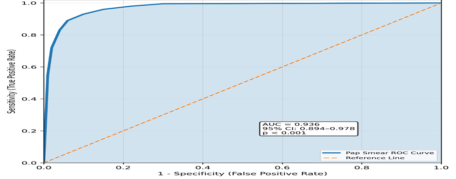
Figure 1: Receiver Operating Characteristic (ROC) Curve Pap Smear vs Histopathology
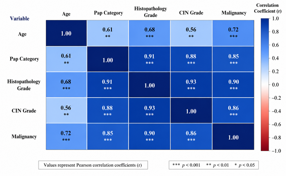
Figure 2: Correlation Heatmap of Clinical and Histopathological Variables
The correlation heatmap demonstrated strong positive associations among the study variables. Pap smear category showed excellent correlation with histopathological grade (r = 0.91), while histopathological grade strongly correlated with CIN grade (r = 0.93). Malignancy was positively associated with histopathological grade (r = 0.90), Pap smear category (r = 0.85), and CIN grade (r = 0.86). Age showed moderate positive correlations with malignancy (r = 0.72) and histopathological grade (r = 0.68). Overall, correlation coefficients ranged from 0.56 to 0.93, indicating consistent positive relationships between cytological abnormalities, histopathological severity, CIN grade, and malignancy. These findings demonstrate excellent concordance between Pap smear and histopathological diagnosis, supporting the reliability of Pap smear as a screening tool and histopathology as the gold standard for definitive diagnosis. No negative correlations were observed.
DISCUSSION:
The present study evaluated the histopathological spectrum of cervical lesions and correlated Pap smear findings with histopathological diagnosis among 150 women. The mean age of the study population was 39.8 ± 10.6 years, with the majority of patients (34.7%) belonging to the 31–40 years age group. A similar age distribution was reported by Ranabhat et al., who observed that abnormal epithelial lesions were most commonly detected in women aged 31–40 years, highlighting this age group as the primary target for cervical cancer screening (10). Patel et al. also reported a predominance of cervical epithelial abnormalities among women in the fourth decade of life, which is consistent with the present findings (11).
In the present study, white discharge per vaginum (38.7%) was the most common presenting complaint, followed by abnormal uterine bleeding (28.0%). Comparable observations were reported by Tailor et al., who found vaginal discharge to be the most frequent indication for Pap smear examination, emphasizing that symptomatic women constitute an important population for opportunistic cervical cancer screening (12).
The present study demonstrated that Negative for Intraepithelial Lesion or Malignancy (NILM) was the predominant Pap smear finding (49.3%), while epithelial abnormalities including ASC-US (5.3%), LSIL (10.7%), HSIL (6.7%), and malignancy (6.6%) accounted for the remaining cases. Similar findings were reported by Bal et al., who observed that the majority of cervical smears were negative for intraepithelial lesions, whereas epithelial abnormalities constituted a smaller but clinically significant proportion requiring further histopathological evaluation (13).
Histopathological examination in the present study revealed that chronic cervicitis (41.3%) was the most common lesion, followed by CIN I (13.3%), squamous metaplasia (12.0%), CIN II (9.3%), CIN III (6.7%), and squamous cell carcinoma (14.7%). These findings are comparable to those reported by Deepika et al., who found chronic cervicitis to be the predominant histopathological diagnosis, with cervical intraepithelial neoplasia representing the commonest premalignant lesion and squamous cell carcinoma being the most frequent malignant lesion (14).
An important finding of the present study was the excellent correlation between Pap smear and histopathological diagnosis, which was statistically significant (χ² = 98.74, p < 0.001). Similarly, Altaf reported a strong concordance between cervical cytology and histopathological examination and emphasized that histopathology remains the definitive method for confirming cervical epithelial abnormalities detected on Pap smear (15). Comparable results were also observed by Kumar et al., who demonstrated good agreement between abnormal cervical cytology and cervical biopsy findings, particularly in patients with high-grade squamous intraepithelial lesions, supporting the diagnostic value of Pap smear as a screening modality (16).
The diagnostic performance of Pap smear in the present study was excellent, with a sensitivity of 89.5%, specificity of 94.3%, positive predictive value of 85.0%, negative predictive value of 96.2%, and an overall diagnostic accuracy of 92.0%. These findings are consistent with the recommendations summarized by Rayner et al., who concluded that although HPV-based screening is increasingly recommended, Pap smear cytology continues to demonstrate high specificity and remains an effective, economical, and widely applicable screening tool, particularly in low- and middle-income countries where organized HPV screening may not be universally available (17).
Furthermore, the present study demonstrated that the mean age increased significantly with increasing severity of cervical lesions, from 37.6 ± 9.4 years in benign lesions to 42.3 ± 8.7 years in premalignant lesions and 52.8 ± 9.1 years in malignant lesions (ANOVA F = 18.62, p < 0.001). The correlation analysis also revealed a strong positive relationship between Pap smear category and histopathological grade (r = 0.91) and between histopathological grade and CIN grade (r = 0.93), indicating excellent agreement between cytological and histopathological assessment. These findings further reinforce the importance of clinicopathological correlation for accurate diagnosis and timely management of cervical lesions.
Overall, the present study demonstrated that inflammatory and benign cervical lesions constituted the majority of cases; however, a substantial proportion of premalignant and malignant lesions was identified through cervical cytology and confirmed histopathologically. The strong concordance between Pap smear and histopathological examination supports the continued use of the Pap smear as an effective primary screening tool, while emphasizing the indispensable role of histopathological examination as the gold standard for definitive diagnosis and appropriate patient management.
CONCLUSION:
The present study demonstrated that chronic cervicitis was the most common histopathological lesion of the cervix, followed by cervical intraepithelial neoplasia and invasive squamous cell carcinoma. The majority of women belonged to the 31–40 years age group, with white discharge per vaginum being the most frequent presenting complaint. Pap smear cytology showed excellent agreement with histopathological diagnosis, with a statistically significant correlation (χ² = 98.74, p < 0.001) and high diagnostic performance, including a sensitivity of 89.5%, specificity of 94.3%, and overall diagnostic accuracy of 92.0%. The study also demonstrated that increasing age was significantly associated with higher grades of cervical lesions and malignancy. These findings reaffirm that the Pap smear is an effective, reliable, and economical screening tool for the early detection of premalignant and malignant cervical lesions, while histopathological examination remains the gold standard for definitive diagnosis. Appropriate correlation between cytological and histopathological findings facilitates timely diagnosis, appropriate treatment, and effective prevention of cervical cancer.
REFERENCES:
1. WHO Classification of Tumours Editorial Board. WHO classification of tumours. Female genital tumours [Internet]. 5th ed. Lyon: International Agency for Research on Cancer; 2020 [cited 2026 Aug 1]. Available from: https://tumourclassification.iarc.who.int
2. Papanicolaou GN, Traut HF. Diagnosis of uterine cancer by the vaginal smear [Internet]. New York: Commonwealth Fund; 1943 [cited 2026 Aug 1]. Available from: https://archive.org/
3. World Health Organization. WHO guideline for screening and treatment of cervical pre-cancer lesions for cervical cancer prevention [Internet]. 2nd ed. Geneva: World Health Organization; 2021 [cited 2026 Aug 1]. Available from: https://www.who.int/publications/i/item/9789240030824
4. Nayar R, Wilbur DC. The Bethesda system for reporting cervical cytology: definitions, criteria, and explanatory notes [Internet]. 3rd ed. Cham: Springer; 2015 [cited 2026 Aug 1]. Available from: https://link.springer.com/book/10.1007/978-3-319-11074-5
5. Sachan PL, Singh M, Patel ML, Sachan R. A Study on Cervical Cancer Screening Using Pap Smear Test and Clinical Correlation. Asia Pac J Oncol Nurs. 2018 Jul-Sep;5(3):337-341. doi: 10.4103/apjon.apjon_15_18. PMID: 29963597; PMCID: PMC5996593.
6. Solomon D, Nayar R, editors. The Bethesda system for reporting cervical cytology: definitions, criteria, and explanatory notes [Internet]. 2nd ed. New York: Springer; 2004 [cited 2026 Aug 1]. Available from: https://link.springer.com/book/10.1007/978-0-387-21759-9
7. Arbyn M, Weiderpass E, Bruni L, de Sanjosé S, Saraiya M, Ferlay J, et al. Estimates of incidence and mortality of cervical cancer in 2018: a worldwide analysis. Lancet Glob Health. 2020;8(2):e191-e203. Available from: https://doi.org/10.1016/S2214-109X(19)30482-6
8. Bruni L, Albero G, Serrano B, Mena M, Gómez D, Muñoz J, et al. Human papillomavirus and related diseases report [Internet]. Barcelona: ICO/IARC Information Centre on HPV and Cancer; 2023 [cited 2026 Aug 1]. Available from: https://hpvcentre.net
9. World Health Organization. Cervical cancer [Internet]. Geneva: World Health Organization; 2024 [cited 2026 Aug 1]. Available from: https://www.who.int/news-room/fact-sheets/detail/cervical-cancer
10. Ranabhat, S., Shrestha, R., & Tiwari, M. (2011). Analysis of abnormal epithelial lesions in cervical Pap smears in Mid-Western Nepal. Journal of Pathology of Nepal, 1(1), 30-33. https://doi.org/10.3126/jpn.v1i1.4447
11. Patel MM, Pandya AN, Modi J. Cervical Pap Smear Study and Its Utility in Cancer Screening, To Specify the Strategy for Cervical Cancer Control. Natl J Community Med [Internet]. 2011 Jun. 30 [cited 2026 Aug. 1];2(01):49-51. Available from: https://njcmindia.com/index.php/file/article/view/1842
12. Tailor HJ, Patel RD, Patel PR, Bhagat VM. Study of cervical Pap smears in a tertiary care hospital of South Gujarat, India. Int J Res Med Sci. 2016;4(1):286-8. Available from: https://www.msjonline.org/index.php/ijrms [cited 2026 Aug 1].
13. Bal MS, Goyal R, Suri AK, Mohi MK. Detection of abnormal cervical cytology in Papanicolaou smears. J Cytol. 2012 Jan;29(1):45-7. doi: 10.4103/0970-9371.93222. PMID: 22438616; PMCID: PMC3307451.
14. Deepika, Khan A, Salmani A. Histopathological spectrum of cervical lesions in women attending a tertiary care hospital. Int J Clin Obstet Gynaecol. 2026;10(2):833-5. Available from: https://www.gynaecologyjournal.com/ [cited 2026 Aug 1].
15. Altaf FJ. Cervical cancer screening with cervical cytology and histopathology correlation. Saudi Med J. 2006;27(9):1348-51. Available from: https://smj.org.sa/Krishnappa R, Prathima S, Revathi R. Correlation of Pap smear with cervical biopsy in epithelial lesions of cervix. J Evol Med Dent Sci. 2013;2(39):7405-12.
16. Kumar SD, Rajeswari K, Krishna R, Meenakshisundaram K. A retrospective study of Pap smear and cervical biopsy correlation in abnormal cervical cytology cases. IP Arch Cytol Histopathol Res. 2021;6(2):87-90. Available from: https://www.achr.co.in/ [cited 2026 Aug 1].
17. Rayner M, Welp A, Stoler MH, Cantrell LA. Cervical cancer screening recommendations: now and for the future. Healthcare (Basel). 2023;11(16):2273. doi:10.3390/healthcare11162273. PMID: 37628471; PMCID: PMC10454304.