Computed Tomography Spectrum of Mediastinal Masses: A Comprehensive Pictorial Essay and Institutional Clinical Study.
- Kiran B Budihal , Department of Radio Diagnosis, K.S.Hospital, Koppal, Karnataka, India.
- Dakshayini H S , Assistant Professor, Department of Radio Diagnosis, Alva’s medical college, Moodbidiri, Karnataka, India.
- Tejeshwini C , Senior resident, Department of Medicine, Koppal Medical College, Karnataka, India.
- Chetan C , Consultant Radiologist, Ecotown Diagnostics, Bengaluru, Karnataka, India.
- Ibrahim Faheem , Department of Radio Diagnosis, K.S.Hospital Koppal, Karnataka, India.
Article Information:
Abstract:
Background: Mediastinal lesions present a diagnostic challenge to clinicians and radiologists due to the complex anatomy of the thoracic cavity and the diverse spectrum of pathologies ranging from benign cysts to highly aggressive primary and metastatic malignancies. Computed Tomography (CT) serves as the indispensable primary imaging modality for anatomical localization, characterization of tissue composition, assessment of vascular invasion, and staging. Objectives: To review the clinical and cross-sectional computed tomography (CT) spectrum of mediastinal masses, establish key radiological features for compartmental differentiation, and analyze a series of pathologically proven primary and secondary mediastinal neoplasms evaluated at a tertiary cancer institute. Materials and Methods: A descriptive retrospective and observational radiological study was conducted in the Department of Radio-Diagnosis at Kidwai Memorial Institute of Oncology, Bangalore from December 2022-July 2023. Contrast-enhanced CT scans of subjects presenting with mediastinal masses were evaluated for spatial compartmentalization (anterior, middle, and posterior mediastinum), attenuation values (fat, soft tissue, fluid, calcification), presence of vascular obstruction, local invasion, and distant organ metastasis. Results: Primary mediastinal masses demonstrated distinct compartmental predilections: 54% occurred in the anterior mediastinum, 20% in the middle mediastinum, and 26% in the posterior mediastinum. Thymic neoplasms, germ cell tumors, and lymphomas dominated the anterior compartment, whereas middle mediastinal lesions predominantly comprised lymphadenopathy, esophageal masses, and congenital cysts. Systemic manifestations such as Superior Vena Cava (SVC) syndrome, tracheoesophageal fistulation, pericardial effusion, and extrathoracic metastases were accurately mapped on multi-detector CT. Conclusion: Cross-sectional CT imaging is vital for precise anatomical localization, histopathological correlation, staging, and therapeutic planning of mediastinal masses.
Keywords:
Article :
INTRODUCTION:
The mediastinum is the central compartment of the thoracic cavity, bounded laterally by the pleural sacs, anteriorly by the sternum, posteriorly by the thoracic vertebral column, superiorly by the thoracic inlet, and inferiorly by the diaphragm.¹ Owing to the dense concentration of vital cardiovascular, respiratory, gastrointestinal, lymphatic, and neural structures within this region, a diverse range of pathological processes may arise.¹ Mediastinal lesions encompass a broad radiological and histopathological spectrum, including primary neoplasms, lymphadenopathy, congenital cystic lesions, infectious collections, and vascular abnormalities.²
The anatomical distribution of primary mediastinal tumors demonstrates a characteristic compartmental pattern. Approximately 54% of these tumors arise in the anterior mediastinum, 20% in the middle mediastinum, and 26% in the posterior mediastinum.³ Accurate localization and characterization of mediastinal lesions are essential because management strategies vary considerably according to the underlying pathology. Benign and early-stage lesions, such as thymomas and mature teratomas, are often managed with surgical resection, whereas malignant conditions including non-seminomatous germ cell tumors, high-grade lymphomas, and advanced esophageal carcinomas typically require multimodal treatment involving systemic chemotherapy, radiotherapy, and, in selected cases, surgical intervention.²˒⁴ Consequently, precise diagnosis and compartment-based evaluation of mediastinal lesions are critical for guiding appropriate therapeutic decisions and optimizing patient outcomes.².
MATERIALS AND METHODS:
MATERIALS AND METHODS
This institutional pictorial essay and observational study was conducted in the Department of Radio-Diagnosis at the Kidwai Memorial Institute of Oncology, Bangalore, Karnataka, India. The cases were collected from December 2022-July 2023. The study sample comprised patients presenting with clinical manifestations of thoracic mass lesions, referred for baseline diagnosis, staging, or therapeutic response assessment.10 such cases were included in this study.
CT Acquisition Protocol
All CT examinations were performed on multi-detector 64 slice siemens CT scanner. Contrast-enhanced CT (CECT) acquisition parameters included tube voltage of 120 kVp, auto-milliampere modulation (150–350 mA), collimation of 0.625 mm, and reconstructed axial slice thickness of 2.5 mm and 1.25 mm for multiplanar coronal and sagittal reformations. Non-ionic iodinated contrast medium (300–370 mg I/mL) was administered intravenously via a power injector at 1.2–1.5 mL/kg body weight.
RESULTS:
The total number of cases was 10. Out of which 6 were males and 4 were females.
The age group of patients were between 12-75 years and most patients were between 20-40 years .
The most common type of mediastinal mass were thymic lesions (40 %), followed by lymphoma (20%), Mediastinal germ cell tumours (20 %) and other variants (Carcinoma oesophagus, Retrosternal goitre). (Table 1).
Table 1: List of cases with CT features and Histopathological diagnosis.
|
CASE # |
AGE / SEX |
PRIMARY LOCATION |
KEY CT CHARACTERISTICS |
HISTOPATHOLOGICAL DIAGNOSIS (HPE) |
|
Case 1 |
22 yr / Male |
Prevascular (Anterior) |
Heterogeneous mass, calcification, bilateral renal lesions |
Mediastinal Germ Cell Tumor with Renal Metastasis |
|
Case 2 |
31 yr / Female |
Superior & Prevascular |
Coarse calcifications, invasive borders, pericardial effusion |
Invasive Thymoma |
|
Case 3
|
45 yr / Male |
Visceral (Middle) |
Large necrotizing mass, tracheoesophageal fistula, lung consolidation |
Esophageal / Mediastinal Poorly Differentiated Carcinoma |
|
Case 4 |
34 yr / Female |
Prevascular & Visceral |
Large mass causing SVC syndrome, lung, liver, and renal metastases |
Systemic Lymphoma with Multiorgan Deposits |
|
Case 5 |
12 yr / Male |
Prevascular & Visceral |
Confluent bulky anterior mediastinal nodal mass encircling great vessels |
Pediatric Mediastinal Lymphoma (Hodgkin Disease) |
|
Case 6 |
24 yr / Male |
Prevascular (Anterior) |
Large multiloculated necrotic mass with calcification and mass effect |
Thymic Carcinoma / Malignancy |
|
Case 7 |
75 yr/ Male |
Prevascular (Anterior) |
Heterogeneous necrotizing mass with pleural/ chest wall invasion |
Thymic Carcinoma / Malignancy |
|
Case 8 |
47 yr / Female |
Superior / Prevascular |
Multi-nodular high-attenuation mass continuous with thyroid, calcification |
Retrosternal Goitre |
|
Case 9 |
63 yr / Male |
Prevascular (Anterior) |
Invasive anterior mass with broad sternal and vascular contact |
Thymic Malignancy |
|
Case 10 |
55 yr / Female |
Prevascular (Anterior) |
Well-circumscribed homogeneous anterior mediastinal soft-tissue mass |
Non-Invasive Thymoma |
DETAILED PICTORIAL ESSAY OF FEATURED CLINICAL CASES
Case 1: Primary Mediastinal Germ Cell Tumor with Renal Metastasis in a 22-Year-Old Male
Clinical Presentation: A 22-year-old young male presented with retrosternal chest pain, weight loss, fever.
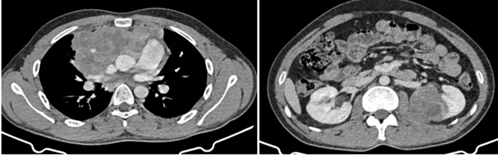
Figure 1 :CT Findings: Thoracic CT reveals a large, highly heterogeneous mass centered in the anterior prevascular mediastinum containing coarse internal calcifications, cystic/necrotic focus, and solid enhancing components. Abdominal cross-sectional acquisition further reveals multiple bilateral hypoattenuating solid mass lesions within the renal parenchyma, representing distant hematogenous renal metastases.
Histopathological Correlation: Elevated serum alpha-fetoprotein (AFP) and beta-human chorionic gonadotropin (β-hCG) supported the histopathological diagnosis of a Primary Non-Seminomatous Mediastinal Germ Cell Tumor with metastatic involvement of both kidneys.
Case 2: Invasive Thymoma in a 31 Year Old Female
Clinical Presentation: A 31-year-old female presented with progressive dyspnea, non-productive cough, chest fullness, and facial puffiness of three months duration.
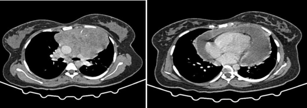
Figure 2:CT Findings: Axial CECT of the chest demonstrates a large, bulky, soft-tissue density mass occupying the superior and anterior (prevascular) mediastinum. The mass exhibits heterogeneous contrast enhancement with internal punctate and linear rim calcifications. There is loss of the fat plane between the mass and the pericardium, accompanied by a moderate to large pericardial effusion. The anterior border of the ascending aorta and pulmonary trunk shows abutment over >180 degrees, indicating local invasion.
Histopathological Correlation: Core needle biopsy confirmed an Invasive Epithelial Thymoma .
Case 3: Middle Mediastinal Malignancy with Tracheoesophageal Fistula in a 45 Year Old Male
Clinical Presentation: A 45-year-old male evaluated for severe dysphagia, recurrent coughing immediately following liquid ingestion, high-grade fever, and marked cachexia.
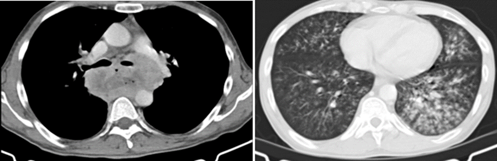
Figure 3:CT Findings: CECT demonstrates an aggressive, deeply necrotic middle (visceral) mediastinal mass originating from the esophageal wall with direct invasion into the posterior wall of the subcarinal trachea, forming a distinct Tracheoesophageal Fistula (TEF). Associated lung window settings reveal extensive patchy consolidation, tree-in-bud nodularities, and ground-glass opacities in the dependent left lung zones, diagnostic of severe secondary aspiration pneumonitis. Enlarged, necrotic cervical and mediastinal lymph nodes are also identified.
Histopathological Correlation: Esophagoscopy and biopsy confirmed a Poorly Differentiated Carcinoma of the esophagus with extensive subcarinal invasion.
Case 4;Mediastinal Lymphoma with lung ,liver and renal deposits in 34 year old female .
Clinical Presentation: A 34 -year-old male evaluated for dyspnea ,weight loss and long standing fever.
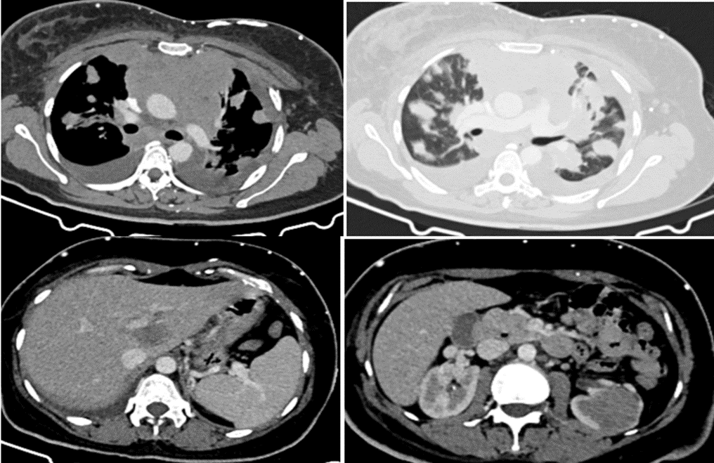
Figure 4:CT Findings :Multiple coalescent lobulated nodal masses in the anterior and middle mediastinum . Characteristic features include a "molded" appearance around major thoracic vascular structures without causing arterial invasion. There is widespread extranodal involvement of the lungs, liver, and kidneys.
Histopathological Correlation: Non-Hodgkins Lymphoma with lung, liver and renal metastasis
DISCUSSION:
The anterior mediastinum is the most common site of primary mediastinal neoplasia, accounting for over 50% of all cases.5 Differential diagnosis in this region is dominated by the classic "4 Ts": thymoma (and other thymic malignancies), teratoma (and other germ cell tumors), "terrible" lymphoma, and thyroid lesions (retrosternal goitre).6˒7
Tomiyana N et al (2009) observed that anterior mediastinal tumor account for 50-60 % of all mediastinal masses including thymoma, teratoma, thyroid disease and lymphoma8.Where as our study shows that almost 90% cases are involving the anterior compartment.
Duwe BV et al (2005) found that masses of middle mediastinum are typically congenital cysts while those arising in posterior medistinum are often neurogenic tumor.9 In our study middle mediastinal masses were lymphomas and oesophageal malignancy with metastasis .Since this study was conducted in regional cancer institute there was this disparity.
Sridhar R et al (2021) concluded that 45.2 % of mediastinal lesions are malignant and the masses were commonly located in middle mediastinum10, whereas in our study 80 % were malignant masses and 20% were thymomas .
Maximum number of patients were seen in 3rd decade of life in the study conducted by Aroor AR et al (2014)11,Our study found 2nd and 3rd decade (40 %) to be predominant age group. Mediastinal germ cell tumours are common in adolescent age group while lymphomas have bimodal age distribution .Hodgkin Lymphomas are common in adults whee as non Hodgkin lymphoma are common in older adults.
Baram A et al (2016) found that male predilection for mediastinal masses was more than female, out of 85 patients 46 were males and 39 were females12, quite similar to our study with male predominance (60%).
CONCLUSION:
Mediastinal masses encompass an extraordinarily wide diagnostic spectrum ranging from incidental benign cystic lesions to rapidly progressive primary and secondary malignancies. Cross-sectional Computed Tomography (CT) serves as the primary and definitive imaging workhorse for thoracic oncological staging. Through precise anatomical.
Compartmentalization, attenuation analysis, multiplanar reconstruction, and evaluation of local invasion and metastases, CECT empowers the multidisciplinary team to form an accurate diagnosis and design optimal treatment algorithms.
REFERENCES:
1. Gray's Anatomy. Standring S, editor. Gray's Anatomy: The Anatomical Basis of Clinical Practice. 41st ed. Philadelphia: Elsevier; 2016.
2. Shields' General Thoracic Surgery. LoCicero J III, Feins RH, Colson YL, Rocco G, editors. Shields' General Thoracic Surgery. 8th ed. Philadelphia: Wolters Kluwer; 2019.
3. Carter BW, Benveniste MF, Madan R, et al. Imaging of anterior mediastinal tumours. Clin Radiol. 2012;67(10):e47-e60.
4. National Comprehensive Cancer Network. NCCN Clinical Practice Guidelines in Oncology: Thymomas and Thymic Carcinomas; Hodgkin Lymphoma; Esophageal and Esophagogastric Junction Cancers. Current versions.
5. Carter BW, Benveniste MF, Madan R, de Groot PM, Truong MT, Marom EM. ITMIG classification of mediastinal compartments and multidisciplinary approach to mediastinal masses. Radiographics. 2017;37(2):413-436.
6. Juanpere S, Cañete N, Ortuño P, Martínez S, Sanchez G, Bernado L. A diagnostic approach to the mediastinal masses. Insights Imaging. 2013;4(1):29-52. doi:10.1007/s13244-012-0201-0.
7. Detterbeck FC, Zeeshan A. Thymoma: current diagnosis and treatment. Chin Med J (Engl). 2013;126(11):2186-2191.
8. Tomiyama N, Honda O, Tsubamoto M, Inoue A, Sumikawa H, Kuriyama K et al. Anterior mediastinal tumors: diagnostic accuracy of CT and MRI. Eur J Radiol. 2009 Feb; 69(2):280-8. doi: 10.1016/j.ejrad.2007.10.002. Epub 2007 Nov 26. PMID: 18023547.
9. Duwe BV, Sterman DH, Musani AI. Tumors of the mediastinum. Chest. 2005 Oct;128(4):2893-909. doi: 10.1378/chest.128.4.2893. PMID: 16236967.
10. Sridhar R, Narasimhan R, Sundararajan L, Singh RB. Clinicoradiopathological Features among Mediastinal Masses. Indian J Respir Care [Internet] 2021; 10 (1):41-46. doi: 10.4103/ijrc.ijrc_48_20
11. Aroor AR, Prakasha S R, Seshadri S, S T, Raghuraj U. A study of clinical characteristics of mediastinal mass. J Clin Diagn Res [Internet]. 2014 Feb;8(2):77-80. doi: 10.7860/JCDR/2014/7622.4013. Epub 2014 Feb 3. PMID: 24701488.
12. Baram A, Tayeb ZA. Mediastinal Masses: Retrospective Single Center Based Study. J Cancer Sci Ther [Internet] 2016 Jan, 8 (10): 252-56. doi: 10.4172/1948-5956.1000422.