ULTRASOUND GUIDED LIVER BIOPSIES: “AS SAFE AS A TORTOISE UNDER ITS SHELL” – 6 MONTHS EXPERIENCE IN A TERTIARY ONCOLOGY CENTER.
- Chetan C , Consultant Radiologist, Ecotown Diagnostics, Bengaluru, Karnataka, India.
- Kiran B Budihal , Department of Radio Diagnosis, K.S.Hospital, Koppal, Karnataka, India.
- Ibrahim Faheem , Department of Radio Diagnosis, K.S.Hospital Koppal, Karnataka, India.
- Dakshayini H S , Assistant Professor, Department of Radio Diagnosis, Alva’s medical college, Moodbidiri, Karnataka, India.
- Tejeshwini C , Senior resident, Department of Medicine, Koppal Institute of Medical Sciences, Koppal, Karnataka, India.
Article Information:
Abstract:
Background: In oncological practice, confirming hepatic focal lesions is critical, as various coincidental neoplastic and non-neoplastic lesions directly alter treatment trajectories. Ultrasound (US)-guided percutaneous core needle biopsy has largely superseded exploratory diagnostic laparotomy due to its minimally invasive nature and rapid recovery profile. Objective: To evaluate the diagnostic yield, safety profile, complications, and technical determinants of US-guided percutaneous core liver biopsies in an oncology setting. Methods: A prospective observational study of 100 consecutive patients undergoing USG guided core liver biopsies was conducted over 6 months (January 1, 2022, to June 30, 2022) at Kidwai Memorial Institute of Oncology, Bengaluru. Procedures were performed using 18-gauge core needles under local anesthesia. Diagnostic adequacy, early (<6 hours) and late (6–24 hours) complications, and causes of inconclusive histopathology were analyzed. Results: Diagnostic adequacy was achieved in 96% (n=96) of cases. Secondary (metastatic) tumors accounted for 84% (n=84) of diagnosed lesions, with biliodigestive primaries being the most prevalent, followed by colorectal, squamous cell, and breast carcinomas. Complications occurred in 4% (n=4) of patients, dominated by hemorrhage (75% of all adverse events, including intraperitoneal hemorrhage ) and transient vasovagal hypotension. No procedural mortality was recorded (0%). Tissue necrosis and extensive fibrosis were the chief drivers of non-diagnostic samples. Conclusion: Real-time US-guided core needle biopsy using an 18-gauge needle provides high diagnostic yield (96%) with an exceptionally favorable safety profile, making it a reliable pillar in oncological staging and treatment planning.
Keywords:
Article :
INTRODUCTION:
Percutaneous liver biopsy remains the definitive diagnostic standard for characterization of focal liver lesions and diffuse hepatic parenchymal diseases. In contemporary oncological care, precise histopathologic verification and immunohistochemical (IHC) profiling are mandatory prior to initiating targeted therapies, neoadjuvant regimens, or radical surgical interventions (1,2).
Although cross-sectional imaging modalities such as multiphasic Contrast-Enhanced Computed Tomography (CECT) and Dynamic Contrast-Enhanced Magnetic Resonance Imaging (MRI) exhibit high sensitivity for detecting hepatic lesions, imaging features alone can remain equivocal. Coincidental benign lesions (e.g., hemangiomas, focal nodular hyperplasia, adenomas) or synchronous secondary malignancies can mimic metastases. Biopsy confirmation avoids inappropriate upstaging or withholding of potentially curative surgical resection (3,4).
Historically, surgical sampling or blind percutaneous biopsies carried higher morbidity and variable diagnostic accuracy. Over the past three decades, real-time ultrasound guidance has transformed percutaneous liver biopsy into a routine outpatient procedure. Ultrasound guidance allows real-time visualization of needle passage, precise targeting of viable tumor zones, avoidance of adjacent vascular structures or biliary trees, and monitoring during breath-hold maneuvers, thereby improving procedural safety and diagnostic yield (5,6).
Literature indicates overall complication rates of approximately 1%–3% and mortality below 0.1% for image-guided percutaneous liver biopsies (5,7). However, procedural dynamics, institutional practices, needle gauge selection, and patient-specific factors in resource-constrained tertiary oncology centers warrant continued evaluation.
Aims & Objectives
Primary Objective
• To prospectively assess the diagnostic yield (efficacy) and overall safety profile (complication rates) of US-guided percutaneous core liver biopsies in a tertiary oncology center.
Secondary Objectives
1. To detail the standardized technical protocol, post-procedural monitoring, and management pathways for adverse events utilized at our institution.
2. To identify key clinicopathologic causes underlying inconclusive or inadequate histopathologic samples.
MATERIALS AND METHODS:
Study Design and Setting
This prospective observational study was conducted over a six-month period from January 1, 2022, to June 30, 2022, within the Department of Radiodiagnosis at the Kidwai Memorial Institute of Oncology, Bengaluru, Karnataka, India.
Patient Selection Criteria
Inclusion Criteria:
• Patients referred for histological characterization of suspected primary or secondary focal hepatic lesions identified on screening abdominal US, CECT, or MRI.
• Patients requiring IHC analysis for therapeutic planning.
• Pre-procedural coagulation profile: International Normalized Ratio (INR) ≤ 1.5, Platelet count ≥ 50,000/μL, and Prothrombin Time (PT) within normal limits.
Exclusion Criteria:
• Uncorrected coagulopathy or ongoing systemic anticoagulation therapy.
• Severe ascites obstructing safe needle access track.
• Inability of the patient to cooperate with breath-hold instructions.
• Hydatid cyst or vascular malformations along the target trajectory.
Pre-Procedural Workup and Procedural Technique
Written informed consent was obtained from all participants. Complete blood counts, platelet counts, and coagulation screens were reviewed. Real-time ultrasound equipment (LOGIQ P9, GE Healthcare) fitted with a 3.5–5.0 MHz broad-band convex transducer was utilized for all procedures.
Under strict aseptic conditions and local anesthesia (5–10 mL of 2% lignocaine hydrochloride), semi-automatic 18-gauge core biopsy gun were used to yield continuous tissue samples. Under direct US visualization, the needle was advanced to the margin of the focal lesion, targeting peripheral lesion zones specifically to avoid central necrotic cores(Figure 1). A minimum of normal liver parenchyma (1–2 cm) was traversed when feasible to create a tamponade track. 2 to 3 core samples (1.5–2.0 cm in length) were obtained per session and fixed in 10% neutral buffered formalin.
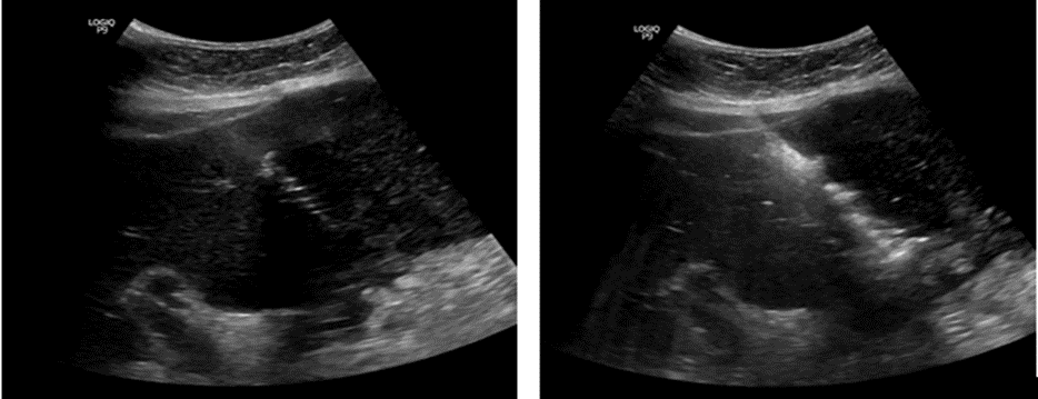
Figure 1 :a and b Shows USG of liver with 18 G biopsy neelde .The lesion is approached through 2-3 cm of normal tissue to create tamponade effect .Tip of the needle is within the lesion. The histopathology confirmed Metastasis from adenocarcinoma colon.
Post-Procedure Protocol
Patients were positioned in the right lateral decubitus position for 2 hours to apply natural compression over the puncture site. Vital signs were monitored every every 30 minutes for the first two hours, and hourly thereafter up to 6 hours. Routine bedside US was performed at 6 hours post-biopsy to check for subcapsular hematomas, intrahepatic hemorrhage, or free fluid in Morrison's pouch(Figure 2).
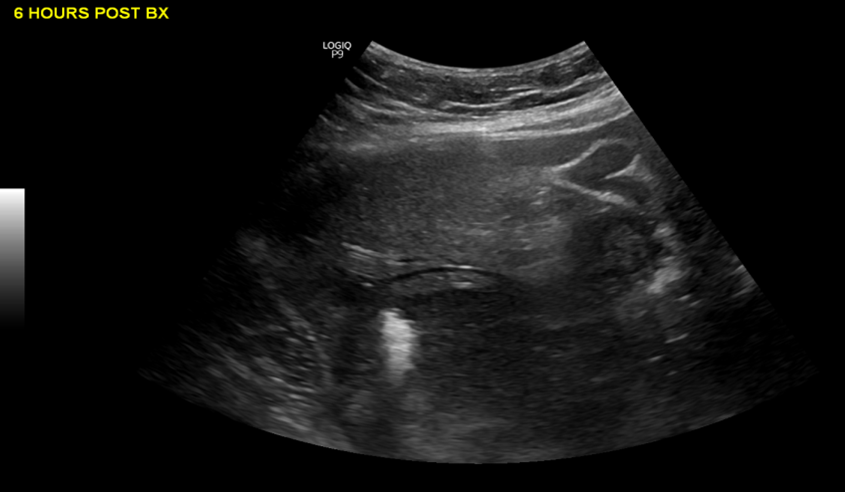
Figure 2: USG screening 6 hours post procedure to look for complications .There was no intra hepatic /subcapsular hematoma,No intraperitoneal hemorrhage .
RESULTS:
Diagnostic Adequacy and Yield
Over the 6-month study period, 100 patients underwent US-guided percutaneous core liver biopsies. High-quality diagnostic tissue samples capable of supporting histological diagnosis and IHC subtyping were obtained in 96% (n=96) of cases. Inconclusive or non-diagnostic tissue samples occurred in 4% (n=4) of cases.
Table 1: Overall diagnostic yield of US-guided core liver biopsy.
|
Diagnostic Parameter |
Count (n=100) |
Percentage (%) |
|
Adequate / Conclusive Specimen |
96 |
96% |
|
Inadequate / Inconclusive Specimen |
4 |
4% |
Histopathological Spectrum and Complications
Primary hepatic malignancies were diagnosed in 12% of cases. The remaining 84% (n=84) of conclusive biopsies demonstrated secondary metastatic lesions. Among secondary malignancies: Biliodigestive primaries accounted for 35.7% (n=30), Colorectal for 26.2% (n=22), Squamous Cell Carcinoma for 20.2% (n=17), Breast Carcinoma for 11.9% (n=10), and Other Primaries for 6.0% (n=5). The overall procedural complication rate was 4% (n=4). Three out of four complications (75%) were hemorrhagic. No procedural or 30-day mortality was observed (0%).
Table 2: Breakdown of procedural complications by onset time.
|
Complication Category |
Early (<6h) |
Late (6–24h) |
Total (n) |
Share of Events (%) |
|
Intraperitoneal Hemorrhage |
1 |
0 |
1 |
25% |
|
Intrahepatic/Subcapsular Hematoma |
1 |
1 |
2 |
50% |
|
Transient Vasovagal Reaction |
1 |
0 |
1 |
25% |
|
Mortality |
0 |
0 |
0 |
0% |
|
Total Complications |
3 |
1 |
4 |
100% |
DISCUSSION:
Diagnostic accuracy in liver mass evaluation is essential for selecting systemic chemotherapy, targeted therapy, immunotherapy, or neoadjuvant treatment strategies. In our tertiary oncology setting, 18G core needle biopsy achieved a 96% diagnostic yield. This finding is consistent with published international literature reporting diagnostic success rates ranging from 93% to 98% for ultrasound-guided core needle biopsies (6,8). Real-time ultrasound guidance facilitates accurate needle placement within viable tumor tissue, minimizing sampling from central necrotic regions.
The choice of needle gauge involves balancing diagnostic adequacy against procedural risk. Although fine-needle aspiration (FNA) is associated with a lower risk of bleeding, it often provides limited architectural information and insufficient tissue for extensive immunohistochemical analysis. Core biopsy using an 18G needle yields intact tissue cores that preserve architecture and allow reliable tumor subtyping and biomarker evaluation. Accordingly, 18G core biopsy is preferred at our institution because contemporary oncological management increasingly depends on comprehensive IHC and molecular profiling (2,6,9).
Inconclusive results occurred in 4% (n = 4) of cases. Analysis demonstrated that fragment length and the number of needle passes were comparable between conclusive and inconclusive groups. Instead, extensive tissue necrosis and intralesional fibrosis emerged as the principal causes of non-diagnostic specimens. To reduce sampling of necrotic tissue, Color Doppler imaging should be used to identify and target vascularized peripheral regions of hypoechoic or heterogeneous lesions, thereby improving diagnostic yield (6,10) .
Hemorrhage accounted for 75% (3/4) of adverse events. Most complications (75%) occurred within the first six hours after the procedure. These findings support a mandatory six-hour post-procedural observation protocol with serial vital sign monitoring and routine bedside ultrasonography before discharge, consistent with published recommendations for image-guided liver biopsy (5,7) .
CONCLUSION:
High Diagnostic Yield: Real-time US-guided percutaneous core liver biopsy using 18-gauge needles provides high diagnostic adequacy (96%) for histological and immunohistochemical characterization.
Safety Profile: The procedure carries low complication rates (4%) and low overall risk, with no procedural mortality (0%) observed.
Primary Complication: Hemorrhage is the most common adverse event (75% of complications), making pre-procedure screening and post-procedure monitoring essential.
Observation Window: A 6-hour post-procedural observation period combined with routine ultrasound screening effectively detects early complications.
Technical Recommendation: Targeting the viable peripheral margin of hepatic lesions helps avoid central necrosis and reduces inconclusive results.
REFERENCES:
1. European Association for the Study of the Liver (EASL). EASL Clinical Practice Guidelines on the management of liver diseases. J Hepatol. 2022;76(1):182–236.
2. Rockey DC, Caldwell SH, Goodman ZD, Nelson RC, Smith AD. Liver biopsy. Hepatology. 2009;49(3):1017–44.
3. Marrero JA, Kulik LM, Sirlin CB, Zhu AX, Finn RS, Abecassis MM, et al. Diagnosis, staging, and management of hepatocellular carcinoma: 2018 practice guidance by the American Association for the Study of Liver Diseases. Hepatology. 2018;68(2):723–50.
4. Vilgrain V, Ronot M, Abdel-Rehim M, Zappa M, d'Assignies G, Bruno O. Hepatic benign lesions: imaging update. Eur Radiol. 2016;26(10):3146–57.
5. Grant A, Neuberger J; British Society of Gastroenterology. Guidelines on the use of liver biopsy in clinical practice. Gut. 1999;45(Suppl IV):IV1–IV11.
6. Patel IJ, Davidson JC, Nikolic B, Salazar GM, Schwartzberg MS, Walker TG, et al. Society of Interventional Radiology Consensus Guidelines for periprocedural management and image-guided biopsy. J Vasc Interv Radiol. 2019;30(8):1155–67.
7. Piccinino F, Sagnelli E, Pasquale G, Giusti G. Complications following percutaneous liver biopsy: a multicentre retrospective study on 68,276 biopsies. J Hepatol. 1986;2(2):165–73.
8. Silva MA, Hegab B, Hyde C, Guo B, Buckels JA, Mirza DF. Needle track seeding following biopsy of liver lesions in the diagnosis of hepatocellular cancer: a systematic review and meta-analysis. Gut. 2008;57(11):1592–6.
9. Aube C, Oberti F, Lonjon J, Pageaux GP, Seror O. Imaging-guided liver biopsy in focal liver lesions: recommendations and current practice. Diagn Interv Imaging. 2017;98(9):619–27.
10. Fornari F, Civardi G, Cavanna L, Di Stasi M, Rossi S, Buscarini E, et al. Value of contrast-enhanced and Doppler ultrasonography in guiding liver biopsy of focal hepatic lesions. Ultrasound Med Biol. 1994;20(6):569–75.