To Study Hepatic Venous Waveforms and Spleno-Portal Index on Ultrasound in Cases of Liver Cirrhosis and Their Correlation with Disease Severity.
- Vivek Yonati , Associate professor, Department of Radiodiagnosis, MGMMC MYH Indore (M.P).
- Prachi Shukla , Assistant Professor, Department of Radiodiagnosis, MGMMC MYH indore (M.P).
- Alka Agrawal , Professor and Head of Department, Department of Radiodiagnosis, MGMMC MYH Indore (M.P).
- Soham Roy Chowdhury , Junior Resident, Department of Radiodiagnosis, MGMMC MYH Indore (M.P).
- Shrikant Shukla , Junior Resident, Department of Radiodiagnosis, MGMMC MYH Indore (M.P).
- Jatin Rawat , Junior Resident, Department of Radiodiagnosis, MGMMC MYH Indore (M.P).
Article Information:
Abstract:
Background: Doppler ultrasonography provides a non-invasive assessment of haemodynamic changes in liver cirrhosis. This study evaluated hepatic venous waveforms, damping index, and spleno-portal index and examined their association with disease severity. Methods: This hospital-based cross-sectional study included 100 adults with clinically diagnosed liver cirrhosis. B-mode and Doppler ultrasonography were performed to assess hepatic venous waveform, damping index, portal venous flow, and spleno-portal index. Disease severity was graded using the Child–Pugh classification. Associations were tested using the chi-square test or Fisher’s exact test. Diagnostic performance for identifying Child–Pugh class C cirrhosis was assessed using receiver operating characteristic analysis. Results: The mean age was 46.3 ± 13.7 years, and 82% of participants were male. Child–Pugh classes A, B, and C accounted for 8%, 35%, and 57% of patients, respectively. Hepatic venous waveforms were biphasic in 54%, monophasic in 34%, and triphasic/tetraphasic in 12%. Hepatic venous waveform was significantly associated with Child–Pugh class (p=0.001); 82.4% of patients with a monophasic waveform belonged to class C. A damping index above 0.6 was also associated with disease severity (p=0.004), whereas the spleno-portal index was not (p=0.089). The damping index showed the highest overall discrimination for class C cirrhosis (AUC=0.66). Conclusion: Hepatic venous waveform and damping index may complement clinical assessment of cirrhosis severity. The spleno-portal index showed limited sensitivity and should not be used alone for severity grading.
Keywords:
Article :
INTRODUCTION:
Liver cirrhosis is a chronic and irreversible condition characterized by diffuse fibrosis, distortion of normal hepatic architecture, and the formation of regenerative nodules. It may be classified morphologically as micronodular or macronodular, with alcohol-related liver disease and chronic viral hepatitis being important underlying causes [1]. Chronic liver disease affects approximately 1.5 billion people worldwide, while cirrhosis and its complications account for substantial morbidity and mortality. In India, the burden is particularly high. Alcohol-related liver disease, chronic hepatitis B and C, and non-alcoholic fatty liver disease are among the leading causes [2].
Progressive fibrosis increases resistance to hepatic blood flow and leads to portal hypertension, which is a major cause of complications such as ascites, splenomegaly, variceal bleeding, and hepatic encephalopathy [3]. Although liver biopsy has traditionally been considered the reference standard for assessing hepatic fibrosis, it is invasive and is limited by sampling errors, subjective interpretation, and procedure-related complications [4]. Clinical scoring systems, including the Child–Pugh and Model for End-Stage Liver Disease scores, are widely used to assess disease severity and prognosis. Serum-based fibrosis markers have also been proposed, but their accuracy may be influenced by extrahepatic factors [5]. Computed tomography and magnetic resonance imaging can demonstrate structural changes; however, they are relatively expensive and provide limited dynamic assessment of hepatic blood flow [6].
Doppler ultrasonography offers a safe, accessible, and non-invasive method for evaluating hepatic haemodynamics. With progressive cirrhosis, the normal triphasic hepatic venous waveform may become biphasic or monophasic. The damping index provides a quantitative measure of this loss of phasicity, with higher values indicating greater waveform flattening. The spleno-portal index incorporates splenic and portal venous parameters and may serve as an indirect indicator of portal hypertension [7]. Therefore, this study evaluates hepatic venous waveforms, damping index, and spleno-portal index in patients with liver cirrhosis and examines their correlation with disease severity assessed using the Child–Pugh score.
MATERIALS AND METHODS:
This hospital-based cross-sectional study was conducted in the Department of Radiodiagnosis,
M.G.M. Medical College, M.Y. Hospital, and Super Speciality Hospital, Indore, Madhya Pradesh, India. The study was carried out over one year after obtaining approval from the Institutional Scientific and Ethics Committee. Written informed consent was obtained from all participants before enrolment.
A total of 100 adult patients referred by the Department of Medicine and associated departments for hepatic and portal venous Doppler ultrasonography were included. Patients aged over 18 years with a clinical diagnosis of liver cirrhosis or its sequelae and who provided informed consent were eligible. Patients with a history of hepatic surgery, liver tumour, abdominal trauma, obesity, veno-occlusive disease, or renal disease were excluded.
A detailed clinical history was obtained, and relevant clinical findings and complications of liver cirrhosis were recorded. Disease severity was assessed using the Child–Pugh classification based on the available clinical and laboratory parameters. Before ultrasonography, patients were instructed to fast for 4–6 hours to reduce interference from bowel gas. All examinations were performed using a 3–5 MHz curvilinear transducer, with the patient in the supine or slight left lateral decubitus position.
B-mode ultrasonography was initially performed to assess liver size, margins, echotexture, portal vein diameter, and splenic dimensions. The craniocaudal length of the right hepatic lobe was measured in the midclavicular line. The right hepatic vein was examined through a subcostal or intercostal approach. The Doppler cursor was aligned with the direction of blood flow, and a sample volume of 5–10 mm was used. Recordings were obtained during suspended respiration at the end of expiration to minimize respiratory variation. Hepatic venous waveforms were classified as triphasic, biphasic, or monophasic. The damping index was determined from the minimum and maximum velocities of the hepatic venous waveform (Figure 1).
For portal venous assessment, the main portal vein was examined at the porta hepatis before its division into the right and left branches. The Doppler sample volume was placed within the longitudinal axis of the vessel, and the insonation angle was maintained below 60°. Patients were asked to briefly suspend respiration during image acquisition. The direction of portal venous flow was categorized as hepatopetal or hepatofugal. The main portal vein diameter and mean blood-flow velocity were also recorded. The spleen was examined in the right lateral decubitus position. Its craniocaudal and transverse dimensions and echotexture were documented. The splenic index was obtained from the maximum craniocaudal and transverse diameters. The spleno-portal index was determined using the splenic index and mean portal vein velocity.
Data were entered into Microsoft Excel and analysed using jamovi software. Continuous variables were summarized as mean and standard deviation, while categorical variables were presented as frequencies and percentages. Associations of hepatic venous waveform, damping index, and spleno-portal index with Child–Pugh class were examined using the chi-square test or Fisher’s exact test, as appropriate. Receiver operating characteristic curve analysis was performed to evaluate their ability to identify Child–Pugh class C cirrhosis. Diagnostic performance was expressed as sensitivity, specificity, positive predictive value, negative predictive value, and area under the curve. All tests were two-sided, and p<0.05 was considered statistically significant.
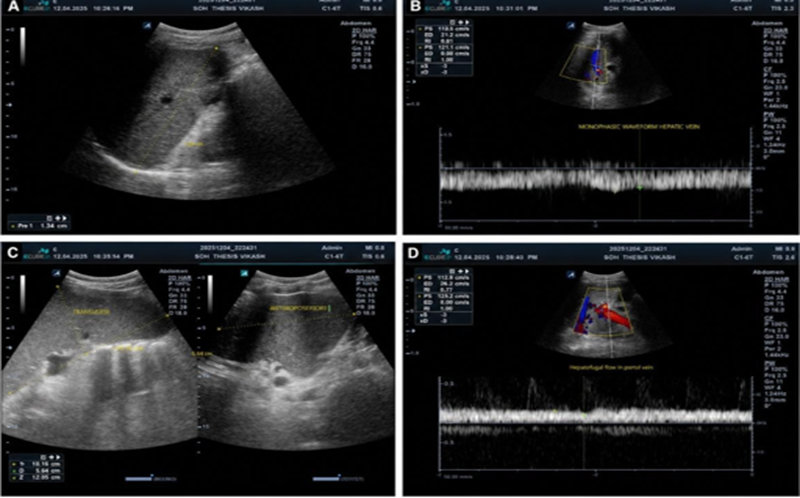
Figure 1. B-mode and Doppler ultrasonographic findings in a patient with Child–Pugh class C liver cirrhosis.
(A) Liver showing a regular surface and heterogeneous echotexture. (B) Monophasic hepatic venous waveform. (C) Splenomegaly. (D) Hepatofugal flow in the portal vein
RESULTS:
Among the 100 participants, the mean age was 46.3 ± 13.7 years, with the largest proportion aged 41–50 years (31%), followed by 31–40 years (24%). Most participants were male (82%). Abdominal pain and distension were the most frequent symptoms (79%), whereas ascites was the most common clinical sign (91%). Alcohol-related cirrhosis was the predominant aetiology (89%) (Table 1).
Table 1. Demographic and clinical characteristics of the study population (N = 100)
|
Variable |
Category |
Number (n) |
Percentage (%) |
|
Age group (years) |
18–20 |
1 |
1.0 |
|
21–30 |
11 |
11.0 |
|
|
31–40 |
24 |
24.0 |
|
|
41–50 |
31 |
31.0 |
|
|
51–60 |
15 |
15.0 |
|
|
61–70 |
12 |
12.0 |
|
|
>70 |
6 |
6.0 |
|
|
Sex |
Male |
82 |
82.0 |
|
Female |
18 |
18.0 |
|
|
Clinical symptoms |
Abdominal pain and distension |
79 |
79.0 |
|
Pedal oedema |
66 |
66.0 |
|
|
Shortness of breath |
43 |
43.0 |
|
|
Haematemesis/melena |
18 |
18.0 |
|
|
Altered sensorium |
15 |
15.0 |
|
|
Clinical signs |
Ascites |
91 |
91.0 |
|
Pedal oedema |
70 |
70.0 |
|
|
Jaundice |
59 |
59.0 |
|
|
Splenomegaly |
37 |
37.0 |
|
|
Hepatomegaly |
14 |
14.0 |
|
|
Aetiology |
Alcohol-related cirrhosis |
89 |
89.0 |
|
Alcohol with hepatitis B |
4 |
4.0 |
|
|
Hepatitis B |
2 |
2.0 |
|
|
Cryptogenic cirrhosis |
5 |
5.0 |
More than half of the participants were classified as Child–Pugh class C (57%), while 35% and 8% belonged to classes B and A, respectively. Gross ascites was present in 32%. Serum albumin below 2.8 g/dL was observed in 67%, bilirubin above 3 mg/dL in 46%, and PT prolongation exceeding six seconds in 66% of patients with available data. On B-mode ultrasonography, ascites, splenomegaly, and irregular liver surface were observed in 91%, 54%, and 51%, respectively (Table 2).
Table 2. Clinical severity, laboratory parameters, and B-mode ultrasonographic findings
|
Parameter |
Category |
n (%) |
|
Child–Pugh class |
A |
8 (8.0) |
|
B |
35 (35.0) |
|
|
C |
57 (57.0) |
|
|
Ascites grade |
None |
9 (9.0) |
|
Minimal |
17 (17.0) |
|
|
Mild |
25 (25.0) |
|
|
Moderate |
17 (17.0) |
|
|
Gross |
32 (32.0) |
|
|
Serum albumin, g/dL |
<2.8 |
67 (67.0) |
|
2.8–3.5 |
17 (17.0) |
|
|
>3.5 |
16 (16.0) |
|
|
Serum bilirubin, mg/dL |
<1.2 |
16 (16.0) |
|
1.2–2.0 |
16 (16.0) |
|
|
2.1–3.0 |
22 (22.0) |
|
|
>3.0 |
46 (46.0) |
|
|
PT prolongation |
<4 seconds |
13 (13.4) |
|
4–6 seconds |
20 (20.6) |
|
|
>6 seconds |
64 (66.0) |
|
|
B-mode findings |
Ascites |
91 (91.0) |
|
Splenomegaly |
54 (54.0) |
|
|
Irregular liver surface |
51 (51.0) |
|
|
Pleural effusion |
48 (48.0) |
|
|
Periportal/perisplenic collaterals |
39 (39.0) |
|
|
Hepatomegaly |
20 (20.0) |
|
|
Dilated portal vein |
16 (16.0) |
|
|
Portal vein thrombosis |
4 (4.0) |
The most frequent hepatic venous waveform was biphasic (54%), followed by monophasic (34%) and triphasic/tetraphasic (12%). A damping index above 0.6 was recorded in 48%. Among 96 patients with a measurable spleno-portal index, 41.7% had values between 4 and 8, while 39.6% had values below 4 (Table 3).
Table 3. Hepatic and portal venous Doppler findings
|
Doppler parameter |
Category |
n (%) |
|
Hepatic venous waveform |
Monophasic |
34 (34.0) |
|
Biphasic |
54 (54.0) |
|
|
Triphasic/tetraphasic |
12 (12.0) |
|
|
Damping index |
>0.6 |
48 (48.0) |
|
≤0.6 |
52 (52.0) |
|
|
Spleno-portal index |
<4 |
38 (39.6) |
|
4–8 |
40 (41.7) |
|
|
8.1–12 |
17 (17.7) |
|
|
>12 |
1 (1.0) |
Hepatic venous waveform was significantly associated with Child–Pugh class (p=0.001), with 82.4% of monophasic waveforms occurring in class C. A damping index above 0.6 was also significantly associated with disease severity (p=0.004). Although increasing spleno-portal index values were more frequent in class C, the association was not statistically significant (p=0.089) (Table 4).
Table 4. Association of Doppler parameters with Child–Pugh class
|
Doppler parameter |
Category |
Class A |
Class B |
Class C |
p-value |
|
Hepatic venous waveform |
Monophasic |
2 (5.9) |
4 (11.8) |
28 (82.4) |
0.001 |
|
Biphasic |
3 (5.6) |
26 (48.1) |
25 (46.3) |
||
|
Triphasic/tetraphasic |
3 (25.0) |
5 (41.7) |
4 (33.3) |
||
|
Damping index |
>0.6 |
4 (8.3) |
9 (18.8) |
35 (72.9) |
0.004 |
|
≤0.6 |
4 (7.7) |
26 (50.0) |
22 (42.3) |
||
|
Spleno-portal index |
<4 |
4 (10.5) |
20 (52.6) |
14 (36.8) |
0.089 |
|
4–8 |
2 (5.0) |
13 (32.5) |
25 (62.5) |
||
|
8.1–12 |
2 (11.8) |
1 (5.9) |
14 (82.4) |
||
|
>12 |
0 |
0 |
1 (100.0) |
Abnormal hepatic venous waveform demonstrated high sensitivity (93.0%) but low specificity (18.6%) for identifying Child–Pugh class C. The damping index provided the highest overall discrimination (AUC=0.66), while an elevated spleno-portal index showed high specificity (93.0%) but low sensitivity (26.3%) (Table 5).
Table 5. Diagnostic performance of Doppler parameters for identifying Child–Pugh class C cirrhosis
|
Parameter |
Sensitivity (%) |
Specificity (%) |
PPV (%) |
NPV (%) |
AUC |
|
Abnormal hepatic venous waveform |
93.0 |
18.6 |
60.2 |
66.7 |
0.56 |
|
Damping index >0.6 |
61.4 |
69.8 |
72.9 |
57.7 |
0.66 |
|
Elevated spleno-portal index |
26.3 |
93.0 |
83.3 |
48.8 |
0.63 |
DISCUSSION:
This study evaluated hepatic venous waveforms, damping index, and spleno-portal index as non-invasive indicators of disease severity in 100 patients with liver cirrhosis. The mean age was 46.3 ± 13.7 years, and 82% of participants were male. This profile was comparable to that reported by Bhutto et al., who observed a mean age of 48.2 years and a predominance of men among patients with chronic liver disease [8]. The marked male predominance in our cohort may be explained by the high frequency of alcohol-related cirrhosis, which accounted for 89% of cases.
Most patients presented with features of advanced disease. Abdominal pain and distension were reported by 79%, while ascites was detected clinically in 91%. Low serum albumin, elevated bilirubin, and prolonged prothrombin time were also common. Similar clinical findings were described by Mahapatra et al., while Runyon et al. identified ascites as an important marker of hepatic decompensation [9], [10]. Furthermore, 57% of our patients belonged to Child–Pugh class C. This proportion was higher than that reported by Mittal et al., possibly because the present study was conducted at a tertiary referral centre receiving patients with more advanced disease [11].
On B-mode ultrasonography, ascites was the most frequent finding, followed by splenomegaly, irregular liver surface, pleural effusion, and portosystemic collaterals. These findings reflect portal hypertension, reduced hepatic synthetic function, and progressive architectural distortion. Similar observations were reported by Berzigotti et al., who emphasized the value of ultrasonography in identifying morphological and haemodynamic changes associated with chronic liver disease [12].
The biphasic hepatic venous waveform was the most common pattern, observed in 54% of patients, followed by the monophasic pattern in 34%. Only 12% retained a triphasic or tetraphasic waveform. Hepatic venous waveform was significantly associated with Child–Pugh class (p=0.001), with 82.4% of patients showing a monophasic waveform belonging to class C. This progressive loss of phasicity may result from hepatic fibrosis and reduced venous compliance. A similar association between waveform abnormalities and cirrhosis severity was reported by Yasmin et al [13]. Kawanaka et al. also observed progressive waveform flattening with worsening liver disease [14]. Likewise, Bolondi et al. found that loss of the normal triphasic pattern was more frequent in advanced cirrhosis [15].
A damping index above 0.6 was found in 48% of patients and was significantly associated with Child–Pugh class (p=0.004). Nearly 73% of patients with an elevated damping index belonged to class C. These findings support the observations of Kim et al., who found that increased damping reflected worsening hepatic dysfunction and reduced venous phasicity [16].
The spleno-portal index was not significantly associated with Child–Pugh class (p=0.089). Piscaglia et al. similarly reported that portal Doppler indices may not consistently parallel clinical severity because they are affected by collateral circulation, respiration, hydration, and compensatory haemodynamic changes [17].
Abnormal hepatic venous waveforms showed high sensitivity but poor specificity for class C cirrhosis. The damping index provided the best overall discrimination, whereas an elevated spleno-portal index had high specificity but low sensitivity. Thus, hepatic venous waveform and damping index may complement clinical assessment, but none of these Doppler parameters should be used alone to grade cirrhosis severity.
This study has certain limitations. Its cross-sectional, single-centre design and relatively small sample may limit the generalizability of the findings. Most participants had alcohol-related and advanced cirrhosis, resulting in an uneven distribution across Child–Pugh classes. Doppler measurements were not compared with hepatic venous pressure gradient or histopathological findings. In addition, interobserver variability and the effects of physiological factors on Doppler parameters were not assessed. Larger prospective studies with longitudinal follow-up are required to validate their prognostic value.
CONCLUSION:
Doppler ultrasonography provides a safe, accessible, and non-invasive approach to assessing haemodynamic alterations in liver cirrhosis. Abnormal hepatic venous waveforms, particularly monophasic patterns, and a damping index above 0.6 were significantly associated with higher Child–Pugh class, supporting their value in severity assessment. The spleno-portal index showed high specificity but limited sensitivity. Incorporating hepatic venous waveform and damping index assessment into routine ultrasonography may improve risk stratification and follow-up. Further prospective studies using hepatic venous pressure measurements are required for validation.
REFERENCES:
1. Sharma S, Prasad Adhikari I, Khadka H. Changes in Doppler Waveform of Hepatic Vein in Liver Cirrhosis. International Journal of Biochemistry & Physiology. 2019;4(2):000152.
2. Swaroop S, Vaishnav M, Arora U, et al. Etiological Spectrum of Cirrhosis in India: A Systematic Review and Meta-analysis. J Clin Exp Hepatol. 2024;14(2):101291.
3. Antil N, Sureka B, Mittal MK, Malik A, Gupta B, Thukral BB. Hepatic Venous Waveform, Splenoportal and Damping Index in Liver Cirrhosis: Correlation with Child Pugh’s Score and Oesophageal Varices. J Clin Diagn Res. 2016;10(2):TC01-TC5.
4. Maharaj B, Maharaj RJ, Leary WP, et al. Sampling variability and its influence on the diagnostic yield of percutaneous needle biopsy of the liver. Lancet. 1986;1(8480):523-525.
5. Simon TG, Schneeweiss S, Wyss R, et al. Development and Validation of a Novel Tool to Predict Model for End-Stage Liver Disease (MELD) Scores in Cirrhosis, Using Administrative Datasets. Clin Epidemiol. 2023;15:349-362.
6. Sartoris R, Vilgrain V. Quantitative Computed Tomography-Based Approaches for Noninvasive Diagnosis of Portal Hypertension in Patients with Cirrhosis. Portal Hypertens Cirrhosis. 2023;2(2):92-7.
7. Saleem S, Rauf MH, Sohail M, Taufiq N, Khan MU. The Assessment of Diagnostic Accuracy of Real-Time Shear Wave Elastography in Detecting Liver Cirrhosis Keeping Histopathology as Reference Standard. Pak Armed Forces Med J. 2022;72(2):590-3.
8. Bhutto AR, Abbasi A, Butt N, Khan A, Munir SM. Hepatic vein waveform in liver cirrhosis: correlation with Child’s class and size of varices. J Pak Med Assoc. 2012;62(8):794-7.
9. Mahapatra GS, Dash S, Mishra D, Sahoo PK, Behera MK. A study on the clinico-etiological profile of cirrhosis of the liver and prognostic value of the MELD score on short-term survival. Bengal Physician Journal. 2020;7(3):94-99.
10. Runyon BA. Introduction to the revised American Association for the Study of Liver Diseases Practice Guideline management of adult patients with ascites due to cirrhosis 2012. Hepatology. 2013;57(4):1651-3.
11. Mittal P, Gupta R, Mittal G, Kalia V. Association between portal vein color Doppler findings and the severity of disease in cirrhotic patients with portal hypertension. Iran J Radiol. 2011;8(4):211-217.
12. Berzigotti A. Ultrasound in portal hypertension—part 1: general aspects and splanchnic hemodynamics; part 2: Doppler and elastography. Ultraschall Med. 2017;38(5):434–455.
13. Yasmin T, Sultana S, Ima MN, Islam MQ, Roy SK, Rafat S. Correlation between hepatic vein wave form changes on Doppler ultrasound and the severity of diseases in cirrhotic patients. Journal of Medicine. 2021;22(2):100-6.
14. Kawanaka H, Kinjo N, Anegawa G, Yoshida D, Migoh S, Konishi K, Ohta M, Yamaguchi S, Tomikawa M, Hashizume M, Maehara Y. Abnormality of the hepatic vein waveforms in cirrhotic patients with portal hypertension and its prognostic implications. Journal of gastroenterology and hepatology. 2008;23(7):e129-36.
15. Bolondi L, Li Bassi S, Gaiani S, Zironi G, Benzi G, Santi V, Barbara L. Liver cirrhosis: changes of Doppler waveform of hepatic veins. Radiology. 1991;178(2):513-6.
16. Kim MY, Baik SK, Suk KT, Yea CJ, Lee IY, Kim JW, et al. Damping index of hepatic vein waveform in cirrhosis: correlation with portal hypertension and liver function. J Gastroenterol Hepatol. 2008;23(10):1521–1526.
17. Piscaglia F, Donati G, Serra C, Muratori R, Solmi L, Gaiani S, et al. Value of splanchnic Doppler ultrasound in the diagnosis of portal hypertension. Ultrasound Med Biol. 2007;33(5):707–715.