Role of Transperineal Ultrasonography in the Evaluation of Anorectal Malformations in Infants.
- Alka Agrawal , Professor and Head of Department , Department of Radiodiagnosis,MGMMC MYH indore (M.P)
- Saurabh Atram , Professor, Department of Radiodiagnosis,MGMMC MYH indore (M.P)
- Suraj Mathur , Assistant Professor , Department of Radiodiagnosis,MGMMC MYH indore (M.P)
- Shankar , Junior Resident , Department of Radiodiagnosis,MGMMC MYH indore (M.P)
- Soham Roy Chowdhury , Junior Resident , Department of Radiodiagnosis,MGMMC MYH indore (M.P)
- Shrikant Shukla , Junior Resident , Department of Radiodiagnosis,MGMMC MYH indore (M.P).
Article Information:
Abstract:
Background: Accurate identification of the level of anorectal malformations (ARMs) is essential for selecting the appropriate surgical approach. Transperineal ultrasonography (TPUS) provides radiation-free evaluation of the distal rectal pouch, fistulous tracts, and sphincter complex. Methods: This prospective cross-sectional study included 32 infants with clinically diagnosed ARM. High-resolution TPUS was performed before definitive surgery using a 7–15 MHz linear transducer. The pouch–perineum distance, puborectalis thickness, sphincter morphology, and presence of fistulous communication were assessed. Intraoperative findings were used as the reference standard. Diagnostic performance and agreement between TPUS and operative classification were evaluated. Results: The mean age was 2.09 ± 0.86 days, and 62.5% of the infants were male. TPUS classified 8 cases (25.0%) as low, 18 (56.3%) as intermediate, and 6 (18.8%) as high ARM. Mean pouch–perineum distance increased from 9.43 ± 3.19 mm in low ARM to 18.15 ± 5.17 mm in intermediate and 33.32 ± 4.64 mm in high ARM. The distance was significantly greater in high than in low/intermediate ARM (p < 0.001). Fistulas were detected in 65.6% of cases. TPUS showed 87.5% overall concordance with operative findings, with strong agreement (κ = 0.810; p < 0.001). Conclusion: TPUS is a reliable, non-invasive, and radiation-free method for preoperative classification of ARM. It can serve as a first-line investigation, while distal colostography remains useful for complex fistulas.
Keywords:
Article :
INTRODUCTION:
Anorectal malformations (ARMs) comprise a spectrum of congenital abnormalities involving the distal rectum and anus, with an estimated incidence of 1 in 4,000–5,000 live births and a slight male predominance.1 They range from relatively simple perineal fistulas to complex cloacal anomalies and may coexist with urogenital or other systemic abnormalities. Accurate classification is essential because the level of the distal rectal pouch determines the surgical approach and influences prognosis.2
Traditionally, prone cross-table lateral radiography, invertography, and distal colostography have been used to assess ARMs. However, these investigations involve ionizing radiation, may require delayed imaging or invasive procedures, and can occasionally provide uncertain anatomical information. Although magnetic resonance imaging offers excellent soft-tissue resolution, its routine use in infants is limited by cost, availability, and the possible need for sedation.3,4
Transperineal ultrasonography is a safe, non-invasive, radiation-free, and readily available alternative. It enables real-time evaluation of the distal rectal pouch, pouch–perineum distance, fistulous communications, and anal sphincter complex. Previous studies have reported good agreement between transperineal ultrasonographic and operative findings in determining the level of ARMs. Nevertheless, evidence regarding its routine clinical utility remains limited, particularly in resource-constrained settings.5,6 Therefore, the present study evaluated the diagnostic utility of high-resolution transperineal ultrasonography in classifying ARMs in infants and compared its findings with surgical observations.
MATERIALS AND METHODS:
This prospective cross-sectional study was conducted in the Department of Radiodiagnosis, M.G.M. Medical College and M.Y. Hospital, Indore, Madhya Pradesh, in collaboration with the Department of Pediatric Surgery/General Surgery. The study was carried out over 12 months after approval from the Institutional Scientific Review Board and Institutional Ethics Committee. Written informed consent was obtained from the parent or legal guardian of every participant. A total of 32 infants who fulfilled the eligibility criteria were enrolled.
Infants with a clinically diagnosed anorectal malformation, with or without fistulous communication, were included if they presented before definitive surgical correction. Infants who had already undergone definitive surgery, were haemodynamically unstable, or were unsuitable for ultrasonography were excluded. Cases with severe perineal infection, wounds, or dressings interfering with examination were also excluded. Infants in whom an adequate transperineal examination could not be performed or who did not undergo or complete posterior sagittal anorectoplasty were excluded from the final analysis.
Relevant clinical information, including age, sex, type of malformation, visible fistulous opening, and associated congenital anomalies, was recorded. Before ultrasonography, the general condition and perineal anatomy of each infant were assessed. Transperineal ultrasonography was performed before surgery using a high-frequency 7–15 MHz linear transducer on an advanced ultrasound system. Infants were examined in the supine frog-leg position. The transducer was placed gently over the perineum, and excessive pressure was avoided to prevent distortion of the anatomy and underestimation of measurements (Figure 1).
Scanning was initially performed in the midsagittal plane, with transverse and coronal views obtained whenever required. The distal rectal pouch was identified as a blind-ending structure. The pouch–perineum distance was measured in millimetres as the shortest distance between the distal end of the rectal pouch and the perineal skin surface. The level of the malformation was then determined using the sonographic findings. The presence, type, and course of any fistulous communication were assessed, including perineal, rectourethral, and rectovaginal fistulas. The anal sphincter and perineal muscular complex were also evaluated for their presence and morphology. All examinations were performed by a single experienced observer to reduce interobserver variation (Figure 2 and 3).
Data were analysed using IBM SPSS Statistics version 27.0. Normality of continuous variables was assessed using the Shapiro–Wilk test. Continuous variables were expressed as mean ± standard deviation, while categorical variables were presented as frequencies and percentages. Intraoperative findings during definitive surgery were considered the reference standard and compared with transperineal ultrasonography findings. The chi-square test was used to assess the association between TPUS and operative classifications, while agreement was evaluated using Cohen’s kappa coefficient. Differences in pouch–perineum distance between high and low/intermediate ARM groups were assessed using the independent-samples t-test. The sensitivity, specificity, positive predictive value, negative predictive value, and diagnostic accuracy of TPUS were calculated for each ARM type. Findings from conventional imaging modalities, including prone cross-table lateral radiography, distal colostography, and voiding cystourethrography, were also reviewed wherever available. A p-value <0.05 was considered statistically significant.
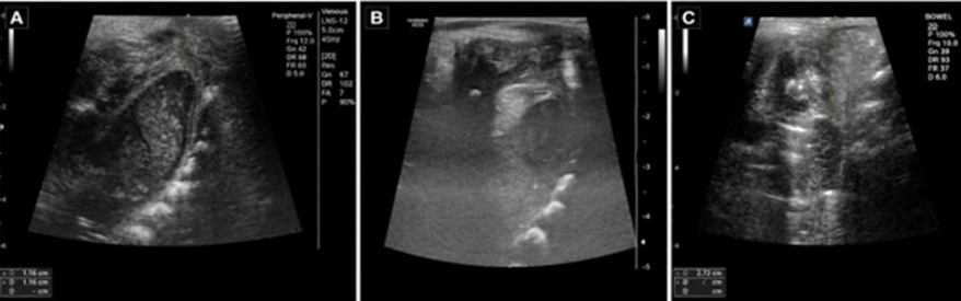
Figure 1. Transperineal ultrasonographic classification of anorectal malformations based on pouch-to-perineum distance. (A) Pouch-to-perineum distance of 9.2 mm, suggestive of low-type anorectal malformation. (B) Pouch-to-perineum distance of 15 mm, suggestive of intermediate-type anorectal malformation. (C) Pouch-to-perineum distance of 27 mm, suggestive of high-type anorectal malformation.
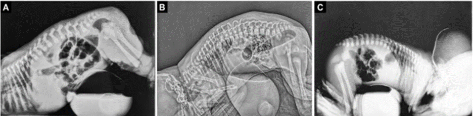
Figure 2. Prone cross-table lateral radiographic findings in anorectal malformations. (A) Distal bowel gas shadow located below the pubococcygeal line but not reaching the perineum. (B) Distal bowel gas shadow located below the pubococcygeal line but not reaching the perineum. (C) Distal bowel gas shadow located above the pubococcygeal line.
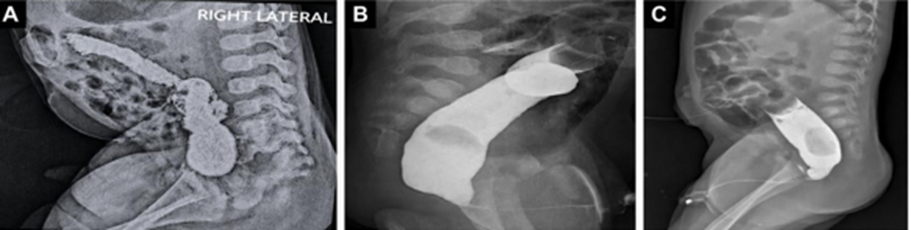
Figure 3. Distal loop colostographic findings in anorectal malformations. (A) Distal rectal pouch ending at a low level without fistulous communication. (B) Distal rectal pouch ending at a low level without fistulous communication. (C) Distal rectal pouch ending at a high level with fistulous communication to the bulbar urethra.
RESULTS:
The study included 32 infants with a mean age of 2.09 ± 0.86 days (range: 1–4 days), of whom 20 (62.5%) were male. Delayed passage of meconium was the most common presentation, followed by abdominal distension. A perineal dimple was present in 62.5% of infants, while 53.1% had no meconium passage (Table 1).
Table 1. Demographic and clinical characteristics of infants with anorectal malformations (N = 32)
|
Characteristics |
n (%) or mean ± SD |
|
|
Demographic Data |
Age (days) |
2.09 ± 0.86 |
|
Age range (days) |
1–4 |
|
|
Male |
20 (62.5) |
|
|
Female |
12 (37.5) |
|
|
Clinical presentation |
Failure/delayed passage of meconium |
10 (31.3) |
|
Abdominal distension |
7 (21.9) |
|
|
Bilious/intermittent vomiting |
5 (15.6) |
|
|
Meconium through an abnormal perineal opening |
4 (12.5) |
|
|
Feeding intolerance/poor feeding |
2 (6.3) |
|
|
Irritability/excessive crying |
2 (6.3) |
|
|
Constipation/straining during defecation |
2 (6.3) |
|
|
Perineal findings |
Perineal dimple present |
20 (62.5) |
|
Absent anal opening |
7 (21.9) |
|
|
Shallow/poorly formed anal dimple |
8 (25.0) |
|
|
Meconium staining of the perineum |
2 (6.3) |
|
|
Ectopic perineal opening |
1 (3.1) |
|
|
Perineal bulge during crying/straining |
2 (6.3) |
|
|
Passage of meconium |
No passage |
17 (53.1) |
|
Perineal passage |
10 (31.3) |
|
|
Urethral passage |
5 (15.6) |
|
|
Clinical presentations and perineal findings were not mutually exclusive. SD: standard deviation. |
||
Associated VACTERL anomalies were identified in 11 infants (34.4%). Vertebral defects were present in 18.8%, while hydronephrosis was the most frequent abdominal ultrasonographic abnormality, observed in 15.6% (Table 2).
Table 2. Associated radiological and congenital abnormalities (N = 32)
|
Finding |
n (%) |
|
VACTERL-associated anomalies present |
11 (34.4) |
|
Vertebral defects |
6 (18.8) |
|
Cardiomegaly |
3 (9.4) |
|
Hydronephrosis |
5 (15.6) |
|
Hydroureteronephrosis |
3 (9.4) |
|
Dilated bowel loops |
4 (12.5) |
|
Mild bowel dilatation |
4 (12.5) |
|
Distended rectal pouch |
4 (12.5) |
|
Minimal distal bowel distension |
4 (12.5) |
|
Moderate distal bowel stasis |
3 (9.4) |
|
Mild bowel gas distension |
2 (6.3) |
|
Thick-walled urinary bladder |
1 (3.1) |
|
Absent rectal gas shadow |
1 (3.1) |
|
Normal abdominal ultrasonography |
1 (3.1) |
TPUS classified 8 lesions (25.0%) as low, 18 (56.3%) as intermediate, and 6 (18.8%) as high ARM. Increasing ARM severity was accompanied by a progressive increase in pouch–perineum distance and reduction in puborectalis thickness. Fistulous communication was detected in 65.6% of infants (Table 3).
Table 3. Transperineal ultrasonographic findings according to ARM classification
|
TPUS classification |
n (%) |
Pouch–perineum distance (mm), Mean ± SD |
Puborectalis thickness (mm), Mean ± SD |
Sphincter complex morphology |
Fistula present, n (%) |
|
Low ARM |
8 (25.0) |
9.43 ± 3.19 |
1.52 ± 0.18 |
Well-developed and symmetrical |
7 (87.5) |
|
Intermediate ARM |
18 (56.3) |
18.15 ± 5.17 |
0.89 ± 0.22 |
Mild hypoplasia/reduced bulk |
10 (55.6) |
|
High ARM |
6 (18.8) |
33.32 ± 4.64 |
0.48 ± 0.12 |
Poorly developed/attenuated |
4 (66.7) |
|
Total |
32 (100.0) |
18.64 ± 9.21 |
— |
— |
21 (65.6) |
Operative examination identified 8 low, 16 intermediate, and 8 high lesions. TPUS classification was concordant with operative findings in 28 of 32 cases (Table 4).
Table 4. Comparison of TPUS findings with operative findings
|
Parameter |
Low ARM |
Intermediate ARM |
High ARM |
Total |
|
TPUS classification, n (%) |
8 (25.0) |
18 (56.3) |
6 (18.8) |
32 (100.0) |
|
Intraoperative classification, n (%) |
8 (25.0) |
16 (50.0) |
8 (25.0) |
32 (100.0) |
|
TPUS–operative concordance |
8/8 |
16/18 |
4/6 |
28/32 |
|
Mean pouch–perineum distance (mm) |
9.43 ± 3.19 |
18.15 ± 5.17 |
33.32 ± 4.64 |
— |
|
Operative management |
Primary anoplasty |
PSARP/staged repair |
Colostomy with definitive repair/PSARP |
— |
Overall concordance was 87.5%, with strong and statistically significant agreement between TPUS and operative findings (κ = 0.810; p < 0.001) (Table 5).
Table 5. Agreement Between Transperineal Ultrasonography and Intraoperative Findings
|
Agreement statistic |
Result |
|
Overall concordance |
87.5% |
|
Mismatched cases |
4 (12.5%) |
|
Chi-square (χ²) |
53.33 |
|
Chi-square p-value |
<0.001 |
|
Cohen’s kappa, κ (SE) |
0.810 (0.083) |
|
Kappa p-value |
<0.001 |
The diagnostic accuracy of TPUS was 100% for low, 87.5% for intermediate, and 90.6% for high ARM (Table 6).
Table 6. Diagnostic performance of TPUS and comparison of pouch–perineum distance
|
ARM classification |
Sensitivity (%) |
Specificity (%) |
PPV (%) |
NPV (%) |
Accuracy (%) |
|
Low ARM |
100.0 |
100.0 |
100.0 |
100.0 |
100.0 |
|
Intermediate ARM |
88.9 |
87.5 |
88.9 |
87.5 |
87.5 |
|
High ARM |
75.0 |
95.8 |
83.3 |
92.0 |
90.6 |
|
Overall |
— |
— |
— |
— |
87.5 |
Pouch–perineum distance was significantly greater in high ARM than in combined low/intermediate ARM (33.32 ± 4.64 vs. 15.25 ± 6.42 mm; p < 0.001) (Table 7).
Table 7. Comparison of Pouch–Perineum Distance Between High and Low/Intermediate Anorectal Malformations
|
Operative group |
n |
Pouch–perineum distance (mm), mean ± SD |
P Value |
|
Low/intermediate ARM |
24 |
15.25 ± 6.42 |
p <0.001* |
|
High ARM |
8 |
33.32 ± 4.64 |
|
|
Independent-samples t-test; *statistically significant (p< 0.05) |
|||
DISCUSSION:
The present study evaluated the diagnostic utility of transperineal ultrasonography (TPUS) in 32 infants with anorectal malformations (ARMs). The mean age was 2.09 ± 0.86 days, and 62.5% were male. This male predominance is consistent with the epidemiological pattern described by Levitt and Peña, while Jardosh et al. also reported similar findings in an Indian population.7,8 Failure or delayed passage of meconium and abdominal distension were the commonest presentations. These findings agree with Holschneider et al., who observed that clinical presentation varies with the level and fistulous communication of the malformation.9
TPUS classified 25.0% of cases as low, 56.3% as intermediate, and 18.8% as high ARM. The pouch–perineum distance increased progressively from low (9.43 ± 3.19 mm) to intermediate (18.15 ± 5.17 mm) and high ARM (33.32 ± 4.64 mm). This difference was statistically significant (p < 0.001), supporting pouch–perineum distance as a useful parameter for determining ARM level. Tanming and Laohapensang reported excellent diagnostic performance using a 10-mm threshold to differentiate low from non-low lesions.10 Similarly, Hosokawa et al. found that combining pouch distance with fistula location improved diagnostic accuracy.11
Puborectalis thickness decreased with increasing ARM severity. Low lesions showed a well-developed sphincter complex, whereas high lesions demonstrated attenuated musculature. Palmisani et al. similarly showed that high-resolution TPUS could delineate the sphincter complex and provide information relevant to operative planning.5 This represents an important advantage over conventional radiography and distal colostography, which provide limited assessment of pelvic soft tissues.
Fistulous communication was detected on TPUS in 21 infants (65.6%). Superficial perineal fistulas were more readily visualized, while deeper rectourinary communications were better demonstrated by distal colostography. Jardosh et al. reported high sensitivity and specificity of TPUS for fistula detection.8 Nevertheless, Stafrace et al. emphasized that distal colostography remains valuable for mapping complex fistulas before definitive repair.12 Thus, the two modalities may have complementary roles.
TPUS findings agreed with intraoperative classification in 87.5% of cases, with significant agreement (κ = 0.810, p < 0.001). Diagnostic accuracy was 100% for low, 87.5% for intermediate, and 90.6% for high ARM. These findings are comparable to the results of Jardosh et al., Tanming and Laohapensang, and Hosokawa et al.8,10,11 Overall, TPUS is a practical, non-invasive, radiation-free technique for initial ARM assessment. However, the small sample size and single-centre design limit generalization. Larger multicentre studies are needed to establish standardized measurement thresholds and further clarify its role in complex lesions.
CONCLUSION:
Transperineal ultrasonography is a safe, reliable, non-invasive and radiation-free modality for the preoperative evaluation of anorectal malformations. It accurately classifies lesions, measures the pouch–perineum distance, and assesses the sphincter complex, showing strong agreement with operative findings. Its overall diagnostic concordance was 87.5%. Although distal colostography remains useful for defining complex fistulous communications, TPUS can serve as the first-line investigation, particularly in neonates and resource-limited settings, thereby supporting timely surgical planning and reducing dependence on radiation-based imaging in routine clinical practice.
REFERENCES:
1. Gangopadhyay AN, Pandey V. Anorectal malformations. J Indian Assoc Pediatr Surg. 2015;20(1):10-15.
2. Rintala RJ. Congenital anorectal malformations: anything new? J Pediatr Gastroenterol Nutr. 2009;48 Suppl 2:S79-82.
3. Morandi A, Maestri F, Ichino M, et al. Labeling male anorectal malformations: objective evaluation of radiologic imaging before surgery. Front Pediatr. 2023;11:1224620.
4. Westgarth-Taylor C, Westgarth-Taylor TL, Wood RJ, Levitt MA. Imaging in anorectal malformations: what does the surgeon need to know? S Afr J Radiol. 2015;19(2):a903.
5. Palmisani F, Krois W, Patsch J, Metzelder M, Reck-Burneo CA. High-Resolution Transperineal Ultrasound in Anorectal Malformations-Can We Replace the Distal Colostogram?. European J Pediatr Surg Rep. 2022;10(1):e84-e88.
6. Ekwunife OH, Umeh EO, Ugwu JO, Ebubedike UR, Okoli CC, Modekwe VI, et al. Comparison of trans-perineal ultrasound-guided pressure augmented saline colostomy distension study and conventional contrast radiographic colostography in children with anorectal malformation. Afr J Paediatr Surg. 2016;13(1):26-31.
7. Levitt MA, Peña A. Anorectal malformations. Orphanet J Rare Dis. 2007;2:33.
8. Jardosh Y, Chavda D, Vadel M. Transperineal ultrasonography in infants with anorectal malformation. Int J Sci Res. 2017;6(3):1653–1655.
9. Holschneider A, Hutson J, Peña A, Beket E, Chatterjee S, Coran A, et al. Preliminary report on the international conference for the development of standards for the treatment of anorectal malformations. J Pediatr Surg. 2005;40:1521–1526.
10. Tanming P, Laohapensang M. The role of transperineal ultrasonography in determination of the level of anorectal malformations. J Med Assoc Thai. 2017;100(4 Suppl):S69–S73.
11. Hosokawa T, Yamada Y, Sato Y, et al. Changes in the Distance Between the Distal Rectal Pouch and Perineum From the Birth Day to the Next Day in Neonates With an Imperforate Anus. J Ultrasound Med. 2017;36(3):601-606.
12. Stafrace S, Lobo L, Augdal TA, Avni FE, Bruno C, Damasio MB, et al. Imaging of anorectal malformations: where are we now? Abdominal imaging task force of the European Society of Paediatric Radiology. Pediatr Radiol. 2022;52:1802–1809.