The relation between the iron storage (Ferritin) protein and glycemic control tracks (hbA1c) in poorly controlled Diabetes Mellitus.
- J. Sudha Rani , Associate Professor, Department of Biochemistry, Malla Reddy Medical College for Women, Malla Reddy Vishwavidyapeeth (Deemed to be University) Suraram, Hyderabad, 500055, India.
Article Information:
Abstract:
Background: Type 2 diabetes mellitus (t2dm) is associated with dysregulated iron metabolism. Elevated serum ferritin is a marker of body iron stores and inflammation and has been implicated in the pathogenesis of insulin resistance and glycemic dysfunction. Unravel the relationship between serum ferritin and glycated hemoglobin (hba1c) is expected to provide understanding the metabolic disturbances in type 2 Diabetes Mellitus. Aim: This study aimed to investigate the role of serum ferritin as an inflammatory marker and to evaluate its correlation with glycemic control in patients with T2DM. Materials And Methods: A total No of 100 cases (Retrospective) were studied by dividing them into two group’s controls (50) and cases 50 diagnosed T2DM patients., blood samples were analyzed for serum ferritin, glycated hemoglobin (HbA1c),The results so obtain were compared with 50 healthy controls Statistical Patients were stratified into two groups based on glycemic control: good control (HbA1c < 7%, n=32) and poor control (HbA1c ≥ 7%, n=42). Statistical significance was measured with Z test and statistical significance measured where p value is <0.0001) Serum ferritin was estimated using chemiluminescent immunoassay and hba1c by turbidometric methode. Conclusion: Serum ferritin may serve as an additional biomarker for assessing metabolic status in poorly controlled diabetes mellitus. Further prospective studies are required to establish its clinical utility in predicting diabetic complications.
Keywords:
Article :
INTRODUCTION:
The research in the past decades has revealed a critical link between metabolic disorders and inflammation, which leads to a concept called “metaflammation.” Metaflammation is a form of low-grade systemic and chronic inflammation related to excess nutrients and energy (1,2). There has been increasing evidence showing diabetes is an inflammatory disease. Type 1 diabetes (T1DM), characterized by autoimmunemediated destruction of pancreatic 𝛽 cells and insufficient secretion of insulin, has long been considered as an inflammatory disease. Not until the early 1990s, however, was type2 diabetes (T2DM) linked to inflammatory response (3). T2DM is manifested by peripheral insulin resistance and aberrant production of insulin, accompanied by chronic low grade inflammation in peripheral tissues such as adipose tissue, liver, and muscle. In the last decades, there has been growing evidence linking obesity and insulin resistance to inflammation (2–4). Given the significant roles inflammation plays in its pathogenesis, T2DM is now being redefined as an immune disorder (5–7). In addition to diabetes, many other metabolic disorders have also been associated with inflammation (2). This special issue showcases a number original research articles and review papers on the topic of inflammatory regulation in metabolic dysfunction.
Serum ferritin, the primary intracellular iron-storage protein, has traditionally been used as a key indicator of body iron stores. However, it is now well-established that serum ferritin is also a robust acute-phase reactant. Its levels can rise dramatically in response to inflammatory stimuli, independent of iron status (8)
Elevated serum ferritin has been epidemiologically linked to an increased risk of developing T2DM and its complications (9). The proposed mechanisms extend beyond its role as an inflammatory marker. Iron is a potent pro-oxidant. Elevated body iron stores can catalyze the formation of highly reactive hydroxyl radicals via the Fenton reaction, leading to increased oxidative stress(10). This oxidative environment can directly damage pancreatic β-cells, impair glucose-stimulated insulin secretion, and worsen insulin resistance by interfering with insulin signaling pathways in hepatocytes and adipocytes. Furthermore, some evidence suggests that ferritin itself may have direct immuno-modulatory effects, potentially acting as a pro-inflammatory mediator.(11)
Measuring the ferritin level in patients can provide core fact regarding their iron status Ferritin is present in every cell type. (12) It serves to store iron in a non-toxic form, to deposit it in a safe form, and to transport it to areas where it is required.(13) Ferritin is an evolutionarily conserved globular protein, composed of 24 poly-peptide chains. It forms a spherical shape that is approximately 8nm in diameter, allowing it to store approximately 4,500 Fe atoms. (14-16) Serum ferritin is heterogeneous due to glycosylation. The glycosylation and direct relationship of serum ferritin concentration to storage in macrophages suggest it is secreted by macrophages in response to changing iron levels.
Distribution of iron in ferritin (17)
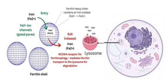
Figure 1
Iron enters ferritin through pores as Fe2+, where it is oxidized to Fe3+ and stored inside the shell. Iron exits ferritin through iron-regulatable NCOA4-mediated autophagic proteolytic degradation of the ferritin shell in lysosomes – a process called ferritinophagy. Inflammation increases ferritin and hepcidin independent of the body’s iron composition. Hepcidin prevents iron egress from cells and increases intracellular ferritin expression.(18)
Serum ferritin has since remained mainstay for evaluation of systemic iron stores despite evidence suggesting that ferritin is elevated during infection and malignancies. These underlying comorbid conditions often confound the interpretation of serum ferritin levels (reviewed in, (19), (20) Until recently, it was assumed that serum ferritin is a leakage product, derived from damaged cells and studies demonstrate that serum levels correlate with disease severity.(21,22,23) While it is possible that damaged cells contribute to an increase in ferritin during disease states, emerging evidence support that ferritin is actively secreted by uninjured cells as a normal physiological process.(24,25) Ferritin transfer from cells to serum in humans; less active secretion, more simply leakage from damaged cells.
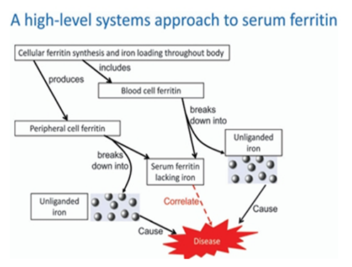
Figure 2
High-level systems approach to serum ferritin. The diagram serves to illustrate why there tend to be correlations between the amount of ferritin in cells, the rate of its excretion by cell damage (involving liberation of unliganded iron) and the levels of serum ferritin. The serum ferritin correlates with disease but the cause is iron, with which it too can correlate. As with any systems biology network, multiple differences indifferent elements of the network can lead to the same overall effects, explaining the lack of a perfect correlation with any individual process. Thus, a first order rate of efflux of ferritin is the product of (and thus contains contributions from) both the internal ferritin concentration and the rate constant for efflux, which may vary independently. For these purposes we do not discriminate the many individual iron species.(26)
Mechanisms of inflammation in diabetes
The mechanisms of inflammation in diabetes are complex and multifaceted, involving a variety of pathways and cellular interactions.(29)Inflammation plays a pivotal role in the pathogenesis, progression, and complications of diabetes, particularly in type 2 diabetes (T2D). In individuals with obesity and insulin resistance, there is often a state of chronic low-grade inflammation. Adipose tissue, particularly visceral fat, serves as a significant source of pro-inflammatory molecules.(27)This chronic inflammation is characterized by elevated levels of proinflammatory cytokines such as tumor necrosis factor-alpha (TNF-α) and interleukin-6 (IL-6).Inflammation in diabetes is closely linked to immune system dysregulation. Macrophages, a type of white blood cell, infiltrate adipose tissue and become activated, producing pro-inflammatory cytokines.(28) This immune response contributes to insulin resistance by interfering with insulin signaling in target tissues.Adipose tissue acts as an endocrine organ, secreting adipokines that influence metabolism and inflammation. In obese individuals, adipose tissue becomes dysfunctional and releases more pro-inflammatory adipokines, such as leptin and less anti-inflammatory adipokines like adiponectin. This imbalance contributes to systemic inflammation.
Inflammation can lead to endothelial dysfunction, characterized by impaired function of the blood vessel lining. This dysfunction reduces nitric oxide production and increases the release of pro-inflammatory factors, contributing to insulin resistance and cardiovascular complications in diabetes.(30) Chronic inflammation is associated with oxidative stress, which involves an imbalance between the production of reactive oxygen species and the body’s ability to neutralize them. Oxidative stress can lead to damage of cells, tissues, and DNA, exacerbating insulin resistance and contributing to beta-cell dysfunction in the pancreas.(31) In type 1 diabetes (T1D), inflammation occurs in the pancreas itself, particularly in the islets of Langerhans.(32) Immune cells infiltrate these islets and attack insulin-producing beta-cells, leading to their destruction.(33) This autoimmune response is a hallmark of T1D.Emerging research suggests that the gut microbiota plays a role in diabetes-related inflammation. An imbalance in the gut microbiome, known as dysbiosis, can lead to increased gut permeability, allowing pro-inflammatory bacterial products to enter the bloodstream and trigger systemic inflammation.High blood glucose levels can lead to the formation of advanced glycation end products, which can stimulate inflammation by binding to receptors on immune cells and promoting the release of pro-inflammatory cytokines.
Significance of HbA1c Test in Diagnosis and Prognosis of Diabetic Patients
HbA1C: Glycated hemoglobin (HbA1c) in blood provides evidence about an individual’s average blood glucose levels during the previous two to three month which is the predicted half-life of red blood cells (RBCs). (34) Historically, HbA1c was first isolated by Huisman et al.(35) in 1958 and characterized by Bookchin and Gallop(36)in 1968, as a glycoprotein. The elevated levels of HbA1c in diabetic patients were reported by Rahbar et al.(37)in 1969. Bunn et al.(38) identified the pathway leading to the formation of HbA1c in 1975. Using the HbA1c as a biomarker for monitoring the levels of glucose among diabetic patients was first proposed by Koenig et al.(39) in 1976.
Proteins are frequently glycated during various enzymatic reactions when the conditions are physiologically favorable. However, in the case of hemoglobin, the glycation occurs by the nonenzymatic reaction between the glucose and the N-terminal end of the β-chain, which forms a Schiff base.(40),(41)During the rearrangement, the Schiff base is converted into Amadori products, of which the best known is HbA1c
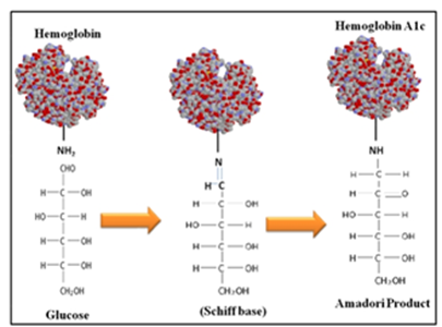
Figure 3
In the primary step of glycated hemoglobin formation, hemoglobin and the blood glucose interact to form aldimine in a reversible reaction. In the secondary step, which is irreversible, aldimine is gradually converted into the stable ketoamine form.(42)The major sites of hemoglobin glycosylation, in the order of prevalence, are β-Val-1, β-Lys-66, and α-Lys-61. Normal adult hemoglobin consists predominantly of HbA (α2β2), HbA2 (α2δ2), and HbF (α2γ2) in the composition of 97%, 2.5%, and 0.5%, respectively. About 6% of total HbA is termed HbA1, which in turn is made up of HbA1a1, HbA1a2, HbA1b, and HbA1c fractions, defined by their electrophoretic and chromatographic properties.
An increase in HbA1c as observed in conditions of poor diabetic control has been associated with increased blood viscosity.(43 )Glycosylation of hemoglobin and increased glucose levels tends to affect RBC properties, lowering the RBC flexibility and increasing their aggregation tendency, leading to increased blood viscosity.(44)Glycosylation of hemoglobin may also affect membrane lipid protein interactions in RBCs, altering their internal viscosity, modifying viscoelastic properties of erythrocyte membranes, and impairing RBC deformability.(45)
There is also evidence that glycosylation of hemoglobin impairs nitric oxide (NO)-related relaxation of human mesenteric vessels.(46)Hemoglobin glycosylation is also reported to alter NO binding with thiols resulting in lowered NO bioavailability and impaired vasodilatation in rabbit aortic rings.(47) Another mechanism by which glycosylation of hemoglobin is proposed to be vasoactive is via the formation of reactive oxygen species.(48) Glycosylation of hemoglobin also lowers oxygen-carrying capacity, thereby promoting hypoxia and its related systemic vascular vasodilatory adaptations and responses.(49)
MATERIALS AND METHODS:
Study Design: Retrospective observational study
Study Population: Patients diagnosed with poorly controlled type 2 diabetes mellitus (HbA1c >7%).
Sample Size: contain 50 cases of DM and 50 normal healthy individuals as control.
Study Setting: single setting, Malla Reddy Medical College for women’s
Inclusion Criteria
• Subjects in the age group 35-60 years both male and female with already diagnosed as type 2 diabetes mellitus, HbA1c above target level who attended diabetic clinic
Exclusion Criteria
• Patient who are below 35years or above 60 years.
• Type 1 diabetes mellitus.
• Patient with Haemochromatosis,Thalassemia, Hemosiderosis.
• Patient with underlying liver, kidney, lung diseases.
Laboratory Tests Done
Serum ferritin was estimated using chemiluminescent immunoassay and hba1c by turbidometric methode.
RESULTS:
Table 1: Distribution of Diabetic Patients by Serum Ferritin Level Stratified by Cases and Controls
|
Groups |
Ferritin (Mean ± SD) |
Z Value |
p value |
|
Control |
75.28 ± 25.79 |
21.73145 |
<0.0001 |
|
Diabetic Patients |
1095.04 ± 330.80 |
|
|
Table 2: Distribution of Diabetic Patients by HbA1c Levels Stratified by Cases and Controls
|
Groups |
HbA1c (Mean ± SD) |
Z Value |
p value |
|
Control |
4.01 ± 0.45 |
36.27 |
<0.001 |
|
Diabetic Patients |
13.10 ± 1.71 |
RESULTS
The ferritin levels were determined using the Fully Automated Biodirectionally Interfaced Chemi Luminescent Immune Assay Method and HbA1c levels were determined by turbidometric methode Table 1 and 2 presents the mean serum Ferritin levels and HbA1c in a sample of 50 individuals, including both males and females. The data indicates that the mean serum Ferritin and HbA1c levels in the cases are greater compared to the mean levels observed in the control group. The observed rise exhibits statistical significance
Summary of Key Insights
There are many theories regarding the role of ferritin in DM. Pancreatic damage due to subclinical hemochromatosis has been considered in some cases of diabetes. These relationships are due to oxidative stress and inflammatory cytokines which potentiates the initiated events.(50) An important role of ferritin during the acute phase response is to restrict the availability of iron by sequestration into the cavity of the ferritin protein sell. High body iron stores that is serum ferritin have been linked to insulin resistance, metabolic syndrome and gestational diabetes. Excess iron damages β-cells of pancreas due to oxidative stress which can contribute to pathogenesis of diabetes mellitus.
Iron deposition in the liver may cause insulin resistance by interfering with the ability of insulin to suppress hepatic glucose production but the mechanism for the association between ferritin and type 2 diabetes is not established.7(52) Iron is auto-oxidized to form highly reactive, lipid soluble iron-oxygen complexes. These free radicals are powerful pro-oxidants, which can change membrane properties and result in tissue damage.(53)Oxidative stress can lead to hyperglycaemic through disturbed glucose metabolism. The insulin extracting capacity of the liver may be interfaced by iron accumulation in hepatocytes and affect insulin synthesis and section in the pancreas. Excess of iron contributes insulin resistance and subsequently insulin secretion is decreased.(53) In the study of Sumeshraj et al. Serum ferritin levels increased as the duration of diabetes increased. (51) The relationship between elevated serum ferritin levels and type 2 diabetes involves an elevation in oxidative stress through the increased formation of free radicals catalysed by iron, which may lead to insulin resistance and hyperglycaemia. (54), (55)
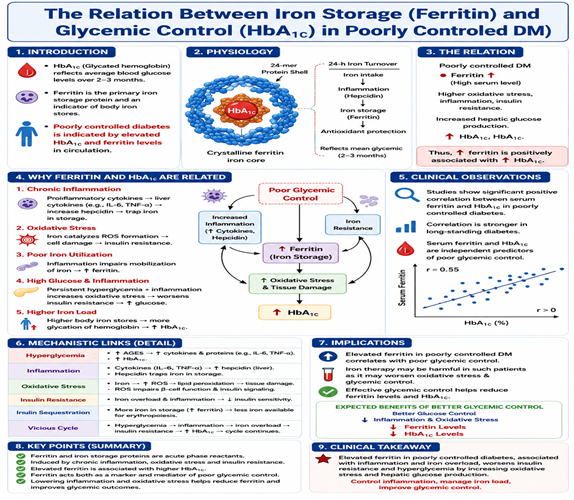
Schematic Diagram of “The relation between the iron storage (Ferritin) protein and glycemic control tracks (hbA1c) in poorly controlled Diabetes Mellitus”
CONCLUSION:
HbA1c (a measure of average blood sugar), ferritin (a marker of iron storage), and diabetes are deeply interlinked. High ferritin contributes to insulin resistance and diabetes risk, while poorly controlled diabetes (high HbA1c) further increases ferritin and inflammation.
1. Ferritin Elevates Diabetes Risk: Insulin Resistance: Excess stored iron (high serum ferritin) is toxic to the pancreas. It causes a spike in oxidative stress (production of free radicals), which damages the cells that produce insulin and stops the body from using insulin effectively.
2. HbA1c and Ferritin Have a Positive Correlation: Glycemic Control: Studies consistently show that patients with high HbA1c levels typically present with significantly elevated serum ferritin. Because uncontrolled diabetes causes systemic inflammation, and inflammation stimulates ferritin production, ferritin often acts as an indicator of how poorly the diabetes is being managed
Although ferritin should not replace HbA1c, it may complement conventional biomarkers in identifying patients at higher risk of disease progression and complications. Large multicenter prospective studies are needed to establish its role in routine clinical practice
The relation between the iron storage (Ferritin) protein and glycemic control tracks (hbA1c) in poorly controlled Diabetes Mellitus.
REFERENCES:
1. M. F. Gregor and G. S. Hotamisligil, “Inflammatory mechanisms in obesity,” Annual Review of Immunology, vol. 29, pp.415–445, 2011.
2. G. S. Hotamisligil, “Inflammation and metabolic disorders,”Nature, vol. 444, no. 7121, pp. 860–867, 2006.
3. G. S. Hotamisligil, N. S. Shargill, and B. M. Spiegelman, “Adipose expression of tumor necrosis factor-𝛼: direct role in obesity-linked insulin resistance,” Science, vol. 259, no. 5091, pp.87–91, 1993.
4. D. A. Winer, S. Winer, L. Shen et al., “B cells promote insulin resistance through modulation of T cells and production of pathogenic IgG antibodies,” Nature Medicine, vol. 17, no. 5, pp.610–617, 2011.
5. S. Tsai, X. Clemente-Casares, X. S. Revelo, S. Winer, and D.A. Winer, “Are obesity-related insulin resistance and type 2diabetes autoimmune diseases?” Diabetes, vol. 64, no. 6, pp.1886–1897, 2015.
6. L. A. Velloso, D. L. Eizirik, and M. Cnop, “Type 2 diabetes mellitus—an autoimmune disease?” Nature Reviews Endocrinology, vol. 9, no. 12, pp. 750–755, 2013.
7. M. Y. Donath and S. E. Shoelson, “Type 2 diabetes as an inflammatory disease,” Nature Reviews Immunology, vol. 11, no. 2, pp.98–107, 2011
8. Wang W, Knovich MA, Coffman LG, Torti FM, Torti SV. Serum ferritin: past, present and future. BiochimBiophys Acta. 2010 Aug;1800(8):760–9
9. Jiang R, Ma J, Ascherio A, Stampfer MJ, Willett WC, Hu FB. Dietary iron intake and blood donations in relation to risk of type 2 diabetes in men: a prospective cohort study. Am J Clin Nutr. 2004 Jan;79(1):70–5.
10. Fernández-Real JM, López-Bermejo A, Ricart W. Cross-Talk between Iron Metabolism and Diabetes. Diabetes. 2002 Aug;51(8):2348–54.
11. Kernan KF, Carcillo JA. Hyperferritinemia and inflammation. Int Immunol. 2017 Sep 1;29(9):401-9
12. Theil EC (1987). "Ferritin: structure, gene regulation, and cellular function in animals, plants, and microorganisms". Annual Review of Biochemistry. 56 (1): 289.315. doi:10.1146/ann urev.bi.56.070187.001445. PMID 3304136
13. Seckback J (1982). "Ferreting out the secrets of plant ferritin - A review". Journal of Plant Nutrition. 5 (4–7): 369– 394. Bibcode:1982JPlaN...5..369S.
14. Crichton RR. Structure and function of ferritin. Angew Chem Int Ed Engl. 1973;12:57–65. [PubMed: 4631281]
15. Lawson DM, Artymiuk PJ, Yewdall SJ, Smith JM, Livingstone JC, Treffry A, et al. Solving the structure of human H ferritin by genetically engineering intermolecular crystal contacts. Nature. 1991;349:541–4. [PubMed: 1992356]
16. Ford GC, Harrison PM, Rice DW, Smith JM, Treffry A, White JL, et al. Ferritin: design and formation of an iron-storage molecule. Philos Trans R Soc Lond B Biol Sci. 1984;304:551– 65. [PubMed: 6142491]
17. Fig.1https://www.thebloodproject.com/wpcontent/uploads/2024/01/NEW-TWEETS-49- 700x394.png
18. Ueda N, and Takasawa K. Impact of Inflammation on Ferritin, Hepcidin and the Management of Iron Deficiency Anemia in Chronic Kidney Disease. Nutrients. 2018;10
19. Kalantar-Zadeh K, Kalantar-Zadeh K, and Lee GH. The fascinating but deceptive ferritin: to measure it or not to measure it in chronic kidney disease? Clin J Am Soc Nephrol. 2006;1 Suppl 1:S9–18. [DOI] [PubMed] [Google Scholar]
20. Wang W, Knovich MA, Coffman LG, Torti FM, and Torti SV. Serum ferritin: Past, present and future. BiochimBiophys Acta. 2010;1800:760– 9. [DOI] [PMC free article] [PubMed] [Google Scholar]
21. Kim T, Streja E, Soohoo M, Rhee CM, Eriguchi R, Kim TW, et al. Serum Ferritin Variations and Mortality in Incident Hemodialysis Patients. Am J Nephrol. 2017;46:120–30. [PubMed: 28704813]
22. Kell DB, and Pretorius E. Serum ferritin is an important inflammatory disease marker, as it is mainly a leakage product from damaged cells. Metallomics. 2014;6:748–73. [PubMed: 24549403]
23. Garcia PC, Longhi F, Branco RG, Piva JP, Lacks D, and Tasker RC. Ferritin levels in children with severe sepsis and septic shock. Acta Paediatr. 2007;96:1829–31. [PubMed: 18001337]
24. Cohen LA, Gutierrez L, Weiss A, LeichtmannBardoogo Y, Zhang DL, Crooks DR, et al. Serum ferritin is derived primarily from macrophages through a nonclassical secretory pathway. Blood. 2010;116:1574–84. [PubMed: 20472835]
25. Truman-Rosentsvit M, Berenbaum D, Spektor L, Cohen LA, Belizowsky-Moshe S, Lifshitz L, et al. Ferritin is secreted via 2 distinct nonclassical vesicular pathways. Blood. 2018;131:342–52. [PubMed: 29074498]
26. Douglas B. Kell a and Etheresia Pretorius b Serum ferritin is an important inflammatory disease marker, as it is mainly a leakage product from damaged cellsMetallomics, 2014,6, 748- 773
27. Tiganis T. Reactive oxygen species and insulin resistance: the good, the bad and the ugly. Trends Pharmacol Sci. 2011;32:82–9.
28. Malenica M, Šilar M, Dujić T, et al. Importance of inflammatory markers and IL-6 for diagnosis and follow up of patients with type 2 diabetes mellitus. Med Glas (Zenica). 2017;14:169–75.
29. Rajendran P, Chen YF, Chen YF, et al. The multifaceted link between inflammation and human diseases. J Cell Physiol. 2018;233:6458–71.
30. Soskić SS, Dobutović BD, Sudar EM, et al. Regulation of inducible nitric oxide synthase (iNOS) and its potential role in insulin resistance, diabetes and heart failure. Open Cardiovasc Med J. 2011;5:153–63.
31. Gerber PA, Rutter GA. The role of oxidative stress and hypoxia in pancreatic beta-cell dysfunction in diabetes mellitus. Antioxid Redox Signal. 2017;26:501–18.
32. Alexandre-Heymann L, Mallone R, Boitard C, Scharfmann R, Larger E. Structure and function of the exocrine pancreas in patients with type 1 diabetes. Rev Endocr Metab Disord. 2019;20:129–49.
33. Peters L, Posgai A, Brusko TM. Islet–immune interactions in type 1 diabetes: the nexus of beta cell destruction. Clin Exp Immunol. 2019;198:326–40
34. Khan MI, Weinstock RS. Chapter 16: Carbohydrates. In: McPherson RA, Pincus MR, eds. Henry’s Clinical Diagnosis and Management by Laboratory Methods. 22nd ed. Philadelphia, PA: Saunders Elsevier; 2011:210–25.
35. Huisman TH, Martis EA, Dozy A. Chromatography of hemoglobin types on carboxymethylcellulose. J Lab Clin Med. 1958;52:312–27.
36. Bookchin RM, Gallop PM. Structure of haemoglobin A1c: nature of the N-terminal beta chain blocking group. Biochem Biophys Res Commun. 1968;32:86–93.
37. Rahbar S, Blumenfeld O, Ranney HM. Studies of an unusual hemoglobin in patients with diabetes mellitus. Biochem Biophys Res Commun. 1969;36:838–43.
38. Bunn HF, Haney DN, Gabbay KH, Gallop PM. Further identification of the nature and linkage of the carbohydrate in haemoglobin A1c. Biochem Biophys Res Commun. 1975;67:103–9.
39. Koenig RJ, Peterson CM, Jones RL, Saudek C, Lehrman M, Cerami A. Correlation of glucose regulation and hemoglobin AIc in diabetes mellitus. N Engl J Med. 1976;295:417–20.
40. Ohtsubo K, Chen MZ, Olefsky JM, Marth JD. Pathway to diabetes through attenuation of pancreatic beta cell glycosylation and glucose transport. Nat Med. 2011;17:1067–75.
41. Marchetti P. Advanced glycation end products (AGEs) and their receptors (RAGEs) in diabetic vascular disease. Medicographia. 2009;31:257–64.
42. Acharya AS, Roy RP, Dorai B. Aldimine to ketoamine isomerization (Amadori rearrangement) potential at the individual nonenzymic glycation sites of hemoglobin A: preferential inhibition of glycation by nucleophiles at sites of low isomerization potential. J Protein Chem. 1991;10:345–58.
43. Leiper JM, Lowe GD, Anderson J, et al. Effects of diabetic control and biosynthetic human insulin on blood rheology in established diabetics. Diabetes Res. 1984;1:27–30.
44. Buhler I, Walter R, Reinhart WH. Influence of D- and L-glucose on erythrocytes and blood viscosity. Eur J Clin Invest. 2001;31:79–85.
45. Watala C, Witas H, Olszowska L, Piasecki W. The association between erythrocyte internal viscosity, protein non-enzymatic glycosylation and erythrocyte membrane dynamic properties in juvenile diabetes mellitus. Int J Exp Pathol. 1992;73:655–63.
46. Vallejo S, Angulo J, Peiro C, et al. Highly glycated oxyhaemoglobin impairs nitric oxide relaxations in human mesenteric microvessels. Diabetologia. 2000;43:83–90.
47. James PE, Lang D, Tufnell-Barret T, Milsom AB, Frenneaux MP. Vasorelaxation by red blood cells and impairment in diabetes: reduced nitric oxide and oxygen delivery by glycated hemoglobin. Circ Res. 2004;94:976–83.
48. Angulo J, Sanchez-Ferrer CF, Peiro C, et al. Impairment of endothelium dependent relaxation by increasing percentages of glycosylated human hemoglobin. Possible mechanisms involved. Hypertension. 1996;28:583–92.
49. Paffett ML, Walker BR. Vascular adaptations to hypoxia: molecular and cellular mechanisms regulating vascular tone. Essays Biochem. 2007;43:105–20.
50. Hramiak IM, Finegood DT, Adams PC. Factors affecting glucose tolerance in hereditary hemochromatosis. Clin Invest Med 1997;20(2):110-118.
51. (5201)Raj S, Rajan GV. Correlation between elevated serum ferritin and HbA1c in type 2 diabetes mellitus. Int J Res Med Sci 2013;1(1):12-15.
52. Oberley LW. Free radicals and diabetes. Free Radiac Biol Med 1988;5(2):113-124
53. Wilson JG, Crook ED, Maher JF, et al. Potential role of increased iron stores in diabetes. Am J Med Sci 2003;325(6):332-339.
54. Oberley LW. Free radicals and diabetes. Free Radic Biol Med 1988;5(2):113-124.
55. Wilson JG, Lindquist JH, Grambow SC, et al. Potential role of increased iron stores in diabetes. Am J Med Sci 2003;325(6):332-339.