Clinicopathological Concordance and Histomorphological Spectrum of Scalp Alopecia Evaluated with Combined Transverse and Vertical Sections: A Prospective Study.
- Heera S , Assistant Professor; Department of Pathology, St. Peter's Medical College Hospital and Research Institute, Hosur, Tamil Nadu, India.
- Jyothi Anantharaj , Professor, Department of Pathology, RajaRajeswari Medical College and Hospital, Bengaluru, Karnataka, India; affiliated to Dr. M.G.R. Educational and Research Institute, Chennai, Tamil Nadu, India.
- Sindura J R , Assistant Professor; Department of Pathology, St. Peter's Medical College Hospital and Research Institute, Hosur, Tamil Nadu, India.
Article Information:
Abstract:
Background: Clinical overlap among alopecias can delay distinction between potentially reversible non-cicatricial disease and irreversible cicatricial disease. Scalp biopsy is most informative when follicular architecture in transverse sections is interpreted together with epidermal, interface and full-depth changes in vertical sections. This study characterised the biopsy spectrum of adult scalp alopecia and quantified agreement between pre-biopsy clinical differentials and final histopathological diagnoses. Methods: This prospective, hospital-based descriptive study included all eligible adult scalp biopsies received at a tertiary-care pathology department in Bengaluru, India, from 1 June 2019 to 31 May 2021. Inadequate and paediatric biopsies were excluded. Four-millimetre punch biopsies were processed using a combined transverse-and-vertical sectioning approach. Follicular counts and ratios, follicular scars and dropout, inflammatory pattern, and epidermal and dermal changes were assessed. Clinicopathological concordance was defined a priori for this analysis as inclusion of the final histopathological diagnosis anywhere in the recorded pre-biopsy clinical differential. Proportions are presented with Wilson 95% confidence intervals (CIs). Results: Of 53 biopsies received, five were inadequate and 48 were analysed. Median age was 33 years (range 18-57), and 26 patients (54.2%) were male. Twenty-six biopsies (54.2%) showed cicatricial alopecia and 22 (45.8%) non-cicatricial alopecia. The leading diagnoses were alopecia areata (14/48, 29.2%), lichen planopilaris (10/48, 20.8%) and psoriasis-associated alopecia (8/48, 16.7%). Follicular count was altered in 45/48 (93.8%) and the terminal-to-vellus ratio in 44/48 (91.7%). Overall clinicopathological concordance was 40/48 (83.3%; 95% CI 70.4-91.3). Concordance was 23/26 (88.5%) for cicatricial and 17/22 (77.3%) for non-cicatricial alopecia (Fisher exact p=0.44). All eight discordant cases were telogen effluvium (n=4), tinea capitis (n=3), or androgenetic alopecia (n=1). Conclusions: Combined transverse and vertical evaluation yielded a broad range of complementary diagnostic findings and resolved clinically misleading diffuse and infection-associated alopecias. The observed concordance supports close clinicopathological integration, while the clustering of discordance identifies telogen effluvium and tinea capitis as important biopsy-resolved mimics. Because there was no single-orientation comparator, the incremental diagnostic yield of combined sectioning requires direct comparative study.
Keywords:
Article :
INTRODUCTION:
Alopecia comprises a heterogeneous group of disorders whose prognosis depends fundamentally on whether the follicular unit is preserved or replaced by scar. Non-cicatricial alopecias retain the potential for regrowth, whereas cicatricial alopecias cause permanent follicular destruction and require timely recognition. Clinical examination and trichoscopy are central to evaluation, but biopsy remains important when the diagnosis is uncertain, when scarring is suspected, or when overlapping patterns prevent confident classification [1-3].
Histopathological interpretation is technically demanding because diagnostic changes may vary with disease activity, biopsy site and level of section. Vertical sections display the entire epidermis, dermoepidermal junction, dermis and subcutis, making them useful for interface change, perifollicular fibrosis and the vertical distribution of inflammation. Transverse sections sample a larger proportion of follicles and permit assessment of follicular density, phase distribution, terminal-to-vellus ratio, miniaturisation and focal perifollicular abnormalities [4-7]. Neither orientation is universally sufficient.
Studies comparing orientation have reached differing conclusions, but collectively support complementary use when tissue permits [8-10]. Single-biopsy protocols such as HoVert and Tyler-type processing aim to retain epidermal/interface information while permitting quantitative transverse assessment [11,12].
Indian studies have also demonstrated the practical feasibility of obtaining both orientations and have highlighted the diagnostic consequences of grossing quality [13,14]. Nevertheless, many published series emphasise sectioning performance or individual disease groups rather than the clinical diagnoses that histology corrects across an unselected biopsy population.
We therefore evaluated adult scalp biopsies received prospectively over two years using combined transverse and vertical sections. The primary objectives were to describe the histomorphological spectrum and determine clinicopathological concordance. A secondary objective was to identify the diagnoses and microscopic features concentrated among clinically discordant cases.
MATERIALS AND METHODS:
Study design and setting
This prospective, hospital-based descriptive study was conducted in the Department of Pathology, RajaRajeswari Medical College and Hospital, Bengaluru, India. All scalp biopsies sent from the Department of Dermatology with a clinical suspicion of alopecia between 1 June 2019 and 31 May 2021 were assessed for eligibility. The report follows the principles of the Strengthening the Reporting of Observational Studies in Epidemiology (STROBE) statement.
Participants
Adult patients undergoing scalp biopsy for alopecia were eligible. Paediatric patients and biopsies inadequate for histomorphological assessment were excluded. A target of 60 biopsies had been planned from prior departmental volume; accrual was curtailed during the COVID-19 pandemic. The analytic cohort was a census of all eligible specimens received during the fixed study period.
Clinical assessment and biopsy processing
Age, sex, clinical presentation, scalp site and one or more pre-biopsy clinical differential diagnoses were recorded from the requisition and study proforma. Scalp tissue was obtained using a 4-mm punch under local anaesthesia and fixed in 10% formalin for 24 hours. Each specimen was processed using a Tyler-type approach to obtain both transverse and vertical sections from the available tissue. Paraffin sections were stained with haematoxylin and eosin. Special stains were used when indicated; periodic acid-Schiff (PAS) staining was used to demonstrate fungal elements in suspected tinea capitis.
Histopathological assessment
The microscopic evaluation included total follicular count; numbers of terminal, vellus, anagen and non-anagen hairs; terminal-to-vellus ratio; depth of follicular bulbs; blank spots; follicular scars and their distribution; follicular dropout; follicular distortion, plugging, miniaturisation, pigment casts, haemorrhage, trichomalacia, fibrous streamers and telogen germinal units; severity, location and predominant cell type of inflammation; and associated epidermal or dermal changes.
Alopecia was classified as non-cicatricial or cicatricial according to preservation versus destruction and fibrous replacement of follicles. Cicatricial alopecia was further classified as primary when the follicle was the principal target and secondary when scarring accompanied another dermatosis or infection.
Outcomes and concordance
The final histopathological diagnosis was based on the combined findings from both orientations and relevant special stains. For the primary outcome, a case was considered concordant when the final histopathological diagnosis appeared anywhere in the recorded clinical differential; it was considered discordant when the final diagnosis was absent from all listed clinical possibilities.
This deliberately reflects agreement with the clinician's differential rather than exact first-choice agreement.
Statistical analysis
De-identified master-chart data were re-tabulated for manuscript preparation. Categorical variables are reported as number and percentage. Age is reported as median and range. The main concordance proportion is accompanied by a two-sided Wilson 95% CI. Concordance in cicatricial and non-cicatricial groups was compared using Fisher's exact test; p<0.05 was considered statistically significant. Given the small disease-specific cell counts, no multivariable modelling was attempted.
Ethics
The Institutional Ethics Committee of RajaRajeswari Medical College and Hospital approved the study (RRMCH-IEC/77/2019-20; approval dated 18 October 2019).
RESULTS:
Participant and biopsy characteristics
Fifty-three scalp biopsies were received during the study period. Five (9.4%) were excluded because tissue was inadequate, leaving 48 biopsies for analysis. The median age calculated from the master chart was 33 years (range 18-57); 26 patients (54.2%) were male and 22 (45.8%) female.
The vertex was the most frequently sampled site (14/48, 29.2%), followed by the frontal (13/48, 27.1%) and occipital scalp (10/48, 20.8%). Twenty-two cases (45.8%) were non-cicatricial, 15 (31.3%) primary cicatricial and 11 (22.9%) secondary cicatricial (Table 1).
Histopathological spectrum
Alopecia areata was the most frequent diagnosis (14/48, 29.2%), followed by lichen planopilaris (10/48, 20.8%) and psoriasis-associated alopecia with cicatricial change (8/48, 16.7%).
Telogen effluvium accounted for four cases (8.3%), tinea capitis for three (6.3%), and trichotillomania, androgenetic alopecia and pseudopelade of Brocq for two cases each (4.2%). Acne keloidalis nuchae, keratosis pilaris and alopecia mucinosa were represented by one case each (2.1%) (Table 2).
Histomorphological findings
Forty-five biopsies (93.8%) had an altered follicular count, and 44 (91.7%) had an altered terminal-to-vellus ratio. Inflammation was mild in 16 (33.3%), moderate in 15 (31.3%), severe in 12 (25.0%) and absent in five (10.4%) biopsies. Lymphocytes were the predominant inflammatory cell in 41 (85.4%); neutrophils predominated in two (4.2%).
Among alopecia areata biopsies, peribulbar inflammation was present in 11/14 (78.6%), perifollicular inflammation in 5/14 (35.7%) and peri-infundibular inflammation in 2/14 (14.3%). The four telogen effluvium biopsies showed telogen germinal units and no inflammation. Both androgenetic alopecia biopsies showed follicular miniaturisation, and both trichotillomania biopsies demonstrated pigment casts, intrafollicular haemorrhage and trichomalacia.
All 10 lichen planopilaris biopsies contained lymphocytic inflammation; perifollicular inflammation and dermal fibrosis were each present in 7/10 (70.0%), dermoepidermal-junction inflammation in 6/10 (60.0%), and fibrous streamers in 5/10 (50.0%).
Psoriasis-associated alopecia showed follicular dropout in 6/8 (75.0%), perifollicular inflammation in 6/8 (75.0%), epidermal hypertrophy in 5/8 (62.5%) and fibrous streamers in 4/8 (50.0%). PAS demonstrated fungal spores in all three tinea capitis biopsies. Representative microscopic findings are shown in Figures 2 and 3.
Clinicopathological concordance
The final histopathological diagnosis was included in the pre-biopsy clinical differential in 40/48 cases, giving an overall concordance of 83.3% (95% CI 70.4-91.3). Concordance was 23/26 (88.5%; 95% CI 71.0-96.0) among cicatricial and 17/22 (77.3%; 95% CI 56.6-89.9) among non-cicatricial cases; the difference was not statistically significant (Fisher exact p=0.44).
Concordance was complete for alopecia areata, lichen planopilaris, psoriasis-associated alopecia, trichotillomania, pseudopelade of Brocq, acne keloidalis nuchae, keratosis pilaris and alopecia mucinosa. All four telogen effluvium cases and all three tinea capitis cases were clinically discordant; one of two androgenetic alopecia cases was also discordant (Figure 1).
Thus, the eight diagnostic revisions comprised telogen effluvium (50.0%), tinea capitis (37.5%) and androgenetic alopecia (12.5%) (Table 4).
Tables
Table 1. Demographic, clinical and histological characteristics of the study cohort (N=48)
|
Characteristic |
Summary |
|
Age, median (range), years |
33 (18-57) |
|
18-20 years |
8 (16.7%) |
|
21-30 years |
13 (27.1%) |
|
31-40 years |
13 (27.1%) |
|
41-50 years |
11 (22.9%) |
|
51-60 years |
3 (6.3%) |
|
Male |
26 (54.2%) |
|
Female |
22 (45.8%) |
|
Vertex biopsy |
14 (29.2%) |
|
Frontal biopsy |
13 (27.1%) |
|
Occipital biopsy |
10 (20.8%) |
|
Parietal biopsy |
6 (12.5%) |
|
Frontotemporal biopsy |
3 (6.3%) |
|
Temporal biopsy |
2 (4.2%) |
|
Non-cicatricial alopecia |
22 (45.8%) |
|
Primary cicatricial alopecia |
15 (31.3%) |
|
Secondary cicatricial alopecia |
11 (22.9%) |
Table 2. Histopathological spectrum and clinicopathological concordance
|
Final histopathological diagnosis |
n (%) |
Concordant, n/N (%) |
|
Alopecia areata |
14 (29.2) |
14/14 (100) |
|
Lichen planopilaris |
10 (20.8) |
10/10 (100) |
|
Psoriasis-associated alopecia |
8 (16.7) |
8/8 (100) |
|
Telogen effluvium |
4 (8.3) |
0/4 (0) |
|
Tinea capitis |
3 (6.3) |
0/3 (0) |
|
Trichotillomania |
2 (4.2) |
2/2 (100) |
|
Androgenetic alopecia |
2 (4.2) |
1/2 (50) |
|
Pseudopelade of Brocq |
2 (4.2) |
2/2 (100) |
|
Acne keloidalis nuchae |
1 (2.1) |
1/1 (100) |
|
Keratosis pilaris |
1 (2.1) |
1/1 (100) |
|
Alopecia mucinosa |
1 (2.1) |
1/1 (100) |
|
Total |
48 (100) |
40/48 (83.3) |
Table 3. Salient histomorphological findings by major diagnostic group
|
Diagnosis |
Salient findings, n/N (%) |
|
Alopecia areata (n=14) |
Peribulbar inflammation 11/14 (78.6); perifollicular inflammation 5/14 (35.7); peri-infundibular inflammation 2/14 (14.3) |
|
Telogen effluvium (n=4) |
Telogen germinal units 4/4 (100); inflammation absent 4/4 (100) |
|
Androgenetic alopecia (n=2) |
Follicular miniaturisation 2/2 (100) |
|
Trichotillomania (n=2) |
Pigment casts, intrafollicular haemorrhage and trichomalacia each 2/2 (100) |
|
Lichen planopilaris (n=10) |
Lymphocytic inflammation 10/10 (100); perifollicular inflammation and dermal fibrosis each 7/10 (70.0); junctional inflammation 6/10 (60.0); fibrous streamers 5/10 (50.0) |
|
Psoriasis-associated alopecia (n=8) |
Follicular dropout and perifollicular inflammation each 6/8 (75.0); epidermal hypertrophy 5/8 (62.5); fibrous streamers 4/8 (50.0) |
|
Tinea capitis (n=3) |
PAS-positive fungal spores 3/3 (100); lymphocytic inflammation 3/3 (100); peri-infundibular inflammation 2/3 (66.7) |
Table 4. Diagnoses and defining findings in the eight clinicopathologically discordant cases
|
Final diagnosis |
n |
Recorded clinical differentials |
Biopsy findings that established diagnosis |
|
Telogen effluvium |
4 |
AA/DLE (2), DLE/AA (1), AA/TC (1) |
Increased telogen hairs and terminal germinal units; absent inflammation |
|
Tinea capitis |
3 |
AA/AM (1), PB/AA (1), AA/PB (1) |
Fungal spores within/around hair shafts on PAS |
|
Androgenetic alopecia |
1 |
AA/TE |
Follicular miniaturisation with altered terminal-to-vellus ratio |
AA, alopecia areata; AGA, androgenetic alopecia; AM, alopecia mucinosa; DLE, discoid lupus erythematosus; PAS, periodic acid-Schiff; PB, pseudopelade of Brocq; TC, tinea capitis; TE, telogen effluvium.
Figure 1
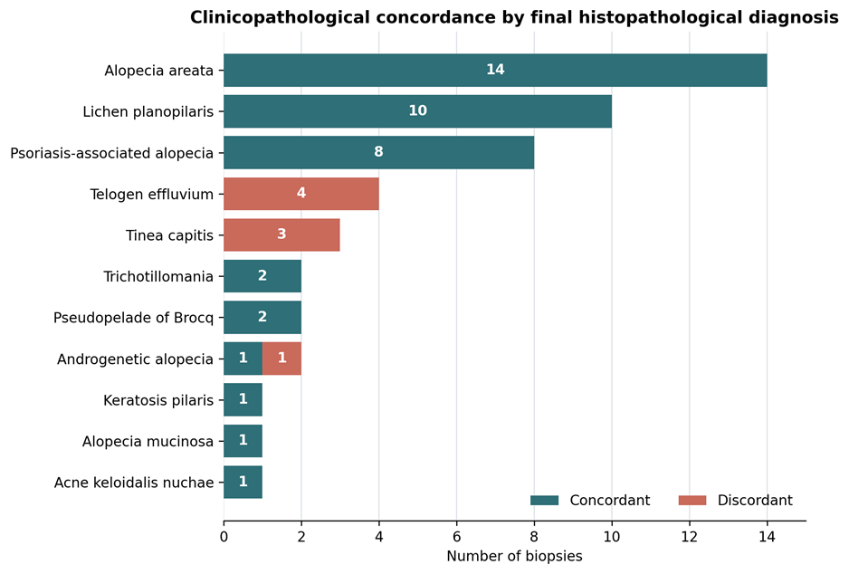
Figure 1. Clinicopathological concordance by final histopathological diagnosis. Concordance means that the final histopathological diagnosis appeared anywhere in the recorded pre-biopsy clinical differential. Teal segments are concordant; coral segments are discordant.
Figure 2
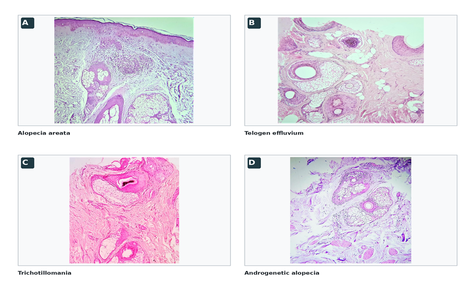
Figure 2. Representative non-cicatricial alopecias. (A) Alopecia areata with dense lymphoplasmacytic inflammation (H&E, ×4). (B) Transverse section of telogen effluvium showing increased telogen follicles and vellus hairs (H&E, ×10). (C) Trichotillomania showing trichomalacia and intrafollicular haemorrhage (H&E, ×10). (D) Androgenetic alopecia with follicular miniaturisation and sebaceous-gland hypertrophy (H&E, ×40).
Figure 3
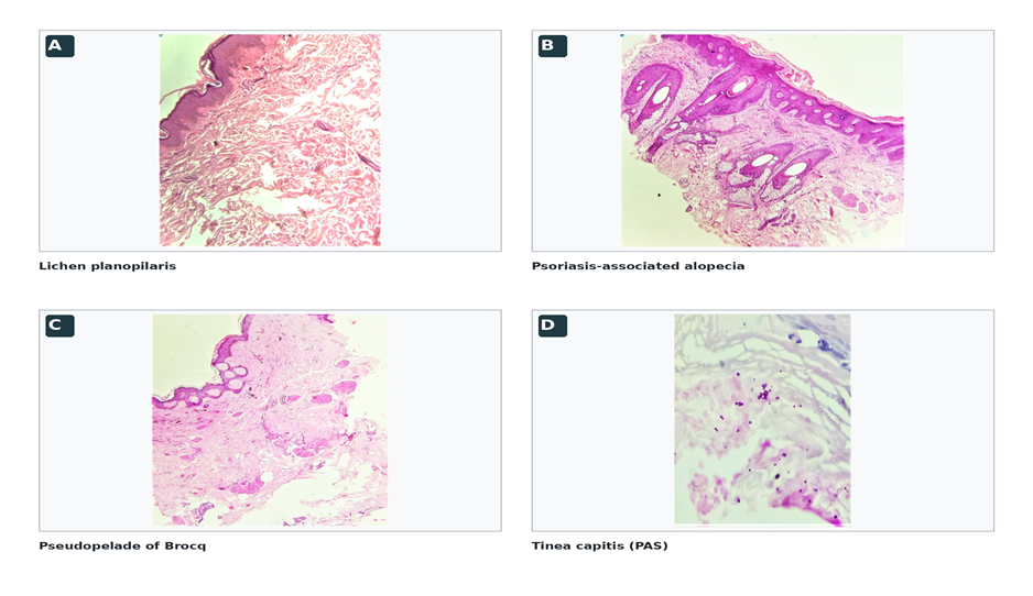
Figure 3. Representative cicatricial alopecias. (A) Lichen planopilaris with diffuse scarring and perivascular inflammation (H&E, ×10). (B) Psoriasis-associated alopecia with psoriasiform epidermal hyperplasia and follicular dropout (H&E, ×4). (C) Pseudopelade of Brocq with complete absence of pilosebaceous units (H&E, ×4). (D) Tinea capitis with fungal spores highlighted by PAS (×40).
DISCUSSION:
Principal findings
This prospective biopsy series demonstrates three clinically relevant findings. First, cicatricial disease constituted slightly more than half of biopsied alopecia, reflecting the enrichment expected in a tertiary-care population selected for histology. Second, combined transverse and vertical examination captured complementary quantitative, inflammatory, interface and fibrosing features across 11 final diagnoses. Third, although overall clinicopathological concordance was high at 83.3%, every discordant case belonged to only three entities: telogen effluvium, tinea capitis and androgenetic alopecia. Histology therefore had its greatest corrective role in diffuse-cycle disorders and an infection that clinically mimicked non-infectious alopecia.
Spectrum and comparison with previous studies
Alopecia areata was the most frequent diagnosis, consistent with the large biopsy series of Palo and Biligi, whereas other selected populations have reported androgenetic alopecia or lichen planopilaris as the leading diagnosis [14-16]. Such differences should not be read as community prevalence. A biopsy series is shaped by referral thresholds, local disease patterns, age exclusions and the clinician's uncertainty. The relatively high cicatricial fraction and the prominence of lichen planopilaris similarly reflect selection of cases in which irreversible hair loss needed histological confirmation. The 83.3% concordance rate is closely comparable to the 85% rate reported by Aslani and colleagues when agreement included either of the first two clinical differential diagnoses; their exact first-diagnosis agreement was lower [15]. This distinction matters. Our concordance definition intentionally measures whether histology confirmed any clinically anticipated diagnosis, not whether the clinician's first choice was correct. It is therefore a liberal but clinically meaningful measure of differential-level agreement. The remaining 16.7% represents cases in which microscopy introduced a diagnosis not previously considered.
Non-cicatricial alopecia
Peribulbar inflammation occurred in 78.6% of alopecia areata biopsies. Its absence in the remainder does not exclude alopecia areata because inflammatory intensity falls with disease stage, while altered hair-cycle proportions, miniaturisation and pigment casts may persist. Whiting's stage-based description and a recent comparison of alopecia areata with pattern hair loss reinforce the value of integrating multiple features rather than requiring a classic "swarm of bees" in every biopsy [17,18]. All four telogen effluvium cases were absent from the clinical differentials. Their combination of increased telogen germinal units and absent inflammation illustrates why a quantitative transverse view is particularly useful when diffuse shedding is clinically attributed to alopecia areata or another inflammatory process. Conversely, both androgenetic alopecia biopsies showed miniaturisation, and one corrected a clinical differential of alopecia areata/telogen effluvium. The two trichotillomania biopsies showed the internally consistent traumatic triad of pigment casts, intrafollicular haemorrhage and trichomalacia, findings that accord with described mechanical-injury signs [19].
Cicatricial alopecia
Primary cicatricial alopecias are conventionally classified by the dominant inflammatory pattern, but late lesions may converge on non-specific follicular loss and fibrosis [20-23]. Lichen planopilaris was the principal primary cicatricial entity in this series. Perifollicular and junctional lymphocytic inflammation, dermal fibrosis and fibrous streamers formed the dominant pattern, consistent with established vertical-section criteria [24]. The availability of transverse levels aided evaluation of follicular distribution and scars, while vertical levels demonstrated epidermal and interface changes. Eight biopsies were classified as psoriasis-associated cicatricial alopecia; six showed follicular dropout. Permanent scarring associated with scalp psoriasis is uncommon and remains debated, with published cases ranging from reversible non-cicatricial loss to destructive follicular change [25,26]. The frequency in this selected cohort should therefore be interpreted cautiously. It may reflect tertiary referral, stringent sampling of persistent plaques, or local diagnostic practice, and it should not be extrapolated as the frequency of scarring among patients with scalp psoriasis. Tinea capitis accounted for three clinically discordant cases. Fungal spores demonstrated by PAS within or around hair shafts established the diagnosis. Although inflammatory tinea capitis is more often emphasised in children, adult infection can mimic alopecia areata or cicatricial disease, particularly when inflammation is muted or lesions have evolved [27]. The finding supports a low threshold for fungal stains when clinical and routine histological findings are not fully aligned.
Implications for practice
The results support a structured clinicopathological workflow. The clinician should select an active margin for suspected cicatricial alopecia and the most involved representative area for non-cicatricial disease, document duration, distribution, activity and trichoscopic findings, and provide a ranked differential. The pathology requisition should explicitly distinguish focal from diffuse loss and record prior treatment. In the laboratory, combined orientation from a single adequate punch can preserve interface assessment while allowing counts and ratios, provided grossing is performed by trained personnel [1-3,11-14,28].
The concentration of discordance also suggests a practical diagnostic checkpoint: when the biopsy differential includes alopecia areata but the section lacks convincing bulb-centred inflammation, the pathologist should systematically reassess hair-cycle shift, miniaturisation, traumatic injury and fungal elements. Multifactorial alopecia must also remain possible; a large consecutive series found more than one diagnosis in 12.5% of scalp biopsies [29]. Comparative work has shown that transverse sections often have higher diagnostic accuracy for quantitative non-cicatricial disorders, while vertical sections add interface and depth information [30].
Strengths and limitations
Strengths include prospective case acquisition, a uniform two-year window, combined orientation in every analysed biopsy, disease-specific recording of inflammatory and structural features, and re-analysis of the de-identified master chart with an explicit concordance rule and confidence interval. The inclusion of both cicatricial and non-cicatricial disease reflects the real diagnostic spectrum encountered by a pathology service. Several limitations temper interpretation. This was a single-centre, biopsy-selected study with only 48 analysable cases; COVID-19 reduced accrual, and paediatric alopecia was excluded. Disease-specific estimates are imprecise, especially for rare entities. There was no arm assessed by vertical or transverse sections alone, so the incremental yield of combined processing cannot be quantified. Histological review was not reported as blinded or independently duplicated, preventing assessment of interobserver reproducibility. Clinical differentials sometimes contained multiple diagnoses, making the primary concordance definition more permissive than exact first-choice agreement. Trichoscopic findings, disease duration, treatment exposure, direct immunofluorescence and longitudinal outcomes were not consistently available. Finally, the psoriasis-associated cicatricial category requires cautious external interpretation because scarring in scalp psoriasis is uncommon and diagnostically controversial.
CONCLUSION:
In this tertiary-care biopsy cohort, combined transverse and vertical evaluation defined a diverse spectrum of scalp alopecia and achieved 83.3% agreement with the recorded clinical differential. Histopathology most often changed the clinical impression in telogen effluvium and tinea capitis, highlighting the importance of quantitative follicular assessment and targeted fungal stains. The findings support close dermatologist-pathologist communication and careful combined processing of an adequate scalp biopsy, while direct comparative studies are needed to quantify the added yield of each orientation.
REFERENCES:
1. Sperling LC. The role of the scalp biopsy in the evaluation of alopecia. J Am Acad Dermatol. 2023;89(2 Suppl):S16-S19. doi:10.1016/j.jaad.2023.05.047.
2. Pinedo-Moraleda F, Tristán-Martín B, Dradi GG. Alopecias: practical tips for the management of biopsies and main diagnostic clues for general pathologists and dermatopathologists. J Clin Med. 2023;12(15):5004. doi:10.3390/jcm12155004.
3. Knopp E. The scalp biopsy for hair loss and its interpretation. Semin Cutan Med Surg. 2015;34(2):57-66. doi:10.12788/j.sder.2015.0144.
4. Stefanato CM. Histopathology of alopecia: a clinicopathological approach to diagnosis. Histopathology. 2010;56(1):24-38. doi:10.1111/j.1365-2559.2009.03439.x.
5. Bernárdez C, Molina-Ruiz AM, Requena L. Histologic features of alopecias-part I: nonscarring alopecias. Actas Dermosifiliogr. 2015;106(3):158-167. doi:10.1016/j.ad.2014.07.006.
6. Bernárdez C, Molina-Ruiz AM, Requena L. Histologic features of alopecias: part II: scarring alopecias. Actas Dermosifiliogr. 2015;106(4):260-270. doi:10.1016/j.ad.2014.06.016.
7. Headington JT. Transverse microscopic anatomy of the human scalp: a basis for a morphometric approach to disorders of the hair follicle. Arch Dermatol. 1984;120(4):449-456. doi:10.1001/archderm.1984.01650400031009.
8. Elston DM, McCollough ML, Angeloni VL. Vertical and transverse sections of alopecia biopsy specimens: combining the two to maximize diagnostic yield. J Am Acad Dermatol. 1995;32(3):454-457. doi:10.1016/0190-9622(95)90068-3.
9. Elston DM, Ferringer T, Dalton S, Fillman E, Tyler W. A comparison of vertical versus transverse sections in the evaluation of alopecia biopsy specimens. J Am Acad Dermatol. 2005;53(2):267-272. doi:10.1016/j.jaad.2005.03.007.
10. Du X, Li Z, Xu W, Zhou X, Tang S, Song C, et al. Diagnostic value of horizontal versus vertical sections for scarring and non-scarring alopecia: a systematic review and meta-analysis. Eur J Dermatol. 2016;26(4):361-369. doi:10.1684/ejd.2016.2797.
11. Nguyen JV, Hudacek K, Whitten JA, Rubin AI, Seykora JT. The HoVert technique: a novel method for the sectioning of alopecia biopsies. J Cutan Pathol. 2011;38(5):401-406.
12. Elston D. The "Tyler technique" for alopecia biopsies. J Cutan Pathol. 2012;39(3):306.
13. Gurusamy U, Venkataswamy C, Sivaraman A. Evaluation of alopecia: a new processing technique combining vertical and transverse sections from a single scalp biopsy specimen. Int J Trichology. 2018;10(1):11-16. doi:10.4103/ijt.ijt_76_17.
14. Palo S, Biligi DS. Utility of horizontal and vertical sections of scalp biopsies in various forms of primary alopecias. J Lab Physicians. 2018;10(1):95-100. doi:10.4103/JLP.JLP_89_17.
15. Aslani FS, Esfahani MH, Sepaskhah M. Non-scarring alopecias in Iranian patients: a histopathological study with hair counts. Iran J Pathol. 2018;13(3):317-324.
16. Goyal M, Khandpur S, Ramam M, Sharma VK, Singh MK. A study of the histopathological features of alopecias on transverse sections of scalp biopsies. Indian J Dermatol. 2019;64(1):47-54. doi:10.4103/ijd.IJD_477_17.
17. Whiting DA. Histopathologic features of alopecia areata: a new look. Arch Dermatol. 2003;139(12):1555-1559. doi:10.1001/archderm.139.12.1555.
18. Plante J, Valdebran M, Forcucci J, Bosland J, Elbendary A, Jaiswal R, et al. A comparative study of histopathologic features in alopecia areata and pattern hair loss. J Cutan Pathol. 2023;50(6):563-567. doi:10.1111/cup.14384.
19. Royer MC, Sperling LC. Splitting hairs: the "hamburger sign" in trichotillomania. J Cutan Pathol. 2006;33(Suppl 2):63-64. doi:10.1111/j.1600-0560.2006.00526.x.
20. Olsen EA, Bergfeld WF, Cotsarelis G, Price VH, Shapiro J, Sinclair R, et al. Summary of North American Hair Research Society-sponsored workshop on cicatricial alopecia, Duke University Medical Center, February 10 and 11, 2001. J Am Acad Dermatol. 2003;48(1):103-110.
21. Tan E, Martinka M, Ball N, Shapiro J. Primary cicatricial alopecias: clinicopathology of 112 cases. J Am Acad Dermatol. 2004;50(1):25-32. doi:10.1016/j.jaad.2003.04.001.
22. Ross EK, Tan E, Shapiro J. Update on primary cicatricial alopecias. J Am Acad Dermatol. 2005;53(1):1-37. doi:10.1016/j.jaad.2004.06.015.
23. Kolivras A, Thompson C. Primary scalp alopecia: new histopathological tools, new concepts and a practical guide to diagnosis. J Cutan Pathol. 2017;44(1):53-69. doi:10.1111/cup.12822.
24. Tandon YK, Somani N, Cevasco NC, Bergfeld WF. A histologic review of 27 patients with lichen planopilaris. J Am Acad Dermatol. 2008;59(1):91-98. doi:10.1016/j.jaad.2008.03.007.
25. Silva CY, Brown KL, Kurban AK, Mahalingam M. Psoriatic alopecia-fact or fiction? A clinicohistopathologic reappraisal. Indian J Dermatol Venereol Leprol. 2012;78(5):611-619.
26. Almeida MC, Romiti R, Doche I, Valente NYS, Donati A. Psoriatic scarring alopecia. An Bras Dermatol. 2013;88(6 Suppl 1):29-31.
27. Arenas R, Toussaint S, Isa-Isa R. Kerion and dermatophytic granuloma: mycological and histopathological findings in 19 children with inflammatory tinea capitis of the scalp. Int J Dermatol. 2006;45(3):215-219.
28. Yeliur IK, Tirumalae R. Histopathologic approach to alopecia. Indian J Dermatopathol Diagn Dermatol. 2018;5(2):79-88. doi:10.4103/ijdpdd.ijdpdd_48_18.
29. Wohltmann WE, Sperling LC. Histopathologic diagnosis of multifactorial alopecia. J Cutan Pathol. 2016;43(6):483-491. doi:10.1111/cup.12698.
30. Kamyab-Hesari K, Aghazadeh N, Nourmohammadpour P, Ghanadan A, Nikoo A, Gholamali F, et al. Diagnostic accuracy measures for vertical and transverse scalp biopsies in cicatricial and non-cicatricial alopecias. Dermatol Sin. 2018;36(1):30-35. doi:10.1016/j.dsi.2017.08.008.