Association of postoperative femoral posterior condylar offset increase with early function after posterior-stabilised total knee arthroplasty: a prospective observational study.
- Kunal , Junior Resident, Department of Orthopaedics, Bangalore Medical College and Research Institute, Bengaluru, Karnataka, India.
- Sharath Kumar P V , Professor, Department of Orthopaedics, Bangalore Medical College and Research Institute, Bengaluru, Karnataka, India.
- Mohammed Anas , Senior Resident, Department of Orthopaedics, Bangalore Medical College and Research Institute, Bengaluru, Karnataka, India.
- Nihit Baranwal , Junior Resident, Department of Orthopaedics, Bangalore Medical College and Research Institute, Bengaluru, Karnataka, India.
Article Information:
Abstract:
Background: Total knee arthroplasty is the treatment of choice for advanced osteoarthritis. In spite of that, postoperative knee flexion and function show a wide variability. Reconstructing the posterior condylar offset (PCO) alters joint geometry and soft-tissue tension, though clinical evidence about the beneficial PCO change in posterior-stabilised TKA remains equivocal. Consequently, This study assesses the effect of the magnitude (≤2 mm vs >2 mm) of increase in postoperative PCO on early knee flexion and clinical scores at 3 months. Objective: To assess whether the magnitude of postoperative femoral posterior condylar offset (PCO) increase is associated with early knee flexion and functional outcome after posterior-stabilised total knee arthroplasty (PS-TKA). Methods: This prospective observational analytical study included 50 patients aged at least 50 years who underwent primary PS-TKA for Kellgren–Lawrence grade 3 or 4 knee osteoarthritis at a tertiary teaching hospital in Bengaluru, India. Native and postoperative PCO were measured on true lateral radiographs. Patients were stratified into a PCO increase ≤2 mm group (n=25) and a PCO increase >2 mm group (n=25). Knee range of motion was recorded immediately after surgery, at 6 weeks and at 3 months. The Hospital for Special Surgery (HSS) knee score was assessed at 3 months. Values and p-values are reported as presented in the source thesis. Results: The groups were comparable at baseline. Mean postoperative PCO increase was 0.86±0.68 mm in the ≤2 mm group and 3.48±0.94 mm in the >2 mm group (p<0.001). At 3 months, mean knee flexion was 118.52±8.34° versus 112.16±9.78° (p=0.012), and mean HSS score was 79.26±7.24 versus 73.56±8.18 (p=0.008), respectively. HSS scores ≥75 were achieved by 18/25 (72%) versus 12/25 (48%) participants (p=0.039), while flexion >115° was achieved by 21/25 (84%) versus 15/25 (60%) participants (p=0.032). Conclusion: A postoperative PCO increase within 2 mm of native anatomy was associated with better early flexion and HSS outcomes after PS-TKA than an increase exceeding 2 mm. The observational design and short follow-up require cautious interpretation, and the proposed threshold should be validated in larger studies with patient- level multivariable analysis.
Keywords:
Article :
INTRODUCTION:
Total knee arthroplasty (TKA) is an established treatment for advanced knee osteoarthritis, but recovery of flexion and function remains variable. Postoperative range of motion affects walking, stair use, transfers and activities requiring deep flexion. In populations where floor sitting, squatting or kneeling is common, even modest limitations in flexion may be functionally important. The geometry of the reconstructed distal femur is one potentially modifiable contributor to this variation.
Posterior condylar offset (PCO) is the perpendicular distance between a line tangent to the posterior femoral cortex and the most posterior point of the femoral condyle. Femoral component size and anteroposterior position alter this distance during TKA. Both loss and excessive increase of PCO may influence the flexion space, posterior soft-tissue tension and tibiofemoral impingement. A network meta-analysis with a sequential cohort study suggested that an increase between 0 and 2 mm was associated with favourable functional recovery, while also demonstrating heterogeneity among PCO categories and outcomes [1].
The effect of PCO may depend on implant design. In posterior-stabilised (PS) TKA, the post-cam mechanism substitutes for the posterior cruciate ligament and facilitates femoral rollback during flexion. Computational modelling shows that changes in PCO alter polyethylene contact stress, collateral ligament forces and extensor-mechanism loading [2]. Experimental work further indicates that small changes in joint-space geometry can modify ligament tension, laxity and the timing of post-cam engagement [3]. These observations provide a plausible mechanism through which excessive posteriorisation or overstuffing could limit functional recovery.
Clinical evidence remains inconsistent. In a cohort of posterior-substituting fixed-bearing TKAs, Chang and colleagues found that changes in PCO and the PCO ratio did not influence postoperative range of motion, and outcomes were comparable between anterior- and posterior-referencing systems [4]. Differences in implant geometry, measurement methods, baseline motion, surgical technique and follow-up may account for discordant findings. Evidence from Indian patients undergoing PS-TKA is limited.
This study therefore assessed the association between the magnitude of postoperative PCO increase and early knee function following primary PS-TKA. The primary objective was to compare knee flexion at 3 months between patients with a PCO increase ≤2 mm and those with an increase >2 mm. Secondary objectives were to compare HSS knee scores, extension-related outcomes and achievement of clinically useful flexion and functional-score thresholds.
MATERIALS AND METHODS:
Study design and setting
This was a prospective observational analytical study conducted over 18 months in the Department of Orthopaedics, Bangalore Medical College and Research Institute and its affiliated hospitals, Bengaluru, India. Reporting was structured with reference to the STROBE framework. The study flow is shown in Figure 1.
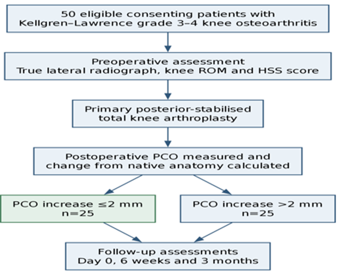
Figure 1. Study flow for the prospective observational cohort. PCO, posterior condylar offset; PS-TKA, posterior-stabilised total knee arthroplasty; HSS, Hospital for Special Surgery.
Participants
Patients of either sex were eligible if they were aged at least 50 years, had Kellgren– Lawrence grade 3 or 4 primary knee osteoarthritis, were scheduled for primary TKA, had a flexion contracture not exceeding 15°, had a body mass index of 20–35 kg/m² and provided written informed consent. Exclusion criteria were severe deformity requiring a constrained implant, rheumatoid arthritis, revision TKA, intra-articular deformity, previous surgery on the study knee and post-traumatic arthritis.
Surgical treatment and radiographic assessment
All participants underwent standard primary PS-TKA. PCO was assessed on true lateral knee radiographs before and after surgery. A line tangent to the posterior femoral cortex was used as the reference, and the perpendicular distance to the most posterior point of the native condyle or femoral component was recorded in millimetres. Postoperative PCO change was calculated as postoperative minus native PCO. Participants were classified after surgery into Group 1, PCO increase ≤2 mm, or Group 2, PCO increase >2 mm.
The posterior condylar offset ratio (PCOR) was also recorded to describe posterior femoral geometry relative to femoral size. The source thesis did not provide patient-level measurement reliability data.
Outcomes and follow-up
The primary outcome was knee flexion at 3 months. Knee flexion and extension lag were recorded immediately after surgery, at 6 weeks and at 3 months. Secondary outcomes included change in flexion, flexion contracture and the total and component HSS knee scores at 3 months. Binary outcomes were HSS score ≥75 and knee flexion >115°.
Sample size
The target sample was 50 participants. The source thesis used a single-proportion calculation based on an anticipated prevalence of PCO difference >2 mm of approximately 8%, a 95% confidence level and an absolute precision of 7.5%.
Statistical analysis
Continuous variables were summarised as mean±standard deviation and compared using the t-test when normally distributed. Skewed data were to be summarised using the median and range and compared using non-parametric methods. Categorical variables were summarised as counts and percentages and compared using the chi-square or an appropriate non-parametric test. Correlations between PCO increase and continuous postoperative outcomes were assessed using correlation coefficients. The significance threshold was p<0.05. Analyses in the source thesis were performed with IBM SPSS Statistics for Windows, version 22.0. Values and p-values in this manuscript are reproduced from the source thesis without independent recalculation because patient-level data were not supplied.
RESULTS:
Table 1. Baseline characteristics. Group 1, PCO increase ≤2 mm; Group 2, PCO increase >2 mm.
|
Characteristic |
Group 1 (n=25) |
Group 2 (n=25) |
p-value |
|
Age, years |
64.88±6.12 |
65.44±5.87 |
0.681 |
|
Sex, male/female |
10/15 |
12/13 |
0.527 |
|
BMI, kg/m² |
27.34±3.15 |
27.68±2.98 |
0.562 |
|
Kellgren– Lawrence grade 3/4 |
14/11 |
13/12 |
0.768 |
|
Preoperative flexion contracture, ° |
6.16±1.02 |
6.32±0.89 |
0.483 |
|
Preoperative knee flexion, ° |
110.36±8.24 |
109.88±7.92 |
0.764 |
|
Preoperative HSS score |
51.28±8.34 |
50.96±7.89 |
0.873 |
The study included 50 patients, with 25 in each PCO-increase group. Complete 3-month results were reported for all 50 participants. Mean age was 64.88±6.12 years in Group 1 and 65.44±5.87 years in Group 2. Women comprised 15/25 and 13/25 participants, respectively. Baseline BMI, Kellgren–Lawrence grade, flexion contracture, knee flexion and HSS score were comparable (Table 1).
Table 2. Posterior condylar geometry. Group 1, PCO increase ≤2 mm; Group 2, PCO increase >2 mm.
|
Outcome |
Group 1 (n=25) |
Group 2 (n=25) |
p-value |
|
Native PCO, mm |
15.68±1.34 |
15.92±1.28 |
0.526 |
|
Postoperative PCO, mm |
16.54±1.16 |
19.40±1.09 |
<0.001 |
|
PCO increase, mm |
0.86±0.68 |
3.48±0.94 |
<0.001 |
|
Native PCOR, % |
95.24±7.18 |
96.18±6.84 |
0.631 |
|
Postoperative PCOR, % |
99.10±6.42 |
114.52±7.96 |
<0.001 |
|
PCOR increase,% |
3.86±4.12 |
18.34±5.71 |
<0.001 |
Native PCO was similar between the groups: 15.68±1.34 mm in Group 1 and 15.92±1.28 mm in Group 2 (p=0.526). Postoperative PCO was 16.54±1.16 mm and 19.40±1.09 mm, respectively (p<0.001). The mean increase from native anatomy was 0.86±0.68 mm in Group 1 and 3.48±0.94 mm in Group 2 (p<0.001). Corresponding PCOR increases were 3.86±4.12% and 18.34±5.71% (p<0.001) (Table 2).
Table 3. Postoperative range of motion. Group 1, PCO increase ≤2 mm; Group 2, PCO increase >2 mm.
|
Outcome |
Group 1 (n=25) |
Group 2 (n=25) |
p-value |
|
Flexion, day 0,° |
73.84±9.26 |
71.36±8.94 |
0.298 |
|
Flexion, 6 weeks, ° |
103.24±10.18 |
98.68±11.42 |
0.087 |
|
Flexion, 3 months, ° |
118.52±8.34 |
112.16±9.78 |
0.012 |
|
Extension lag, 3 months, ° |
1.28±0.92 |
1.56±0.86 |
0.287 |
|
Flexion improvement, day 0 to 3 months, ° |
44.68±9.12 |
40.80±10.24 |
0.156 |
Immediate postoperative flexion was 73.84±9.26° in Group 1 and 71.36±8.94° in Group 2
(p=0.298). At 6 weeks, flexion was 103.24±10.18° and 98.68±11.42° (p=0.087). At 3
months, Group 1 achieved greater flexion than Group 2 (118.52±8.34° versus 112.16±9.78°, p=0.012). Extension lag at 3 months was 1.28±0.92° and 1.56±0.86° (p=0.287). Flexion improvement from day 0 to 3 months was 44.68±9.12° and 40.80±10.24° (p=0.156) (Table3)
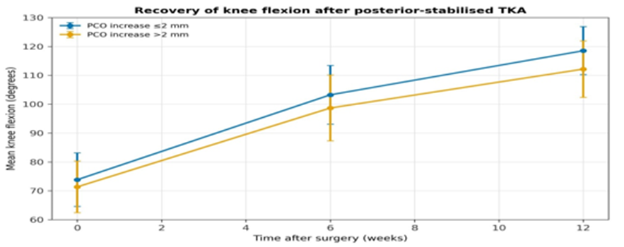
Figure 2. Mean knee flexion after PS-TKA. Values are source-reported means at day 0, 6 weeks and 3 months.
Table 4. HSS knee outcomes at 3 months. Group 1, PCO increase ≤2 mm; Group 2, PCO increase >2 mm.
|
Outcome |
Group 1 (n=25) |
Group 2 (n=25) |
p-value |
|
HSS pain |
26.84±2.68 |
25.16±3.12 |
0.033 |
|
HSS function |
19.28±2.14 |
17.92±2.68 |
0.048 |
|
HSS range of motion |
14.82±1.86 |
13.02±1.94 |
0.006 |
|
HSS muscle strength |
8.64±1.18 |
8.28±1.34 |
0.287 |
|
HSS flexion deformity |
9.76±0.52 |
9.68±0.59 |
0.583 |
|
HSS instability |
9.84±0.37 |
9.80±0.41 |
0.714 |
|
HSS total score |
79.26±7.24 |
73.56±8.18 |
0.008 |
|
HSS ≥75, n (%) |
18 (72) |
12 (48) |
0.039 |
|
Flexion >115°, n (%) |
21 (84) |
15 (60) |
0.032 |
At 3 months, mean HSS total score was higher in Group 1 than Group 2 (79.26±7.24 versus 73.56±8.18, p=0.008). Group 1 also had higher pain (26.84±2.68 versus 25.16±3.12, p=0.033), function (19.28±2.14 versus 17.92±2.68, p=0.048) and HSS range-of-motion component scores (14.82±1.86 versus 13.02±1.94, p=0.006). Muscle strength, flexion deformity and instability components were similar (Table 4).
An HSS score ≥75 was attained by 18/25 (72%) participants in Group 1 and 12/25 (48%) in Group 2 (p=0.039). Flexion >115° was attained by 21/25 (84%) and 15/25 (60%), respectively (p=0.032).
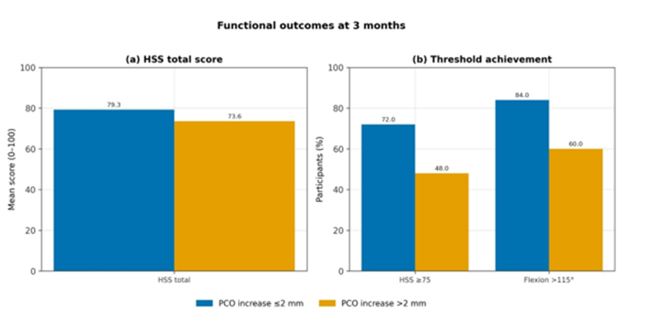
Figure 3. Three-month functional outcomes by magnitude of PCO increase: (a) mean HSS total score and (b) proportions reaching the prespecified HSS and flexion thresholds.
Table 5. Source-reported correlations between PCO increase and postoperative outcomes (n=50).
|
Outcome |
Correlation coefficient, r |
p-value |
|
Knee flexion at 3 months |
0.687 |
0.008 |
|
Flexion improvement, day 0 to 3 months |
0.652 |
0.014 |
|
HSS total score |
0.714 |
0.004 |
|
HSS pain component |
0.518 |
0.042 |
|
HSS function component |
0.592 |
0.021 |
|
HSS range-of-motion component |
0.756 |
<0.001 |
|
Extension lag |
−0.412 |
0.087 |
|
Flexion contracture |
−0.384 |
0.118 |
The source thesis reported positive correlations between PCO increase and knee flexion at 3 months (r=0.687, p=0.008), flexion improvement (r=0.652, p=0.014), HSS total score (r=0.714, p=0.004), HSS pain (r=0.518, p=0.042), HSS function (r=0.592, p=0.021) and the HSS range-of-motion component (r=0.756, p<0.001). It reported non-significant negative correlations with extension lag (r=−0.412, p=0.087) and flexion contracture (r=−0.384, p=0.118) (Table 5).
DISCUSSION:
In this prospective cohort of 50 primary PS-TKAs, a postoperative PCO increase within 2 mm of native anatomy was associated with better early flexion and HSS outcomes than an increase exceeding 2 mm. The between-group difference in mean flexion at 3 months was 6.36°, and the difference in mean HSS total score was 5.70 points. The proportions attaining HSS ≥75 and flexion >115° were also greater in the ≤2 mm group. Extension lag and flexion contracture were similar, suggesting that the observed difference was concentrated in flexion and functional scoring rather than extension.
The principal group comparison is consistent with Zhang and colleagues, whose network meta-analysis and sequential cohort study identified a 0–2 mm PCO increase category as favourable for several functional outcomes [1]. Their work also illustrates the wider inconsistency of the evidence: rankings differed by outcome, and the source studies included varied prosthetic designs and populations. The present study adds a prospectively observed Indian PS-TKA cohort using a clinically simple two-group threshold.
Several biomechanical mechanisms may explain why a limited increase could be preferable to a larger increase. PCO affects the posterior femoral contour, flexion space and the relationship of the femoral component to posterior soft tissues. Finite-element modelling has shown that altering PCO changes polyethylene contact stress and collateral ligament loading in PS-TKA [2]. Hybrid simulator work further demonstrated that 2 mm changes in joint-space geometry altered ligament tension, joint contact force, laxity and post-cam engagement [3]. Excess posteriorisation may therefore overstuff the flexion space or alter kinematics even when the post-cam mechanism remains functional.
Not all clinical studies have found a relationship between PCO change and postoperative motion. Chang and colleagues examined 184 knees treated with a posterior-substituting fixed-bearing implant and found that PCO and PCOR changes did not affect postoperative range of motion; outcomes were similar with anterior and posterior referencing [4]. Differences from the present findings may relate to the distribution of PCO change, implant geometry, surgical technique, baseline flexion, follow-up duration and adjustment for other predictors. Evidence from revision TKA also suggests that PCO may independently predict functional improvement, although that setting has different bone loss, constraint and soft- tissue conditions and cannot be directly extrapolated to primary TKA [5].
The HSS component findings support a functional association beyond flexion alone. Pain, function and the HSS range-of-motion component were higher in the ≤2 mm group, whereas strength, deformity and instability scores were similar. This pattern is compatible with an effect mediated mainly through flexion mechanics rather than gross coronal or sagittal instability. Nevertheless, group allocation occurred after surgery according to the achieved PCO change; the exposure was not randomised. Component sizing, femoral position, soft- tissue balance, rehabilitation and unmeasured patient characteristics could therefore account for part of the association. The source thesis also reported positive correlations between increasing PCO change and better flexion and HSS outcomes, although the stratified means showed poorer outcomes in the >2 mm group. Because patient-level data were not available, the direction and coding of those correlation analyses could not be independently checked. They are reproduced as reported and should not be used to infer a monotonic dose-response relationship. The more directly interpretable finding is the prespecified comparison of the two PCO-increase categories.Clinically, the findings support careful attention to femoral component sizing and anteroposterior positioning. They do not establish that surgically forcing PCO into a particular range will improve outcome, and they do not justify compromising component fit, rotation, joint-line restoration or ligament balance. Rather, a postoperative increase >2 mm may be a marker of a reconstruction that warrants closer evaluation. Larger cohorts with patient-level multivariable analysis should test whether the threshold remains associated with outcome after adjustment for preoperative flexion, implant size, alignment and rehabilitation.
CONCLUSION:
Among patients undergoing primary PS-TKA, a postoperative femoral PCO increase within 2 mm of native anatomy was associated with greater knee flexion and higher HSS scores at 3 months than an increase exceeding 2 mm. This threshold may be a useful radiographic and technical target, but the findings remain observational and require confirmation with longer follow-up, reproducible measurement methods and patient-level adjusted analysis.
REFERENCES:
1. Zhang Y, Wang J, Zhang M, Xu Y. Effect of femoral posterior condyle offset on knee joint function after total knee replacement: a network meta-analysis and a sequential retrospective cohort study. J Orthop Surg Res. 2021 Feb 10;16(1):126. doi: 10.1186/s13018- 021-02233-8. PMID: 33568164; PMCID: PMC7877059.
2. Kang KT, Koh YG, Son J, Kwon OR, Lee JS, Kwon SK. Biomechanical effects of posterior condylar offset and posterior tibial slope on quadriceps force and joint contact forces in posterior-stabilized total knee arthroplasty. Biomed Res Int. 2017;2017:4908639. doi: 10.1155/2017/4908639. Epub 2017 Nov 19. PMID: 29349074; PMCID: PMC5733759.
3. Sekeitto AR, McGale JG, Montgomery LA, Vasarhelyi EM, Willing R, Lanting BA. Posterior-stabilized total knee arthroplasty kinematics and joint laxity: a hybrid biomechanical study. Arthroplasty. 2022 Dec 15;4(1):53. doi: 10.1186/s42836-022-00153-4. PMID: 36522686; PMCID: PMC9753369.
4. Chang MJ, Kang SB, Chang CB, Han DH, Park HJ, Hwang K, et al. Posterior condylar offset changes and its effect on clinical outcomes after posterior-substituting, fixed-bearing total knee arthroplasty: anterior versus posterior referencing. Knee Surg Relat Res. 2020 Jan 1;32(1):10. doi: 10.1186/s43019-019-0022-2. PMID: 32660651; PMCID: PMC7219226.
5. Clement ND, MacDonald DJ, Hamilton DF, Burnett R. Posterior condylar offset is an independent predictor of functional outcome after revision total knee arthroplasty. Bone Joint Res. 2017 Mar;6(3):172-178. doi: 10.1302/2046-3758.63.BJR-2015-0021.R1. PMID: 28360083; PMCID: PMC5376661.