STUDY ON FACIAL NERVE PALSY IN CASES WITH CEREBELLOPONTINE SCHWANNOMA PRESENTING TO A TERTIARY HEALTHCARE CENTRE IN JHARKHAND, INDIA.
- Rashmi Kullu , Senior Resident (Academic), Department of Neurosurgery, RIMS, Ranchi, India.
- Anand Prakash , Additional Professor & HOD, Department of Neurosurgery, RIMS, Ranchi, India.
- Gautam Dutta , Associate Professor, Department of Neurosurgery, RIMS, Ranchi, India.
- Saurav Kumar Besra , Assistant Professor, Department of Neurosurgery, RIMS, Ranchi, India.
- Shiwangi Kashyap , Senior Resident, Department of Neurosurgery, RIMS, Ranchi, India.
Article Information:
Abstract:
Background: Cerebellopontine schwannoma, also known as vestibular schwannoma (VS) or acoustic neuroma is a slow-growing, benign intracranial tumor that originates from the Schwann cells of the eighth cranial nerve's vestibular division. Aims & objectives: The aim of this study is to evaluate the clinical profile and severity of facial nerve palsy in patients with cerebellopontine schwannoma presenting to a tertiary healthcare centre in Jharkhand, India, and to assess the association between the severity of facial nerve palsy and tumour characteristics, including tumour size and duration of symptoms. Materials & Methods: This was a hospital-based observational cross-sectional study conducted in the Department of Neurosurgery, Rajendra Institute of Medical Sciences (RIMS), Ranchi, over a period of one year. A total of 100 patients diagnosed with cerebellopontine schwannoma presenting with facial nerve palsy were included in the study. Result: Among 100 patients, females (58%) and those aged 41–50 years (31%) predominated. HB Grade II was the most common presentation (28%). Larger tumor size, prolonged symptom duration, brainstem compression, and hydrocephalus were significantly associated with severe facial nerve palsy (all p<0.05), whereas hearing loss showed no significant association (p=0.094). Conclusion: We concluded that larger tumor size, longer symptom duration, brainstem compression, and hydrocephalus are all strongly correlated with the severity of facial nerve palsy in cerebellopontine schwannoma. To maintain facial nerve function, lessen neurological impairments, and enhance overall patient outcomes, early diagnosis, quick MRI examination, and prompt surgical intervention are crucial.
Keywords:
Article :
INTRODUCTION:
The Schwann cells of the vestibular division of the eighth cranial nerve give rise to the benign, slowly developing intracranial tumor known as cerebellopontine schwannoma, also known as vestibular schwannoma (VS) or acoustic neuroma [1]. Vestibular schwannomas are the most prevalent tumors in the cerebellopontine angle (CPA), accounting for almost 80–90% of lesions in this anatomical region and 6–8% of all cerebral cancers [1,2]. These tumors are histologically benign, but their gradual enlargement and compression of nearby cerebellar, brainstem, and cranial nerves can cause serious neurological morbidity [2]. The size, development rate, and architecture of the tumor all affect how vestibulocochlear schwannoma manifests clinically. The most frequent presenting symptoms include imbalance, tinnitus, and progressive unilateral sensorineural hearing loss [3].
On the other hand, facial nerve dysfunction is a significant clinical symptom, especially in individuals who present at advanced stages of the disease or have larger tumors [4]. Tumor growth may cause compression, stretching, ischemia, or direct involvement of the facial nerve, resulting in variable degrees of facial weakness or paralysis because the facial nerve passes via the internal auditory canal in close proximity to the vestibular nerve [5]. Both functional and psychosocial well-being are greatly impacted by facial nerve palsy. A patient's quality of life may be negatively impacted by facial asymmetry, poor eye closure due to exposure keratitis, drooling, trouble articulating speech, poor mastication, and altered facial expression [6].
The House–Brackmann (HB) grading system, which ranks facial nerve function from Grade I (normal function) to Grade VI (total paralysis), is frequently used to gauge the severity of facial nerve dysfunction. For assessing preoperative status, postoperative results, and long-term healing, this uniform grading system is commonly used [7]. One of the main objectives in the treatment of cerebellopontine schwannoma is now the preservation of facial nerve function. Neuronavigation, stereotactic radiosurgery, intraoperative facial nerve monitoring, and microsurgical techniques have significantly increased the rates of facial nerve preservation and tumor control. This study aims to evaluate the clinical profile and severity of facial nerve palsy in patients with cerebellopontine schwannoma who present to a tertiary healthcare facility in Jharkhand, India. It also aims to determine whether there is a correlation between the severity of facial nerve palsy and tumor characteristics, such as tumor size and symptom duration.
MATERIALS AND METHODS:
Type of Study: Hospital-based observational cross-sectional study.
Place of Study: Department of Neurosurgery, Rajendra Institute of Medical Sciences,Ranchi
Study Duration: 1 year
Sample Size: 100 Patients diagnosed with cerebellopontine schwannoma presenting with facial nerve palsy
Inclusion Criteria:
• Patients aged 18 years and above.
• Patients with radiologically confirmed cerebellopontine schwannoma (MRI brain with contrast).
• Patients presenting with facial nerve palsy before treatment.
• Patients of either sex willing to participate and providing written informed consent.
• Patients with complete clinical and radiological records.
Exclusion Criteria:
• Patients with facial nerve palsy due to causes other than cerebellopontine schwannoma.
• Patients with recurrent cerebellopontine schwannoma who had undergone previous surgery or radiotherapy.
• Patients with multiple intracranial tumors or neurofibromatosis type 2 (NF2).
• Patients with incomplete clinical, radiological, or follow-up data.
• Patients unwilling to participate in the study.
Study Variables:
• Age (years)
• Sex
• Side of lesion (Right/Left)
• Duration of symptoms
• House–Brackmann (HB) grade of facial nerve palsy
• Hearing loss
• Tinnitus
• Vertigo
• Tumor size (cm)
• Tumor laterality
• Brainstem compression (Present/Absent)
• Hydrocephalus (Present/Absent)
• Internal auditory canal involvement
Statistical Analysis:
Data were entered into Excel and subsequently analyzed using SPSS and GraphPad Prism. Continuous variables were summarized as means with standard deviations, while categorical variables were presented as counts and percentages. Comparisons between independent groups were performed using two-sample t-tests, and paired t-tests were applied for correlated (paired) data. Categorical data were compared using chi-square tests, with Fisher’s exact test applied when expected cell counts were small. A p-value of ≤ 0.05 was considered statistically significant.
RESULTS:
The demographic characteristics of the 100 patients included in the study. The largest proportion of patients belonged to the 41–50 years age group (31%), followed by 31–40 years (27%), 18–30 years (18%), 51–60 years (16%), and >60 years (8%). Female patients constituted 58%, while male patients accounted for 42%. The lesion was observed on the left side in 53% of patients and on the right side in 47% of patients.( Table 1)
Table: 1 Demographic Characteristics of the Study Population
|
Variable |
Number |
Percentage (%) |
|
|
Age Group (years) |
18–30 |
18 |
18 |
|
31–40 |
27 |
27 |
|
|
41–50 |
31 |
31 |
|
|
51–60 |
16 |
16 |
|
|
>60 |
8 |
8 |
|
|
Gender |
Male |
42 |
42 |
|
Female |
58 |
58 |
|
|
Side of Lesion |
Right |
47 |
47 |
|
Left |
53 |
53 |
The clinical presentation according to the House–Brackmann (HB) grading system among the 100 patients. Grade II facial nerve palsy was the most common presentation, affecting 28 patients (28%), followed by Grade III in 24 patients (24%) and Grade I in 22 patients (22%). More severe facial nerve dysfunction was observed in 16 patients (16%) with Grade IV, 8 patients (8%) with Grade V, and 2 patients (2%) with Grade VI.( Table 2)
Table: 2 Clinical Presentations according to Facial Nerve Palsy
|
House-Brackmann Grade |
Patients (n) |
Percentage (%) |
|
Grade I |
22 |
22 |
|
Grade II |
28 |
28 |
|
Grade III |
24 |
24 |
|
Grade IV |
16 |
16 |
|
Grade V |
8 |
8 |
|
Grade VI |
2 |
2 |
The association between tumor size and severity of facial nerve palsy among the patients. Among patients with tumors <2 cm, the majority (20 patients) had mild facial nerve palsy (HB Grade I–II), whereas only 5 patients had moderate-to-severe palsy (HB Grade III–VI). In contrast, patients with tumors ≥4 cm predominantly exhibited moderate-to-severe facial nerve palsy (27 patients) compared with only 8 patients having mild palsy. This association was highly statistically significant (p<0.001), indicating that larger tumor size was significantly associated with more severe facial nerve dysfunction.( Table 3)
Table: 3 Association between Tumor Size and Severity of Facial Nerve Palsy
|
Tumor Size |
HB Grade I–II |
HB Grade III–VI |
p-value |
|
<2 cm |
20 |
5 |
<0.001 |
|
2–3.9 cm |
22 |
18 |
|
|
≥4 cm |
8 |
27 |
The relationship between symptom duration and severity of facial nerve palsy in the patients. Among patients with symptoms lasting less than 6 months, 24 patients had mild palsy compared to 10 patients with moderate-to-severe palsy. However, in patients with symptom duration greater than 12 months, 20 patients had moderate-to-severe palsy, while only 8 patients had mild palsy. The association between prolonged symptom duration and increased severity of facial nerve palsy was statistically significant (p=0.003).( Table 4)
Table: 4 Association between Duration of Symptoms and Facial Nerve Palsy
|
Duration of Symptoms |
Mild (HB I–II) |
Moderate–Severe (HB III–VI) |
p-value |
|
<6 months |
24 |
10 |
0.003 |
|
6–12 months |
18 |
20 |
|
|
>12 months |
8 |
20 |
The association between MRI characteristics and facial nerve palsy among the patients. Among patients without brainstem compression, 28 patients had mild facial nerve palsy and 10 patients had moderate-to-severe palsy. Conversely, in patients with brainstem compression, 40 patients exhibited moderate-to-severe facial nerve palsy compared to 22 patients with mild palsy. This difference was highly statistically significant (p<0.001), suggesting that brainstem compression on MRI is strongly associated with severe facial nerve involvement.( Table 5)
Table: 5 MRI Characteristics and Facial Nerve Palsy
|
MRI Finding |
Mild (HB I–II) |
Moderate–Severe (HB III–VI) |
p-value |
|
No Brainstem Compression |
28 |
10 |
<0.001 |
|
Brainstem Compression Present |
22 |
40 |
The factors associated with severe facial nerve palsy (HB Grade III–VI) among the patients. Severe facial nerve palsy was significantly more common in patients with tumor size ≥4 cm (54.0% vs. 16.0%, p<0.001), brainstem compression (80.0% vs. 44.0%, p<0.001), symptom duration greater than 12 months (40.0% vs. 16.0%, p=0.007), and hydrocephalus (28.0% vs. 10.0%, p=0.021). Although hearing loss was more frequently observed among patients with severe facial nerve palsy (84.0%) than those with mild palsy (70.0%), the association did not reach statistical significance (p=0.094). Overall, larger tumor size, brainstem compression, prolonged symptom duration, and hydrocephalus were identified as significant factors associated with severe facial nerve palsy in the patients.( Table 6)
Table: 6 Factors Associated with Severe Facial Nerve Palsy (HB Grade III–VI)
|
Variable |
Severe Palsy |
Mild Palsy |
p-value |
|
Tumor ≥4 cm |
27 (54.0) |
8 (16.0) |
<0.001 |
|
Brainstem Compression |
40 (80.0) |
22 (44.0) |
<0.001 |
|
Symptom Duration >12 months |
20 (40.0) |
8 (16.0) |
0.007 |
|
Hydrocephalus Present |
14 (28.0) |
5 (10.0) |
0.021 |
|
Hearing Loss Present |
42 (84.0) |
35 (70.0) |
0.094 |
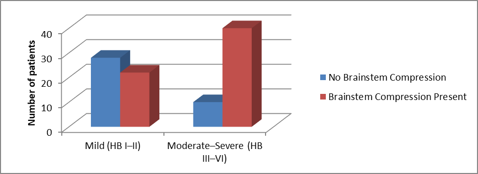
Figure: 1 MRI Characteristics and Facial Nerve Palsy
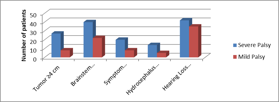
Figure: 2 Factors Associated with Severe Facial Nerve Palsy (HB Grade III–VI)
DISCUSSION:
The present study evaluated the clinical profile and factors associated with facial nerve palsy in 100 patients with cerebellopontine schwannoma. The majority of patients were aged 41–50 years (31%), followed by 31–40 years (27%), with a female predominance (58%). Similar age distributions have been reported in vestibular schwannoma studies, where patients commonly present during the fourth and fifth decades of life, although sex distribution varies among different populations [8, 9]. These findings indicate that cerebellopontine schwannomas predominantly affect middle-aged adults and frequently remain undiagnosed until neurological symptoms become clinically significant.In the present study, House–Brackmann (HB) Grade II facial nerve palsy was the most common presentation (28%), followed by Grade III (24%) and Grade I (22%). Severe facial nerve dysfunction (HB Grades IV–VI) was observed in 26% of patients. Mooney et al. reported that patients presenting with preoperative facial nerve weakness generally had larger tumors and poorer neurological status [8].
The comparatively higher proportion of moderate-to-severe facial nerve palsy observed in the present study may therefore reflect delayed diagnosis and late presentation.Tumor size showed a highly significant association with facial nerve dysfunction in the present study. Patients with tumors <2 cm predominantly had mild facial nerve palsy, whereas those with tumors ≥4 cm commonly exhibited HB Grades III–VI (p<0.001). Similar findings were reported by Hobson et al., who demonstrated that increasing tumor size significantly reduced facial nerve preservation because of greater stretching and compression of the facial nerve [9]. Khan NR et al. also identified tumor volume as an independent predictor of poor facial nerve outcome following microsurgical excision [10]. These observations support the present findings that larger tumors are associated with more severe facial nerve impairment.Symptom duration was another important determinant of facial nerve severity. Patients with symptoms lasting more than 12 months had significantly greater frequencies of moderate-to-severe facial nerve palsy than those presenting within 6 months (p=0.003).
Previous studies have similarly shown that delayed diagnosis permits progressive tumor enlargement, resulting in chronic facial nerve compression and irreversible neural injury [8,10]. Early diagnosis and referral therefore remain essential to improve facial nerve preservation.MRI findings in the present study demonstrated that brainstem compression was strongly associated with severe facial nerve palsy (p<0.001). Patients with radiological evidence of brainstem compression were significantly more likely to present with HB Grades III–VI than patients without compression. Similar observations have been reported by .Khan NR et al., who found that advanced tumors causing brainstem displacement were associated with significantly poorer facial nerve function [10]. Leonetti et al. also emphasized that extensive cerebellopontine angle tumors produce greater neural compression, resulting in more severe cranial nerve deficits [11].
Multivariate analysis identified tumor size ≥4 cm, brainstem compression, prolonged symptom duration (>12 months), and hydrocephalus as significant predictors of severe facial nerve palsy, whereas hearing loss did not achieve statistical significance. Similar predictors have been reported by Hobson et al. and Khan NR et al., who concluded that large tumor size, brainstem compression, and advanced radiological stage independently predict poor facial nerve function [9,10]. Although hearing loss was more frequent among patients with severe palsy, its lack of statistical significance in the present study may be explained by the earlier involvement of the cochlear nerve compared with the facial nerve during tumor progression [11].The findings of the present study are consistent with recent international literature demonstrating that delayed presentation with large cerebellopontine schwannomas substantially increases the risk of facial nerve dysfunction. Early neuroimaging, timely diagnosis, and appropriate neurosurgical intervention before the development of brainstem compression or hydrocephalus may improve facial nerve preservation and long-term neurological outcomes.
CONCLUSION:
We concluded that the features of the tumor and the length of the illness have a major impact on facial nerve palsy in patients with cerebellopontine schwannoma. The majority of patients were middle-aged, slightly more likely to be female, and more likely to have lesions on the left side. The most prevalent clinical presentation was House-Brackmann Grade II, suggesting that mild-to-moderate facial nerve dysfunction predominated at diagnosis. Tumors larger than 4 cm had the highest percentage of moderate-to-severe facial nerve dysfunction, and larger tumors were substantially linked to more severe facial nerve palsy.
The effects of delayed presentation were further highlighted by the substantial correlation between severe facial nerve damage and symptom persistence longer than 12 months. Severe facial nerve palsy was found to be strongly predicted by MRI evidence of brainstem compression, and hydrocephalus also showed a substantial correlation. Patients with severe facial nerve palsy were more likely to have hearing loss, but this link was not statistically significant.These results imply that hydrocephalus, brainstem compression, longer symptom duration, and greater tumor size are significant predictors of severe facial nerve palsy. To maintain facial nerve function, reduce neurological impairments, and enhance overall patient outcomes, early diagnosis, timely neuroimaging, and fast surgical care are crucial.
REFERENCES:
1. Goldbrunner R, Weller M, Regis J, Lund-Johansen M, Stavrinou P, Reuss D, Evans DG, Lefranc F, Sallabanda K, Falini A, Axon P. EANO guideline on the diagnosis and treatment of vestibular schwannoma. Neuro-oncology. 2020 Jan 11;22(1):31-45.
2. Carlson ML, Link MJ. Vestibular schwannomas. New England Journal of Medicine. 2021 Apr 8;384(14):1335-48.
3. Propp JM, McCarthy BJ, Davis FG, Preston-Martin S. Descriptive epidemiology of vestibular schwannomas. Neuro-oncology. 2006 Jan 1;8(1):1-1.
4. Samii M, Draf W. Surgery of the skull base: an interdisciplinary approach. Springer Science & Business Media; 2012 Dec 6.
5. House JW, Brackmann DE. Facial nerve grading system. Otolaryngology—Head and neck surgery. 1985 Apr;93(2):146-7.
6. Sughrue ME, Kane AJ, Shangari G, Rutkowski MJ, McDermott MW, Berger MS, Parsa AT. The relevance of Simpson Grade I and II resection in modern neurosurgical treatment of World Health Organization Grade I meningiomas. Journal of neurosurgery. 2010 Nov 1;113(5):1029-35.
7. Ansari SF, Terry C, Cohen-Gadol AA. Surgery for vestibular schwannomas: a systematic review of complications by approach. Neurosurgical focus. 2012 Sep 1;33(3):E14.
8. Mooney MA, Hendricks B, Sarris CE, Spetzler RF, Almefty KK, Porter RW. Long-term facial nerve outcomes after microsurgical resection of vestibular schwannomas in patients with preoperative facial nerve palsy. Journal of Neurological Surgery Part B: Skull Base. 2018 Jun;79(03):309-13.
9. Hobson CE, Saliba J, Vorasubin N, Lyles RH, Mastrodimos B, Cueva RA. Vestibular schwannoma cerebellopontine angle position impacts facial outcome. The Laryngoscope. 2022 May;132(5):1093-8.
10. Khan NR, Elarjani T, Jamshidi AM, Chen SH, Brown CS, Abecassis J, Silva MA, Lu VM, Wu E, Diaz-Kanelidis M, Bhatia R. Microsurgical management of vestibular schwannoma (acoustic neuroma): facial nerve outcomes, radiographic analysis, complications, and long-term follow-up in a series of 420 surgeries. World neurosurgery. 2022 Dec 1;168:e297-308
11. Leonetti JP, Bartindale MR, Heiferman J, Joyce C, Balasubramanian N. The Natural History of Facial Schwannomas: A Meta-analysis of Case Series. Journal of Neurological Surgery Part B: Skull Base. 2018 Feb;79(S 01):A094.