Early right ventricular–pulmonary vascular uncoupling in cirrhosis: an echocardiographic severity-based analysis.
- Farah Deeba Jehangir , Post DM Senior Resident, Department of Gastroenterology, Sher-I- Kashmir Institute of Medical Sciences, Srinagar, Jammu and Kashmir, India
- Sameer Purra , Post DM Senior Resident, Department of Cardiology, Sher-I- Kashmir Institute of Medical Sciences, Srinagar, Jammu and Kashmir, India
- Shaheena Parveen , Assistant Professor, Department of Gastroenterology, Sher-I- Kashmir Institute of Medical Sciences, Srinagar, Jammu and Kashmir, India.
- Shaheen Nazir , Assistant Professor, Department of Gastroenterology, Sher-I- Kashmir Institute of Medical Sciences, Srinagar, Jammu and Kashmir, India.
- Mushtaq Ahmad Khan , Professor and Head, Department of Gastroenterology, Sher-I- Kashmir Institute of Medical Sciences, Srinagar, Jammu and Kashmir, India.
- Jaswinder Singh Sodhi , Former Professor and Head, Department of Gastroenterology, Sher-I- Kashmir Institute of Medical Sciences, Srinagar, Jammu and Kashmir, India.
- Ajaz Ahmad Lone , Former Professor and Head, Department of Cardiology, Sher-I- Kashmir Institute of Medical Sciences, Srinagar, Jammu and Kashmir, India.
- G M Gulzar , Former Professor and Head, Department of Gastroenterology, Sher-I- Kashmir Institute of Medical Sciences, Srinagar, Jammu and Kashmir, India.
- Altaf Shah , Former Professor and Head, Department of Gastroenterology, Sher-I- Kashmir Institute of Medical Sciences, Srinagar, Jammu and Kashmir, India.
Article Information:
Abstract:
Background: Cirrhosis is associated with a hyperdynamic circulatory state and progressive pulmonary vascular alterations. While overt portopulmonary hypertension is well recognized, early cardiopulmonary interaction abnormalities preceding established pulmonary hypertension remain poorly characterized. Right ventricular–pulmonary vascular coupling reflects the ability of the right ventricle to adapt to increasing pulmonary load and may provide early insight into subclinical cardiopulmonary dysfunction in cirrhosis. Objectives: To evaluate right ventricular–pulmonary vascular coupling in patients with cirrhosis using echocardiographic indices and to assess its relationship with liver disease severity. Methods: In this cross-sectional echocardiographic study, patients with cirrhosis were compared with age-and sex-matched healthy controls. Pulmonary pressures were estimated using tricuspid regurgitant velocity–derived right ventricular systolic pressure (RVSP). Right ventricular systolic performance was assessed using tricuspid annular plane systolic excursion (TAPSE), right ventricular fractional area change (RV FAC), and right ventricular longitudinal strain. RV–pulmonary vascular coupling was evaluated using the TAPSE/RVSP ratio. Associations with MELD-Na score were analyzed. Results: Cirrhotic patients demonstrated significantly higher RVSP compared with controls, while conventional right ventricular systolic indices remained largely within normal limits. Despite preserved TAPSE, the TAPSE/RVSP ratio was significantly reduced in cirrhosis, indicating early RV–pulmonary uncoupling. Progressive deterioration of coupling indices was observed with increasing MELD-Na scores, suggesting severity-dependent cardiopulmonary stress. Conclusions: Cirrhosis is associated with early right ventricular–pulmonary vascular uncoupling that precedes overt pulmonary hypertension or right ventricular systolic failure. Echocardiographic coupling indices may identify a preclinical cardiopulmonary phenotype with important implications for risk stratification in advanced liver disease.
Keywords:
Article :
INTRODUCTION:
Cirrhosis is characterized by profound circulatory and pulmonary vascular alterations that extend beyond the liver. The hyperdynamic circulatory state of cirrhosis, driven by splanchnic vasodilation, increased cardiac output, and neurohumoral activation, exposes the pulmonary vasculature to chronically elevated flow and shear stress [1,2]. While overt portopulmonary hypertension represents a well-defined complication with important implications for liver transplantation, subtler pulmonary vascular changes may develop earlier and remain clinically silent [3,4].
The right ventricle plays a central role in adapting to changes in pulmonary vascular load. Right ventricular–pulmonary vascular coupling reflects the balance between right ventricular contractile performance and afterload and is increasingly recognized as a key determinant of outcomes in pulmonary vascular disease [5,6]. Non-invasive echocardiographic indices, particularly the ratio of tricuspid annular plane systolic excursion to right ventricular systolic pressure (TAPSE/RVSP), have emerged as validated surrogates of RV–pulmonary coupling and are associated with prognosis in pulmonary hypertension and heart failure populations [7–9].
In cirrhosis, most echocardiographic studies have focused on estimating pulmonary pressures or identifying overt portopulmonary hypertension. However, the relationship between rising pulmonary load and right ventricular functional adaptation has not been systematically explored. We hypothesized that early RV–pulmonary vascular uncoupling develops in cirrhosis before overt pulmonary hypertension or right ventricular systolic failure becomes apparent, and that this uncoupling worsens with increasing liver disease severity.
MATERIALS AND METHODS:
Study design and population
This was a cross-sectional echocardiographic study conducted at a tertiary care center after institutional ethics committee approval. Adult patients with clinically and radiologically confirmed cirrhosis were consecutively enrolled. Age- and sex-matched healthy volunteers without known cardiopulmonary or liver disease served as controls.
Patients with known pulmonary hypertension, ischemic heart disease, cardiomyopathy, significant valvular disease, uncontrolled systemic hypertension, atrial fibrillation, or inadequate echocardiographic windows were excluded.
Clinical assessment
Baseline demographic parameters, etiology of cirrhosis, resting oxygen saturation, and blood pressure were recorded. Liver disease severity was assessed using the MELD-Na score.
Echocardiographic assessment
Transthoracic echocardiography was performed in accordance with current ASE/EACVI guidelines [10]. Pulmonary pressures were estimated from tricuspid regurgitation jet velocity using the modified Bernoulli equation, with right atrial pressure estimated from inferior vena cava diameter and respiratory variation. Right ventricular systolic function was assessed using TAPSE, RV fractional area change, and right ventricular free wall longitudinal strain. RV–pulmonary vascular coupling was evaluated using the TAPSE/RVSP ratio, an established non-invasive surrogate of coupling efficiency [7]. Statistical analysis Continuous variables are expressed as mean ± standard deviation. Between-group comparisons were performed using independent t-tests or Mann–Whitney U tests as appropriate. Associations between coupling indices and MELD-Na score were assessed using Pearson correlation. A p value <0.05 was considered statistically significant.
RESULTS:
Baseline characteristics
Baseline demographic and clinical characteristics are shown in Table 1. Cirrhotic patients and controls were comparable in age and sex distribution. Cirrhotic patients demonstrated lower systemic blood pressures and higher resting heart rates. MELD-Na scores spanned a wide range, allowing analysis across disease severity.
Pulmonary hemodynamic estimates
Echocardiographic estimates of pulmonary hemodynamics are summarized in Table 2. Cirrhotic patients exhibited significantly higher tricuspid regurgitant velocity and RVSP compared with controls, although values remained below thresholds typically associated with established pulmonary hypertension in most patients.
Right ventricular systolic performance
Right ventricular systolic function parameters are presented in Table 3. TAPSE and RV FAC remained within normal reference ranges in cirrhotic patients, although mean values were modestly lower than in controls. Right ventricular longitudinal strain demonstrated mild but significant impairment, suggesting early systolic involvement.
Right ventricular–pulmonary vascular coupling
Indices of RV–pulmonary vascular coupling are shown in Table 4. Despite preserved TAPSE, the TAPSE/RVSP ratio was significantly reduced in cirrhotic patients compared with controls, indicating impaired coupling efficiency. This finding reflects an inability of the right ventricle to proportionally augment contractile performance in response to rising pulmonary load.
Relationship with liver disease severity
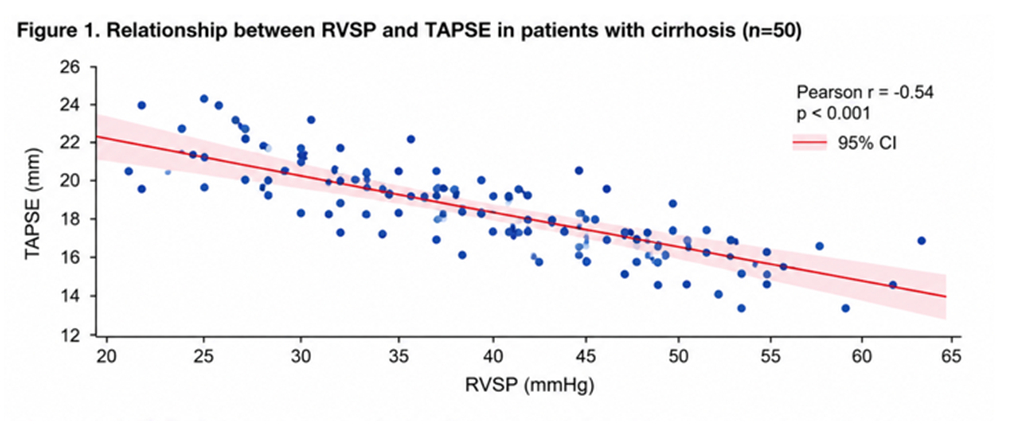
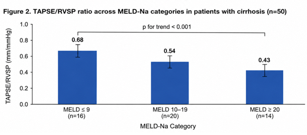
Severity-wise analysis demonstrated progressive deterioration of RV–pulmonary coupling with increasing MELD-Na categories (Table 5). RVSP increased steadily with worsening liver disease, while TAPSE showed only modest decline. Consequently, the TAPSE/RVSP ratio decreased significantly across MELD-Na strata. A scatter plot illustrating the inverse relationship between RVSP and TAPSE is shown in Figure 1, and coupling deterioration across severity categories is depicted in Figure 2.
Table 1. Baseline demographic and clinical characteristics
|
Parameter |
Cirrhosis (n=50) |
Control (n=50) |
p value |
|
Age (years) |
49.1 ± 9.0 |
48.2 ± 8.7 |
0.62 |
|
Male sex, n (%) |
34 (68) |
33 (66) |
0.81 |
|
Heart rate (beats/min) |
87.2 ± 11.8 |
73.4 ± 9.4 |
<0.001 |
|
Mean arterial pressure (mmHg) |
78.6 ± 10.4 |
92.1 ± 9.6 |
<0.001 |
|
Oxygen saturation (%) |
96.2 ± 2.1 |
97.4 ± 1.6 |
0.01 |
|
MELD-Na score |
16.8 ± 7.4 |
— |
— |
Table 2. Echocardiographic estimates of pulmonary hemodynamics
|
Parameter |
Cirrhosis |
Controls |
p value |
|
TR velocity (m/s) |
2.7 ± 0.4 |
2.3 ± 0.3 |
<0.001 |
|
Estimated RVSP (mmHg) |
38.6 ± 8.2 |
28.4 ± 5.6 |
<0.001 |
|
IVC diameter (mm) |
20.1 ± 3.8 |
15.6 ± 2.9 |
<0.001 |
|
Estimated RAP (mmHg) |
8.2 ± 2.6 |
5.1 ± 1.8 |
<0.001 |
Table 3. Right ventricular systolic function parameters
|
Parameter |
Cirrhosis |
Controls |
p value |
|
TAPSE (mm) |
20.9 ± 2.6 |
22.1 ± 2.4 |
0.02 |
|
RV fractional area change |
0.54 ± 0.07 |
0.57 ± 0.06 |
0.04 |
|
RV free wall strain (%) |
−21.3 ± 4.7 |
−23.8 ± 3.1 |
0.01 |
Table 4. Right ventricular–pulmonary vascular coupling indices
|
Parameter |
Cirrhosis |
Controls |
p value |
|
TAPSE/RVSP (mm/mmHg) |
0.55 ± 0.14 |
0.79 ± 0.18 |
<0.001 |
Table 5. Severity-wise analysis of RV–pulmonary coupling
|
MELD-Na Category |
RVSP (mmHg) |
TAPSE (mm) |
TAPSE/RVSP |
|
≤9 (n=22) |
32.4 ± 6.1 |
22.1 ± 2.4 |
0.68 ± 0.12 |
|
10–19 (n=12) |
38.7 ± 6.8 |
21.0 ± 2.3 |
0.54 ± 0.10 |
|
≥20 (n=16) |
44.9 ± 7.3 |
19.6 ± 2.7 |
0.43 ± 0.09 |
p for trend <0.01
DISCUSSION:
The present study demonstrates that patients with cirrhosis exhibit early right ventricular–pulmonary vascular uncoupling detectable by echocardiography, even in the absence of overt pulmonary hypertension or advanced right ventricular systolic failure. While conventional indices such as TAPSE and RV fractional area change remained largely within normal limits, coupling indices integrating right ventricular performance with pulmonary afterload revealed a distinct impairment that worsened with increasing liver disease severity.
The pulmonary circulation in cirrhosis is subjected to a unique hemodynamic milieu characterized by chronically elevated cardiac output, reduced pulmonary vascular resistance in early disease, and progressive endothelial dysfunction with advancing cirrhosis. Experimental and clinical studies have demonstrated increased pulmonary blood flow and shear stress in cirrhosis, leading to altered nitric oxide signaling, endothelin imbalance, and vascular remodeling over time [1–3]. These changes may not immediately translate into diagnostic thresholds for portopulmonary hypertension but nonetheless increase right ventricular afterload in a gradual and insidious manner.
Right ventricular–pulmonary vascular coupling reflects the ability of the right ventricle to augment contractile performance in proportion to increasing afterload. When coupling is preserved, right ventricular systolic function adapts efficiently, maintaining stroke volume and cardiac output. In contrast, uncoupling occurs when afterload rises disproportionately to contractile reserve, resulting in reduced efficiency and increased vulnerability to decompensation [4,5]. In pulmonary arterial hypertension and left heart disease, reduced TAPSE/ RVSP ratio has consistently been associated with worse functional capacity and adverse outcomes [6–8].
In the present cohort, cirrhotic patients demonstrated a clear dissociation between right ventricular systolic indices and pulmonary load. Although TAPSE remained preserved, RVSP was significantly elevated compared with controls, resulting in a marked reduction in TAPSE/RVSP ratio. This pattern suggests that the right ventricle is functioning closer to the limits of its adaptive capacity, even before overt systolic dysfunction becomes apparent. The observation that RV longitudinal strain was modestly impaired further supports the presence of early myocardial involvement that may not be captured by excursion-based indices alone.
A particularly important finding of this study is the progressive deterioration of RV–pulmonary coupling with increasing MELD-Na scores. As liver disease severity advanced, pulmonary pressures increased steadily, while right ventricular systolic performance showed only limited compensatory augmentation. Consequently, coupling efficiency declined in a stepwise fashion across MELD-Na categories. This severity-dependent relationship underscores the close interplay between hepatic dysfunction and cardiopulmonary stress and suggests that RV–pulmonary uncoupling represents a continuum rather than a binary phenomenon.
From a clinical perspective, these findings have important implications. Patients with cirrhosis frequently experience acute hemodynamic challenges during infections, large-volume paracentesis, transjugular intrahepatic portosystemic shunt placement, and liver transplantation. Subclinical RV–pulmonary uncoupling may predispose such patients to acute right ventricular failure under stress, even in the absence of established portopulmonary hypertension.
Incorporating RV–pulmonary coupling assessment into routine echocardiographic evaluation may therefore enhance risk stratification and inform peri-procedural decision-making.
This study deliberately focuses on functional phenotyping rather than diagnostic classification. While right heart catheterization remains the gold standard for diagnosing pulmonary hypertension, it is neither practical nor indicated for routine screening of all cirrhotic patients. Echocardiographic coupling indices offer a pragmatic, non-invasive approach to identify patients in whom cardiopulmonary reserve may already be compromised.
This study demonstrates that cirrhosis is associated with early right ventricular–pulmonary vascular uncoupling detectable by echocardiography, even in the absence of overt pulmonary hypertension or advanced right ventricular systolic failure. The principal finding is a significant reduction in the TAPSE/RVSP ratio among cirrhotic patients, reflecting impaired RV–pulmonary coupling despite largely preserved conventional RV systolic indices.
The pulmonary circulation in cirrhosis is exposed to chronically increased flow, altered vasoactive mediator balance, and endothelial dysfunction [11,12]. These changes may lead to a gradual increase in pulmonary vascular load long before diagnostic thresholds for portopulmonary hypertension are reached. Our findings suggest that the right ventricle initially maintains systolic performance, as reflected by preserved TAPSE, but at the cost of declining coupling efficiency as pulmonary pressures rise.
The progressive decline in coupling indices with increasing MELD-Na scores highlights the close interaction between hepatic disease severity and cardiopulmonary stress. This observation is clinically relevant, as patients with advanced cirrhosis frequently encounter hemodynamic challenges during infections, large-volume paracentesis, TIPS insertion, and liver transplantation.
Subclinical RV–pulmonary uncoupling may predispose these patients to acute right ventricular decompensation during such stressors.
Importantly, this study shifts the focus from identifying established portopulmonary hypertension to recognizing an earlier, potentially reversible cardiopulmonary phenotype. Echocardiographic assessment of RV–pulmonary coupling may therefore serve as a valuable adjunct in the cardiovascular evaluation of cirrhotic patients, particularly those being considered for invasive procedures or transplantation.
Limitations
This study is limited by its cross-sectional design and reliance on echocardiographic surrogates rather than invasive hemodynamic measurements. However, the aim was functional phenotyping rather than definitive diagnosis of pulmonary hypertension. Prospective studies incorporating right heart catheterization and outcome data are warranted.
CONCLUSION:
Patients with cirrhosis exhibit early right ventricular–pulmonary vascular uncoupling characterized by rising pulmonary pressures and declining coupling efficiency despite preserved right ventricular systolic function. This uncoupling worsens with advancing liver disease severity and precedes overt pulmonary hypertension. Non-invasive echocardiographic coupling indices may provide clinically meaningful insight into cardiopulmonary vulnerability in cirrhosis.
REFERENCES:
1. Mukherjee M, Rudski LG, Addetia K, Afilalo J, D'Alto M, Freed BH, et al. Guidelines for the Echocardiographic Assessment of the Right Heart in Adults and Special Considerations in Pulmonary Hypertension: Recommendations from the American Society of Echocardiography. J Am Soc Echocardiogr. 2025;38(3):141-186. doi:10.1016/j.echo.2025.01.006.
2. Humbert M, Kovacs G, Hoeper MM, Badagliacca R, Berger RMF, Brida M, et al. 2022 ESC/ERS Guidelines for the diagnosis and treatment of pulmonary hypertension. Eur Heart J. 2022;43:3618-3731.
3. Krowka MJ, Fallon MB, Kawut SM, et al. International Liver Transplant Society Practice Guidelines: Diagnosis and Management of Portopulmonary Hypertension. Liver Transpl. 2016.
4. Simonetto DA, Shah VH, Kamath PS. Portopulmonary hypertension and hepatopulmonary syndrome. Lancet. 2019.
5. Tello K, Richter MJ, Axmann J, et al. More on TAPSE/PASP ratio as a non-invasive estimate of right ventricular–arterial coupling in pulmonary hypertension. Am J Respir Crit Care Med. 2018.
6. Guazzi M, Bandera F, Pelissero G, et al. Tricuspid annular plane systolic excursion and pulmonary arterial systolic pressure relationship in heart failure. J Am Coll Cardiol. 2013.
7. Lang RM, Badano LP, Mor-Avi V, et al. Recommendations for Cardiac Chamber Quantification by Echocardiography in Adults: An Update from the American Society of Echocardiography and the European Association of Cardiovascular Imaging. J Am Soc Echocardiogr. 2015;28:1-39.e14.
8. Rudski LG, Lai WW, Afilalo J, et al. Guidelines for the Echocardiographic Assessment of the Right Heart in Adults. J Am Soc Echocardiogr. 2010;23:685-713.
9. Møller S, Henriksen JH. Cardiovascular complications of cirrhosis. Gut. 2008;57:268-278.
10. Bernardi M, Moreau R, Angeli P, Schnabl B, Arroyo V. Mechanisms of decompensation and organ failure in cirrhosis. J Hepatol. 2015;63:743-752.
11. European Association for the Study of the Liver (EASL). EASL Clinical Practice Guidelines on the management of patients with decompensated cirrhosis. J Hepatol. 2018.
12. Voelkel NF, Quaife RA, Leinwand LA, et al. Right ventricular function and failure: report of a National Heart, Lung, and Blood Institute working group. Circulation. 2006;114:1883-1891.