Echocardiographic Evaluation Among Patients With Chronic Kidney Disease With And Without Haemodialysis: A Comparative Study.
- Arijit Saha , Senior Resident Cardiology, MBBS, MD General Medicine, DM Cardiology(N.R.S), Department of Cardiology, IPGMER and SSKM Hospital, Kolkata, West Bengal 700020.
Article Information:
Abstract:
Introduction: Chronic kidney disease (CKD) is a major contributor to global morbidity and mortality. Cardiovascular disease is the leading cause of death in CKD, particularly in patients with advanced renal dysfunction and those receiving maintenance haemodialysis. Haemodialysis is life-sustaining but may be associated with repeated volume shifts, pressure overload, inflammation, endothelial dysfunction and myocardial stress, resulting in adverse cardiac remodelling. Aims and objective: To evaluate echocardiographic changes in cardiac morphology and function among CKD patients receiving haemodialysis and to compare them with CKD patients not receiving haemodialysis in an Indian tertiary-care population. Materials and methods: This was a comparative, analytical, prospective, cross-sectional, single-centre, hospital-based study conducted from February 2023 to July 2024 in the Department of Cardiology, N.R.S. Medical College and Hospital. A total of 195 patients were included: 117 patients on maintenance haemodialysis and 78 CKD patients not on haemodialysis. Clinical profile and conventional echocardiographic parameters, including left-sided chamber dimensions, left ventricular mass, systolic function, diastolic function and global longitudinal strain (GLS), were analysed. Results: The haemodialysis group had a lower mean age than the non-haemodialysis group (46.62 vs 57.46 years), while male predominance was similar in both groups. Hypertension was more frequent among haemodialysis patients (71.79% vs 50.00%, p=0.02). Haemodialysis patients had significantly higher prevalence of left atrial enlargement, left ventricular dilatation, increased interventricular septal and posterior wall thickness, higher EPSS, lower LVEF and reduced fractional shortening. LV mass >=200 g was more common in haemodialysis patients (53.85% vs 7.69%). Advanced diastolic dysfunction, mitral regurgitation, pericardial effusion and pulmonary hypertension were also more frequent in the haemodialysis group. Among patients with preserved LVEF, mean GLS was significantly lower in haemodialysis patients (18.29% vs 19.64%, p=0.0014), suggesting subclinical myocardial dysfunction. Conclusion: Haemodialysis is associated with significant adverse cardiac remodelling characterized by left ventricular systolic and diastolic dysfunction, increased left ventricular mass, pulmonary hypertension, valvular abnormalities, infective endocarditis and impaired myocardial deformation as assessed by GLS. Routine echocardiographic surveillance, including strain imaging where available, may improve early cardiovascular risk stratification in patients with advanced CKD.
Keywords:
Article :
INTRODUCTION:
Chronic kidney disease (CKD) is a major public health problem and an important contributor to global morbidity and mortality. The Global Burden of Disease collaboration has identified CKD as a growing cause of disease burden worldwide, with increases in both prevalence and mortality over recent decades.1 In India, deaths attributable to renal failure have also increased, reflecting the rising burden of diabetes, hypertension and limited early detection of kidney disease.2
CKD is frequently asymptomatic in its early stages and is often detected incidentally during evaluation for other medical conditions or during routine screening. Many patients present late, when complications have already developed or when renal replacement therapy is required.3
Diabetes mellitus and hypertension remain the dominant contributors to CKD in both Western and Indian populations.4,5 The increasing prevalence of diabetes and hypertension in India is therefore expected to further increase the burden of CKD and associated cardiovascular complications.6,7
CKD is defined by the presence of kidney damage, manifested by abnormal albumin excretion or reduced kidney function, quantified by measured or estimated glomerular filtration rate (eGFR), persisting for more than three months.8-10 Severity is commonly stratified according to eGFR categories.
Stage 1 is defined by eGFR >=90 mL/min/1.73 m2 with evidence of kidney damage; stage 2 by eGFR 60-89 mL/min/1.73 m2; stage 3 by eGFR 30-59 mL/min/1.73 m2; stage 4 by eGFR 15-29 mL/min/1.73 m2; and stage 5 by eGFR <15 mL/min/1.73 m2 or end-stage renal disease.11,12
Cardiovascular disease is the principal cause of death in patients with CKD and end-stage renal disease. Traditional risk factors such as hypertension, diabetes, dyslipidaemia and older age interact with CKD-specific factors including anaemia, uremic toxins, inflammation, oxidative stress, volume overload, mineral-bone disorder and endothelial dysfunction.
These factors result in left ventricular hypertrophy, myocardial fibrosis, chamber dilatation, systolic and diastolic dysfunction, arrhythmias, valvular calcification, pulmonary hypertension and heart failure.13-20
Haemodialysis improves survival in advanced CKD but is associated with repeated haemodynamic stress. Intermittent ultrafiltration, intradialytic hypotension, rapid electrolyte shifts, arteriovenous fistula-related high-output physiology and catheter-related infections may all contribute to cardiac structural and functional abnormalities. Echocardiography remains the most accessible tool for evaluating cardiovascular involvement in CKD.
Conventional echocardiography provides assessment of chamber dimensions, wall thickness, ventricular systolic function, diastolic function, valvular disease, pericardial effusion and pulmonary pressures. In addition, global longitudinal strain (GLS) can detect subclinical myocardial dysfunction even when left ventricular ejection fraction (LVEF) is preserved.
The present study was undertaken to compare echocardiographic morphology and function in CKD patients undergoing haemodialysis with CKD patients not receiving haemodialysis. The objective was to identify the burden and pattern of cardiac abnormalities in this high-risk Indian population and to highlight the value of echocardiographic surveillance for early cardiovascular risk stratification.
MATERIALS AND METHODS:
Study design: This was a comparative, analytical, prospective, cross-sectional, single-centre, hospital-based study.
Place of study: The study was conducted at N.R.S. Medical College and Hospital. Study period: February 2023 to July 2024, over a period of 18 months.
Sample size: A total of 195 patients were enrolled in the study.
Inclusion criteria
• Patients aged 18 years or older with CKD stage 5 on haemodialysis and CKD patients not on haemodialysis attending the NRSMCH haemodialysis unit, inpatient services or outpatient services, defined according to KDIGO criteria, were included.
• Patients receiving maintenance haemodialysis three sessions per week, 3.5-4 hours per session, using polysulfone dialyser and bicarbonate dialysate, with all types of vascular access, were included in the haemodialysis group.
Exclusion criteria
• Patients not giving consent and patients aged below 18 years were excluded.
• Patients with major systemic illnesses including severe liver insufficiency, untreated thyroid dysfunction and extreme obesity (BMI >45 kg/m2) were excluded.
• Patients with significant pre-existing cardiovascular disease including prior heart failure, cardiomyopathy, myocardial infarction with PCI/CABG, congenital heart disease, rheumatic heart disease, prosthetic valves or previous cardiac surgery were excluded.
• Patients with active alcohol intake, smoking history, recent infection within the last three weeks or advanced heart failure requiring device support were excluded.
Study variables
The study variables included age, residence, duration of disease, compliance to therapy, haemodialysis status, type of vascular access and echocardiographic measures of cardiac structure and function.
Advanced age, longer disease duration, poor therapeutic compliance and haemodialysis exposure were considered clinically relevant variables likely to influence renal and cardiovascular dysfunction.
Echocardiographic assessment
All patients underwent transthoracic echocardiographic assessment. Conventional two-dimensional, M-mode and Doppler parameters were recorded.
Measurements included aortic diameter, aortic valve excursion, left atrial dimension, left ventricular internal diameter in diastole (LVIDd), left ventricular internal diameter in systole (LVIDs), interventricular septal thickness in diastole (IVSd), left ventricular posterior wall thickness in diastole (LVPWd), right ventricular basal diameter, E-point septal separation (EPSS), LVEF, fractional shortening, left ventricular mass, diastolic function category, mitral regurgitation, pericardial effusion and estimated pulmonary artery systolic pressure (ePASP). In patients with preserved LVEF, GLS was assessed to identify subclinical systolic dysfunction.
Statistical analysis
Data were initially entered into Microsoft Excel and analysed using SPSS version 27.0 (SPSS Inc., Chicago, IL, USA) and GraphPad Prism version 5. Numerical variables were summarised as means and standard deviations, while categorical variables were described as counts and percentages.
Two-sample t-tests were used to compare independent groups. Paired t-tests were used where appropriate for paired data. Chi-square tests, including Fisher exact test for small sample sizes, were used for comparison of categorical variables. A p-value <=0.05 was considered statistically significant.
RESULTS:
Table 1. Baseline demographic and clinical profile.
|
Variable |
HD (n=117) |
NHD (n=78) |
p value |
|
Mean age, years |
46.62 |
57.46 |
- |
|
Median age, years |
51 |
59.5 |
- |
|
Male sex |
78 (66.67%) |
51 (65.38%) |
- |
|
Female sex |
39 (33.33%) |
27 (34.62%) |
- |
|
Hypertension |
84 (71.79%) |
39 (50.00%) |
0.02 |
Table 2. Conventional echocardiographic measurements and abnormalities by group.
|
Parameter |
HD |
NHD |
p value |
|
Aortic diameter, mean |
30.89 mm |
30.63 mm |
0.61 |
|
Aortic valve excursion, mean |
17.78 mm |
17.06 mm |
NS |
|
LA >=40 mm |
33 (28.21%) |
9 (11.54%) |
0.0055 |
|
LVIDd >=50 mm |
63 (53.85%) |
15 (19.23%) |
<0.0001 |
|
LVIDs >40 mm |
21 (17.95%) |
6 (7.69%) |
0.04 |
|
IVSd >=12 mm |
54 (46.15%) |
6 (7.69%) |
<0.0001 |
|
LVPWd >=12 mm |
27 (23.08%) |
9 (11.54%) |
0.04 |
|
RV base >=30 mm |
30 (25.64%) |
12 (15.38%) |
NS |
|
EPSS, mean |
8.84 mm |
6.32 mm |
<0.0001 |
|
LVEF, mean |
56.62% |
62.88% |
<0.0001 |
|
LVEF <50% |
30 (25.64%) |
9 (11.54%) |
- |
|
Fractional shortening, mean |
30.59% |
34.49% |
<0.0001 |
Table 3. Left ventricular mass category distribution by group.
|
LV mass category |
HD (n=117) |
NHD (n=78) |
|
<100 g |
3 (2.56%) |
9 (11.54%) |
|
100-199 g |
51 (43.59%) |
63 (80.77%) |
|
>=200 g |
63 (53.85%) |
6 (7.69%) |
Table 4. Diastolic function category by group.
|
Diastolic function category |
HD (n=117) |
NHD (n=78) |
|
Normal |
6 (5.13%) |
18 (23.08%) |
|
Grade I |
39 (33.33%) |
39 (50.00%) |
|
Grade II |
45 (38.46%) |
9 (11.54%) |
|
Grade III |
27 (23.08%) |
12 (15.38%) |
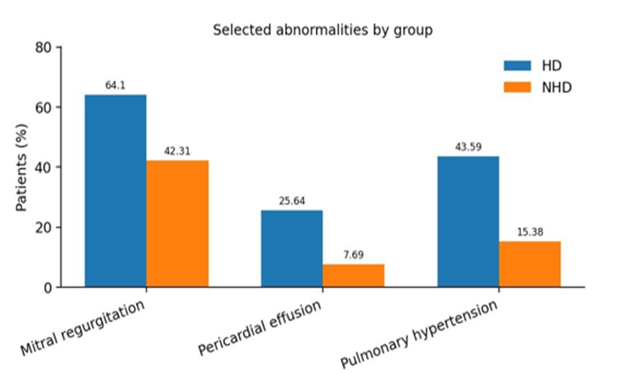
Figure 1. Prevalence of mitral regurgitation, pericardial effusion and pulmonary hypertension in haemodialysis and non-haemodialysis CKD patients.
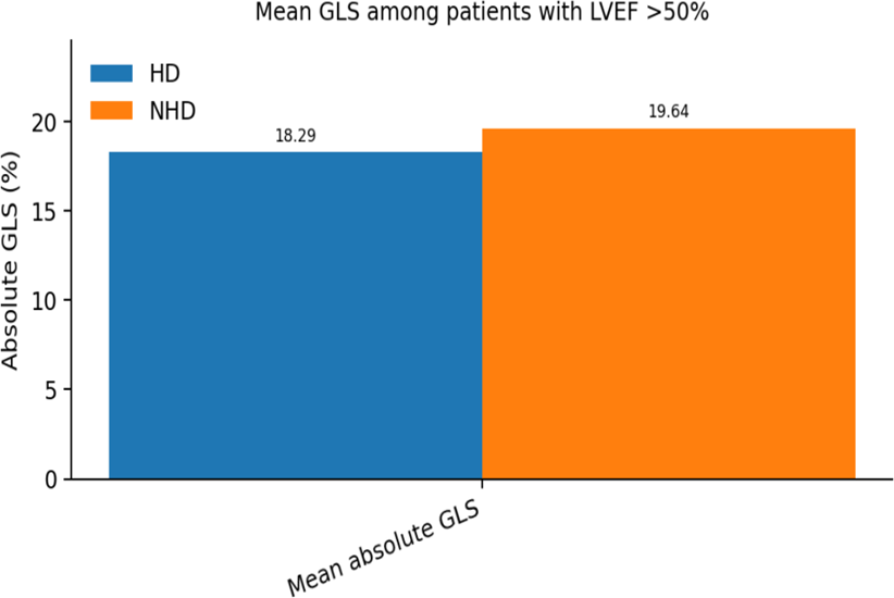
Figure 2. Comparison of global longitudinal strain in CKD patients with preserved left ventricular ejection fraction.
The study included 195 patients, of whom 117 were in the haemodialysis (HD) group and 78 were in the non-haemodialysis (NHD) group. The mean age was lower in the HD group than in the NHD group (46.62 vs 57.46 years), while the median age was 51 years and 59.5 years, respectively. Male predominance was similar in both groups, with 78 males (66.67%) in the HD group and 51 males (65.38%) in the NHD group. Hypertension was significantly more frequent in the HD group (84 patients, 71.79%) compared with the NHD group (39 patients, 50.00%; p=0.02) (Table 1).
Conventional echocardiographic parameters demonstrated significant structural and functional differences between the two groups. Mean aortic diameter was comparable in the HD and NHD groups (30.89 mm vs 30.63 mm; p=0.61), and mean aortic valve excursion also showed no significant difference (17.78 mm vs 17.06 mm). Left atrial enlargement (LA >=40 mm) was significantly more common in the HD group (28.21% vs 11.54%; p=0.0055). Left ventricular internal dimensions were also significantly greater in the HD group, with LVIDd >=50 mm present in 53.85% of HD patients compared with 19.23% of NHD patients (p<0.0001), and LVIDs >40 mm present in 17.95% versus 7.69% (p=0.04).
Markers of left ventricular hypertrophy were more frequent among haemodialysis patients. IVSd
>=12 mm was observed in 46.15% of HD patients compared with 7.69% of NHD patients (p<0.0001), while LVPWd >=12 mm was present in 23.08% versus 11.54% (p=0.04). Right ventricular basal diameter >=30 mm was observed in 25.64% of HD patients and 15.38% of NHD patients. EPSS was significantly higher in the HD group (8.84 mm vs 6.32 mm; p<0.0001). Mean LVEF was significantly lower among HD patients (56.62% vs 62.88%; p<0.0001), and reduced LVEF <50% was present in 25.64% of HD patients compared with 11.54% of NHD patients. Fractional shortening was also significantly lower in the HD group (30.59% vs 34.49%; p<0.0001) (Table 2).
The distribution of left ventricular mass showed a marked difference between groups. In the HD group, only 2.56% had LV mass <100 g, 43.59% had LV mass between 100 and 199 g, and 53.85% had LV mass >=200 g. In contrast, among NHD patients, 11.54% had LV mass <100 g, 80.77% had LV mass between 100 and 199 g, and only 7.69% had LV mass >=200 g. These findings indicate a substantially higher burden of increased LV mass among haemodialysis patients (Table 3).
Diastolic function analysis demonstrated more advanced dysfunction in the HD group. Normal diastolic function was present in only 5.13% of HD patients compared with 23.08% of NHD patients. Grade I diastolic dysfunction was observed in 33.33% of HD patients and 50.00% of NHD patients. Grade II and Grade III diastolic dysfunction were more frequent among HD patients, occurring in 38.46% and 23.08%, respectively, compared with 11.54% and 15.38% in the NHD group. These findings suggest a higher prevalence of advanced diastolic dysfunction among haemodialysis patients (Table 4).
Mitral regurgitation, pulmonary hypertension and pericardial effusion were more frequent in the HD group. Mitral regurgitation was present in 64.10% of HD patients and 42.31% of NHD patients. Pericardial effusion was observed in 25.64% of HD patients compared with 7.69% of NHD patients. Pulmonary hypertension, defined by ePASP >30 mmHg, was present in 43.59% of HD patients compared with 15.38% of NHD patients (Figure 1).
Infective vegetations were observed exclusively among haemodialysis patients in the available results. Nine haemodialysis patients had vegetations; three were associated with femoral venous dialysis catheters and six with internal jugular venous catheters. Thus, two-thirds of vegetations were associated with internal jugular venous catheters. Among patients with preserved LVEF, GLS was significantly worse among haemodialysis patients. Mean GLS was 18.29% in the HD group compared with 19.64% in the NHD group (p=0.0014), indicating subtle systolic impairment not captured by ejection fraction alone (Figure 2).
DISCUSSION:
The present study demonstrated a significantly higher burden of structural and functional cardiac abnormalities among CKD patients undergoing maintenance haemodialysis compared with CKD patients not receiving haemodialysis. Haemodialysis patients exhibited greater left ventricular hypertrophy, chamber dilatation, increased LV mass, impaired systolic and diastolic function, pulmonary hypertension, mitral regurgitation, pericardial effusion and reduced GLS. These findings highlight the profound cardiovascular impact of advanced renal dysfunction and dialysis-related haemodynamic stress.
Cardiovascular disease remains the leading cause of morbidity and mortality in CKD. The pathophysiology is multifactorial. Traditional cardiovascular risk factors such as hypertension, diabetes and dyslipidaemia coexist with non-traditional CKD-related factors including anaemia, uremic toxins, chronic inflammation, oxidative stress, endothelial dysfunction, calcium-phosphate imbalance and volume overload. Together, these mechanisms promote myocardial fibrosis, left ventricular hypertrophy, chamber enlargement, impaired relaxation and progressive ventricular dysfunction.13,17
The high prevalence of LV hypertrophy and increased LV mass among haemodialysis patients in the present study is clinically important. LV hypertrophy in CKD develops due to chronic pressure overload from hypertension, chronic volume overload from sodium and fluid retention, anaemia-related high-output physiology and arteriovenous access-related increases in cardiac output. Over time, these changes lead to concentric remodelling, myocardial fibrosis and reduced ventricular compliance. Previous studies in patients with end-stage renal disease have similarly shown a high prevalence of echocardiographic abnormalities at the initiation of renal replacement therapy.15
Left atrial enlargement was significantly more frequent in the haemodialysis group. Left atrial size reflects chronic exposure to elevated LV filling pressures and is closely related to diastolic dysfunction. In haemodialysis patients, recurrent fluid accumulation between dialysis sessions and rapid ultrafiltration during dialysis may cause repeated fluctuations in preload, contributing to atrial stretch and remodelling. Left atrial enlargement also has prognostic relevance because it is associated with atrial arrhythmias, heart failure and adverse cardiovascular outcomes.
The lower mean LVEF, higher EPSS and reduced fractional shortening observed among haemodialysis patients suggest impaired systolic performance. Although many patients retained relatively preserved ejection fraction, conventional systolic parameters were worse in the haemodialysis group. Dialysis-induced myocardial stunning, coronary microvascular dysfunction, rapid intradialytic haemodynamic shifts, endothelial dysfunction and myocardial fibrosis may explain this observation. Heart failure-related hospitalization is common in chronic dialysis populations and is closely linked to structural cardiac abnormalities.16
A key finding of this study was the reduction in GLS among haemodialysis patients despite preserved LVEF. LVEF is load-dependent and may remain normal until significant myocardial dysfunction has already developed. GLS is more sensitive for detecting early longitudinal fibre dysfunction and subclinical systolic impairment. The lower GLS in the haemodialysis group therefore suggests occult myocardial injury even before overt systolic failure. This observation supports the incorporation of strain imaging into echocardiographic assessment of advanced CKD patients whenever available.
Diastolic dysfunction was substantially more severe among haemodialysis patients, with a higher proportion of Grade II and Grade III dysfunction. Diastolic dysfunction in CKD is largely driven by LV hypertrophy, interstitial fibrosis, vascular stiffness, hypertension and chronic volume overload. Progressive worsening of diastolic function increases left atrial pressure, predisposes to pulmonary venous hypertension and contributes to symptoms of heart failure despite preserved ejection fraction. The findings of the present study are in agreement with previous observations that declining kidney function is associated with subclinical cardiac abnormalities and progressive LV structural changes.18,19
Pulmonary hypertension was markedly more frequent in haemodialysis patients. Several mechanisms may contribute, including chronic volume overload, elevated left-sided filling pressures, endothelial dysfunction, pulmonary vascular remodelling and increased cardiac output associated with arteriovenous fistulae. In the present cohort, patients with arteriovenous fistulae had higher pulmonary artery systolic pressures, supporting the haemodynamic role of high-flow vascular access. Recognition of pulmonary hypertension is important because it may worsen exercise capacity, right ventricular function and overall prognosis.
Mitral regurgitation was more prevalent in the haemodialysis group. This may be explained by left ventricular dilatation, annular dilatation, papillary muscle displacement, mitral annular calcification and chronic volume overload. CKD-mineral bone disorder accelerates valvular and annular calcification, predisposing to valvular dysfunction. Pericardial effusion was also more frequent in haemodialysis patients and may reflect inadequate dialysis, persistent volume overload, uremic inflammation or associated comorbidity.
Infective vegetations were observed only in the haemodialysis group, with the majority associated with temporary internal jugular venous catheters. This finding is clinically relevant because catheter-dependent dialysis increases the risk of bloodstream infection and infective endocarditis. Early creation and use of permanent vascular access, meticulous catheter care and avoidance of prolonged temporary catheter dependence are important preventive strategies.
Overall, the findings of this study reinforce the concept of uremic cardiomyopathy and demonstrate that maintenance haemodialysis patients have a higher burden of cardiac structural and functional abnormalities than non-haemodialysis CKD patients. Routine echocardiographic surveillance can help identify high-risk patients with LV hypertrophy, impaired systolic or diastolic function, pulmonary hypertension, valvular abnormalities, pericardial effusion and subclinical myocardial dysfunction. These findings may guide optimisation of volume status, blood pressure control, anaemia management, dialysis prescription and vascular access planning.
The clinical implication of this study is that echocardiography should not be reserved only for symptomatic haemodialysis patients. Even in the absence of overt heart failure, significant structural remodelling and strain abnormalities may be present. Periodic echocardiographic evaluation, including GLS where feasible, may improve early cardiovascular risk stratification and facilitate timely intervention in this vulnerable population. The findings may also support closer coordination between nephrologists and cardiologists for optimisation of dry weight, antihypertensive therapy, anaemia correction, mineral metabolism control and vascular access strategy. Such integrated care may help reduce the progression of uremic cardiomyopathy and identify patients requiring closer follow-up.
LIMITATIONS
The present study is limited by its single-centre, cross-sectional design with lack of long-term follow-up and advanced imaging, which restricts generalizability and causal inference. Larger multicentre prospective studies with longitudinal data are needed to validate these findings and assess their prognostic significance.
CONCLUSION:
Patients with CKD receiving maintenance haemodialysis exhibit significantly greater structural and functional cardiac abnormalities than non-haemodialysis CKD patients. Haemodialysis is associated with increased left ventricular hypertrophy, higher LV mass, chamber dilatation, impaired systolic and diastolic function, pulmonary hypertension, valvular abnormalities, infective endocarditis, pericardial effusion and reduced GLS. These findings suggest advanced uremic cardiomyopathy and highlight the importance of routine echocardiographic surveillance, including strain imaging where available, for early cardiovascular risk stratification and optimisation of care in this high-risk population.
REFERENCES:
1. Bikbov B, Purcell CA, Levey AS, Smith M, Abdoli A, Abebe M, et al. Global, regional, and national burden of chronic kidney disease, 1990-2017: a systematic analysis for the Global Burden of Disease Study 2017. Lancet. 2020;395(10225):709-733.
2. Dare AJ, Fu SH, Patra J, Rodriguez PS, Thakur JS, Jha P. Renal failure deaths and their risk factors in India 2001-13: nationally representative estimates from the Million Death Study. Lancet Glob Health. 2017;5(1):e89-e95.
3. Jha V, Ur-Rashid H, Agarwal SK, Akhtar SF, Kafle RK, Sheriff R. The state of nephrology in South Asia. Kidney Int. 2019;95(1):31-37.
4. Snyder S, Pendergraph B. Detection and evaluation of chronic kidney disease. Am Fam Physician. 2005;72:1723-1732.
5. Rajapurkar MM, John GT, Kirpalani AL, Abraham G, Agarwal SK, Almeida AF, et al. What do we know about chronic kidney disease in India: first report of the Indian CKD Registry. BMC Nephrol. 2012;13:10.
6. Raman R, Ganesan S, Pal SS, Kulothungan V, Sharma T. Prevalence and risk factors for diabetic retinopathy in rural India: Sankara Nethralaya Diabetic Retinopathy Epidemiology and Molecular Genetic Study III (SN-DREAMS III), report no. 2. BMJ Open Diabetes Res Care. 2014;2(1):e000005.
7. Anjana RM, Pradeepa R, Deepa M, Datta M, Sudha V, Unnikrishnan R, et al. Prevalence of diabetes and prediabetes in urban and rural India: phase I results of the Indian Council of Medical Research-INdia DIABetes (ICMR-INDIAB) study. Diabetologia. 2011;54(12):3022-3027.
8. Coresh J, Selvin E, Stevens LA, Manzi J, Kusek JW, Eggers P, et al. Prevalence of chronic kidney disease in the United States. JAMA. 2007;298(17):2038-2047.
9. KDOQI Clinical Practice Guidelines and Clinical Practice Recommendations for Anemia in Chronic Kidney Disease. Am J Kidney Dis. 2006;47(5 Suppl 3):S11-S145.
10. Levey AS, Eckardt KU, Tsukamoto Y, Levin A, Coresh J, Rossert J, et al. Definition and classification of chronic kidney disease: a position statement from Kidney Disease: Improving Global Outcomes (KDIGO). Kidney Int. 2005;67(6):2089-2100.
11. Coresh J, Astor BC, Greene T, Eknoyan G, Levey AS. Prevalence of chronic kidney disease and decreased kidney function in the adult US population: Third National Health and Nutrition Examination Survey. Am J Kidney Dis. 2003;41(1):1-12.
12. Stevens PE. Evaluation and management of chronic kidney disease: synopsis of the Kidney Disease: Improving Global Outcomes 2012 clinical practice guideline. Ann Intern Med. 2013;158(11):825-830.
13. Muntner P, He J, Astor BC, Folsom AR, Coresh J. Traditional and nontraditional risk factors predict coronary heart disease in chronic kidney disease. J Am Soc Nephrol. 2005;16(2):529-538.
14. Port FK, Hulbert-Shearon TE, Wolfe RA, Bloembergen WE, Golper TA, Agodoa LY, et al. Predialysis blood pressure and mortality risk in a national sample of maintenance hemodialysis patients. Am J Kidney Dis. 1999;33(3):507-517.
15. Foley RN, Parfrey PS, Harnett JD, Kent GM, Martin CJ, Murray DC, et al. Clinical and echocardiographic disease in patients starting end-stage renal disease therapy. Kidney Int. 1995;47(1):186-192.
16. Trespalacios FC, Taylor AJ, Agodoa LY, Bakris GL, Abbott KC. Heart failure as a cause for hospitalization in chronic dialysis patients. Am J Kidney Dis. 2003;41(6):1267-1277.
17. Franczyk-Skora B, Gluba A, Olszewski R, Banach M, Rysz J. Heart function disturbances in chronic kidney disease - echocardiographic indices. Arch Med Sci. 2014;10(6):1109-1116.
18. Park M, Hsu CY, Li Y, Mishra RK, Keane M, Rosas SE, et al. Associations between kidney function and subclinical cardiac abnormalities in CKD. J Am Soc Nephrol. 2012;23(10):1725-1734.
19. Matsushita K, Kwak L, Sang Y, Ballew SH, Skali H, Shah AM, et al. Kidney disease measures and left ventricular structure and function: the Atherosclerosis Risk in Communities Study. J Am Heart Assoc. 2017;6(9):e006259.
20. Jovanovich A, Ix JH, Gottdiener J, McFann K, Katz R, Kestenbaum B, et al. Fibroblast growth factor 23, left ventricular mass, and left ventricular hypertrophy in community-dwelling older adults. Atherosclerosis. 2013;231(1):114-119.