Immunohistochemical Expression of p53 in Breast Carcinoma in Women from Khyber Pakhtunkhwa, Pakistan: A Cross-Sectional Study.
- Sidra Mashal , Lady Reading Hospital, Peshawar, Khyber Pakhtunkhwa, Pakistan.
- Umer , Lady Reading Hospital, Peshawar, Khyber Pakhtunkhwa, Pakistan.
- Aiman Ajmeer , Peshawar Medical College, Riphah International University, Peshawar Campus, Peshawar, Khyber Pakhtunkhwa, Pakistan.
- Nazli Gul , Lady Reading Hospital, Peshawar, Khyber Pakhtunkhwa, Pakistan.
- Khalid Javed , Khyber Girls Medical College, Peshawar, Khyber Pakhtunkhwa, Pakistan.
- Fozia Rauf , Peshawar Medical College, Riphah International University, Peshawar Campus, Peshawar, Khyber Pakhtunkhwa, Pakistan.
- Abdal Ahmad , Peshawar Medical College, Riphah International University, Peshawar Campus, Peshawar, Khyber Pakhtunkhwa, Pakistan.
Article Information:
Abstract:
Background: Aberrant p53 immunohistochemical expression may reflect aggressive breast carcinoma biology, but data from Khyber Pakhtunkhwa, Pakistan, are limited. Objective: To determine the frequency of p53 expression patterns and their associations with clinicopathological variables in breast carcinoma. Methods: This analytical cross-sectional study included 70 formalin-fixed, paraffin-embedded breast carcinoma specimens archived at a tertiary care histopathology department between January and June 2023. Haematoxylin-and-eosin sections were reviewed for histological type, grade, lymph node status, margin status and stage. Immunohistochemistry was performed for oestrogen receptor, progesterone receptor, HER2 and p53. p53 staining was classified as negative, wild-type/low expression or mutant-type/aberrant. Associations were assessed using chi-square or Fisher's exact tests, with p<0.05 considered significant. Results: The mean age was 45±10 years, and 52.9% of patients were younger than 50 years. Invasive ductal carcinoma, grade 2 tumours, stage III disease and luminal A subtype predominated. Mutant-type/aberrant p53 expression occurred in 49 cases (70.0%). p53 expression was associated with age group (p=0.003), molecular subtype (p=0.001), surgery type (p<0.001), tumour grade (p=0.008) and lymph node category (p=0.001), but not deep-margin involvement (p=0.177). Conclusions: Aberrant p53 immunohistochemical expression was frequent and associated with several clinicopathological variables. These findings are associative and require validation using molecular TP53 testing and outcome-based studies.
Keywords:
Article :
INTRODUCTION:
Breast carcinoma remains one of the most frequently diagnosed malignancies among women and continues to be a major contributor to cancer-related morbidity and mortality worldwide. Recent GLOBOCAN 2022 estimates reported approximately 2.3 million new female breast cancer cases globally, confirming breast cancer as one of the leading cancers affecting women across both developed and developing regions [1]. In Pakistan, breast cancer represents a major public health challenge because of its high disease burden, delayed diagnosis, sociocultural barriers, variable awareness, limited access to organized screening, and incomplete regional cancer registration [2-4]. These factors may contribute to the high proportion of locally advanced disease encountered in tertiary care hospitals, particularly in resource-limited settings.
The prognosis of breast carcinoma is determined by a combination of anatomical, histopathological, and biological factors. Tumour size, axillary lymph node status, histological type, histological grade, lymphovascular invasion, margin status, TNM stage, estrogen receptor (ER), progesterone receptor (PR), human epidermal growth factor receptor 2 (HER2), Ki-67 proliferative index, and molecular subtype are routinely used for prognostic assessment and treatment planning. Conventional prognostic tools, including the Nottingham Prognostic Index, remain clinically useful because they integrate tumour size, lymph node involvement, and histological grade; however, these clinicopathological parameters alone do not fully explain the biological heterogeneity and variable clinical behaviour of breast carcinoma [5,6].
Molecular classification has substantially improved the understanding of breast cancer heterogeneity. Gene-expression profiling initially identified biologically distinct intrinsic subtypes, including luminal A, luminal B, HER2-enriched, and basal-like tumours [7,8]. In routine pathology practice, particularly in low- and middle-income countries, immunohistochemical surrogates using ER, PR, HER2, and Ki-67 are commonly used to approximate these molecular groups [9]. This classification has direct clinical relevance because luminal tumours are generally more endocrine-responsive, whereas HER2-enriched and triple-negative/basal-like tumours are more commonly associated with higher grade, aggressive behaviour, early recurrence, and specific systemic treatment requirements [10].
TP53 is one of the most frequently altered tumour-suppressor genes in human malignancy, and its encoded p53 protein plays a central role in maintaining genomic integrity. Under cellular stress, p53 regulates cell-cycle arrest, DNA repair, apoptosis, senescence, metabolism, and other anti-tumour responses [11]. Loss of normal p53 function or acquisition of mutant p53 gain-of-function activity may promote genomic instability, tumour progression, metastatic potential, and treatment resistance. In breast carcinoma, TP53 alterations are not uniformly distributed across molecular subtypes; they are more frequently reported in triple-negative and HER2-enriched tumours, although recent Asian breast cancer data suggest that the biological impact of TP53 mutation may also be subtype-specific and population-dependent [12].
Immunohistochemistry is a practical, cost-effective, and widely available method for assessing p53 protein expression in formalin-fixed paraffin-embedded breast carcinoma tissue. Aberrant p53 immunostaining patterns, including diffuse strong nuclear overexpression, complete absence/null pattern, cytoplasmic staining, or abnormal heterogeneous expression, may act as surrogate indicators of underlying TP53 alteration [13-15]. However, p53 immunohistochemistry should not be interpreted as direct molecular proof of TP53 mutation because not all TP53 mutations produce identical staining patterns, and not every abnormal staining pattern confirms a genetic mutation. Therefore, p53 IHC is best interpreted together with ER, PR, HER2, Ki-67, histological grade, tumour stage, and clinicopathological findings.
Several studies have linked p53 overexpression or aberrant p53 immunohistochemical expression with adverse clinicopathological features, including younger age at diagnosis, high tumour grade, hormone receptor negativity, HER2 positivity, triple-negative phenotype, increased proliferative activity, lymph node involvement, recurrence risk, and poor clinical outcome [12-15]. Despite the growing international evidence, local data from Khyber Pakhtunkhwa (KPK), Pakistan, remain limited. Regional evaluation is important because breast cancer biology, age distribution, stage at presentation, access to diagnostic services, and treatment patterns may differ across populations.
Considering the prognostic significance of p53 and the scarcity of regional data from Khyber Pakhtunkhwa (KPK), Pakistan, this study aimed to evaluate p53 immunohistochemical expression in breast carcinoma cases diagnosed at a tertiary care setting. The primary objective was to determine the frequency and pattern of p53 expression in breast carcinoma. The secondary objectives were to assess the association of p53 expression with molecular subtypes and histological grades of breast carcinoma. Findings from this study may contribute to regional data on breast cancer biology and may support improved clinicopathological risk stratification in this patient population.
MATERIALS AND METHODS:
Study design and setting
This was an analytical cross-sectional study conducted in a tertiary care histopathology setting in Khyber Pakhtunkhwa (KPK), Pakistan. Formalin-fixed paraffin-embedded (FFPE) tissue blocks and corresponding clinicopathological data of breast carcinoma cases were retrieved from the electronic archives of the Histopathology Division, Department of Pathology, Peshawar Medical College, Riphah International University, Peshawar Campus. Histopathological review and immunohistochemical evaluation were performed in the Department of Pathology, Peshawar Medical College.
Study duration
The study was conducted over six months from January 2023 to June 2023.
Sample size and sampling technique
The sample size was duration-based. All eligible breast carcinoma cases available in the histopathology archives during the study period and fulfilling the inclusion criteria were included. A total of 70 FFPE tissue blocks of histopathologically confirmed breast carcinoma were selected by consecutive sampling.
A formal sample size calculation was not performed because this was an archival, duration-based study and all eligible cases available during the defined study period were included. Cases were selected only if adequate representative invasive tumour tissue was available for histopathological review and immunohistochemical assessment.
Inclusion criteria
Female patients aged 25–60 years with histopathologically confirmed primary invasive breast carcinoma from the KPK region were included. Cases were eligible if the tissue was obtained through biopsy, lumpectomy/partial resection, mastectomy, or modified radical mastectomy and if adequate FFPE tumour tissue was available for H&E review and immunohistochemical staining. Cases with available essential clinicopathological data, including age, histological type, tumour grade, lymph node status where applicable, molecular marker status, and p53 immunohistochemical expression, were included.
Exclusion criteria
Female patients younger than 25 years or older than 60 years, patients from regions other than KPK, cases with inadequate or poorly preserved tissue, cases with insufficient invasive tumour component for immunohistochemical assessment, cases with incomplete essential clinicopathological data, and patients who had received chemotherapy before tissue sampling were excluded. Recurrent tumours and cases showing only in situ carcinoma without an invasive component were also excluded where identified from the pathology record.
Histopathological evaluation
Sections of 5 μm thickness were cut from selected FFPE tissue blocks and stained with hematoxylin and eosin. All H&E-stained slides were reviewed independently by two expert histopathologists who were blinded to p53 immunohistochemical status and relevant clinical outcome information. Discordant findings were resolved by joint review and consensus.
Tumour type was classified on routine histomorphology. Histological grading was performed according to the modified Bloom–Richardson/Nottingham grading system. This grading system evaluates three parameters: tubule formation, nuclear pleomorphism, and mitotic count. Each parameter is scored from 1 to 3. The total score is used to categorize tumours as Grade I/well differentiated, Grade II/moderately differentiated, or Grade III/poorly differentiated. Tumour grade, lymph node status, margin status, and other available clinicopathological variables were recorded on a structured proforma.
Lymph node status was assessed from available surgical pathology records and categorized according to the extent of nodal involvement. Margin status was recorded as negative when no tumour was identified at the resection margin and positive when tumour involvement of the margin was documented. TNM stage was recorded from available histopathological and clinical data.
Immunohistochemistry
Immunohistochemical staining was performed on selected tumour sections for estrogen receptor (ER), progesterone receptor (PR), HER2/neu, and p53 according to the departmental laboratory protocol. Appropriate positive and negative controls were used.
ER and PR expression were assessed as nuclear staining in invasive tumour cells. Cases with nuclear staining in at least 1% of invasive tumour cells were considered receptor-positive according to standard breast biomarker interpretation principles. HER2/neu was evaluated according to membranous staining intensity and completeness in invasive tumour cells. HER2 scores of 0 and 1+ were considered negative, 2+ equivocal, and 3+ positive. Equivocal HER2 cases should be reported according to institutional policy and confirmed by in situ hybridization where available.
Molecular subtype classification
Molecular subtype was assigned using immunohistochemical surrogate markers based on ER, PR, and HER2/neu status. Cases were categorized as luminal A, luminal B, HER2-positive, or triple-negative breast carcinoma according to the available receptor profile. ER-positive and/or PR-positive tumours were classified as luminal-type tumours, HER2-positive tumours were classified according to HER2 expression status, and tumours negative for ER, PR, and HER2 were categorized as triple-negative breast carcinoma. Because Ki-67 was not available in the study dataset, subtype classification was based on ER, PR, and HER2/neu status.
p53 immunohistochemical evaluation
p53 immunohistochemical expression was assessed by light microscopy in invasive tumour cells. Nuclear staining intensity and the percentage of stained tumour nuclei were evaluated. p53 scoring was performed independently by two expert histopathologists who were blinded to the clinicopathological variables being compared. Any discrepancy in interpretation was resolved by consensus.
For this study, p53 expression was categorized into three groups: negative, wild-type/low expression pattern, and mutant-type/aberrant expression pattern. Cases showing less than 10% nuclear staining were categorized as negative. Cases showing 10–30% nuclear staining were categorized as wild-type/low expression pattern. Cases showing more than 30% nuclear staining, particularly when strong and diffuse, were categorized as mutant-type/aberrant p53 expression. In this manuscript, the term “mutant-type p53 expression” refers to an immunohistochemical staining pattern suggestive of aberrant p53 expression. It should not be interpreted as molecular confirmation of TP53 gene mutation unless confirmed by molecular testing or DNA sequencing.
Data collection
Data were recorded on a structured proforma. The recorded variables included age, histological type, tumour grade, molecular subtype, type of surgical procedure, lymph node status, margin status, TNM stage, and p53 immunohistochemical category. Patient names and direct identifiers were not included in the final analysis dataset.
Statistical analysis
Data were analyzed using descriptive and inferential statistics. Categorical variables were presented as frequencies and percentages. Quantitative variables, such as age, were summarized as mean with standard deviation or as age groups where appropriate. Associations between p53 expression and categorical clinicopathological variables were assessed using the chi-square test or Fisher’s exact test where expected cell counts were small. A p-value of <0.05 was considered statistically significant. For exploratory subgroup visualization, proportions of mutant-type/aberrant p53 expression were plotted with 95% confidence intervals calculated using the Wilson method for binomial proportions.
Ethical considerations
Ethical approval was obtained from Khyber Medical University, Peshawar (approval code: DIR/KMU-AS&RB/PE/002077). Written permission was obtained from the head of the Histopathology Department, Peshawar Medical College, Riphah International University, Peshawar Campus, Peshawar, to use the study laboratory, archival tissue blocks, laboratory records, and study data. Patient confidentiality was maintained by anonymizing all records and excluding direct identifiers from the analysis dataset. Because the study used archival pathology material and retrospective laboratory data, the requirement for individual informed consent was addressed in accordance with the ethics committee decision and institutional policy.
RESULTS:
Baseline clinicopathological characteristics
A total of 70 breast carcinoma cases were included in the study. The cohort showed a slight predominance of patients aged below 50 years. Modified radical mastectomy was the most common surgical procedure. Invasive ductal carcinoma was the predominant histological type, followed by invasive lobular carcinoma. Most tumours were Grade 2, and Stage III was the most frequently recorded TNM stage. Luminal A was the most common molecular subtype. The baseline clinicopathological characteristics of the study cohort are summarized in Table 1.
Table 1. Baseline clinicopathological characteristics of the study cohort (N = 70).
|
Characteristic |
Category |
n |
% |
|
Age group |
<50 years |
37 |
52.9 |
|
≥50 years |
33 |
47.1 |
|
|
Histological type |
Invasive ductal carcinoma |
56 |
80.0 |
|
Invasive lobular carcinoma |
14 |
20.0 |
|
|
Surgery type |
Modified radical mastectomy |
47 |
67.1 |
|
Lumpectomy/partial resection |
20 |
28.6 |
|
|
Total mastectomy |
3 |
4.3 |
|
|
Tumour grade |
Grade 1 |
1 |
1.4 |
|
Grade 2 |
55 |
78.6 |
|
|
Grade 3 |
14 |
20.0 |
|
|
Molecular subtype |
Luminal A |
31 |
44.3 |
|
Luminal B |
13 |
18.6 |
|
|
HER2-positive |
10 |
14.3 |
|
|
Triple-negative |
16 |
22.9 |
|
|
Lymph node status |
N0 |
26 |
37.1 |
|
N1 |
16 |
22.9 |
|
|
N2 |
23 |
32.9 |
|
|
N3 |
5 |
7.1 |
|
|
Deep margin |
Not involved |
46 |
65.7 |
|
Involved |
24 |
34.3 |
|
|
TNM stage |
Stage I |
0 |
0.0 |
|
Stage II |
10 |
14.3 |
|
|
Stage III |
45 |
64.3 |
|
|
Stage IV |
15 |
21.4 |
P53 immunohistochemical expression
Mutant-type/aberrant p53 expression was the predominant immunohistochemical pattern in the study cohort. Wild-type/low-expression and negative patterns were less frequent. The distribution of p53 expression patterns is shown in Figure 1.
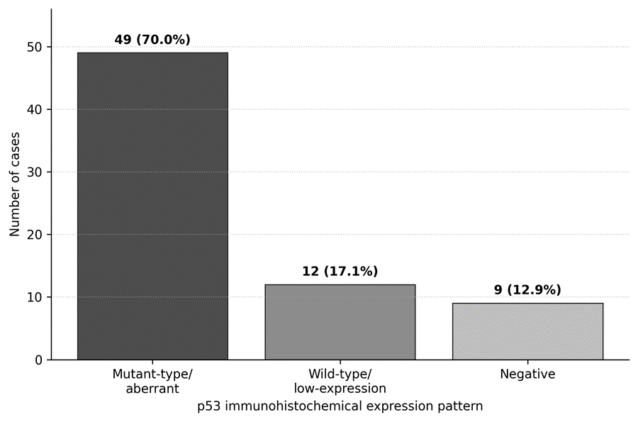
Figure 1. Distribution of p53 immunohistochemical expression patterns (N = 70).
Footnote. Bars show the number and percentage of cases in each p53 immunohistochemical category. “Mutant-type/aberrant” denotes an abnormal immunostaining pattern and does not confirm a TP53 gene mutation without molecular testing.
Association of p53 expression with clinicopathological variables
p53 expression showed statistically significant associations with age group, molecular subtype, surgery type, tumour grade, and lymph node status. Mutant-type/aberrant expression was more frequent among younger patients and was particularly evident in higher-grade tumours. A significant association was also observed across molecular subtypes and nodal categories. Deep margin status did not show a statistically significant association with p53 expression. The detailed cross-tabulation of p53 expression with clinicopathological variables is presented in Table 2. Representative p53 immunohistochemical staining patterns across the molecular subtypes are shown in Figure 2.
Table 2. Association of p53 immunohistochemical expression with clinicopathological variables.
|
Variable |
Category |
Negative |
Wild type/low expression |
Mutant type/aberrant |
Total |
p-value |
|
Age group |
<50 years |
0 |
7 |
30 |
37 |
0.003 |
|
≥50 years |
9 |
5 |
19 |
33 |
||
|
Molecular subtype |
Luminal A |
0 |
9 |
22 |
31 |
0.001 |
|
Luminal B |
0 |
2 |
11 |
13 |
||
|
HER2-positive |
0 |
1 |
9 |
10 |
||
|
Triple-negative |
9 |
0 |
7 |
16 |
||
|
Surgery type |
Modified radical mastectomy |
9 |
2 |
36 |
47 |
<0.001 |
|
Lumpectomy/partial resection |
0 |
10 |
10 |
20 |
||
|
Total mastectomy |
0 |
0 |
3 |
3 |
||
|
Tumour grade |
Grade 1 |
1 |
0 |
0 |
1 |
0.008 |
|
Grade 2 |
8 |
12 |
35 |
55 |
||
|
Grade 3 |
0 |
0 |
14 |
14 |
||
|
Deep margin |
Not involved |
4 |
10 |
32 |
46 |
0.177 |
|
Involved |
5 |
2 |
17 |
24 |
||
|
Lymph node status |
N0 |
4 |
11 |
11 |
26 |
0.001 |
|
N1 |
0 |
1 |
15 |
16 |
||
|
N2 |
3 |
0 |
20 |
23 |
||
|
N3 |
2 |
0 |
3 |
5 |
Footnote: Values are presented as number of cases. p-values were calculated using chi-square or Fisher’s exact test, as appropriate. p<0.05 was considered statistically significant. “Mutant type/aberrant” indicates p53 immunohistochemical expression pattern and does not confirm TP53 mutation without molecular testing.
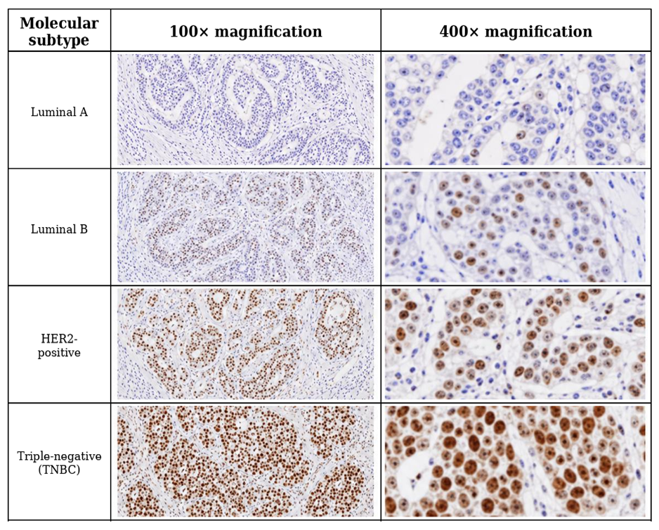
Figure 2. Representative p53 immunohistochemical staining across molecular subtypes of breast carcinoma at 100× and 400× magnification.
Note. Brown nuclear staining indicates p53 immunoreactivity. The photomicrographs are illustrative and should not be interpreted as establishing a subtype-specific p53 pattern or confirming TP53 mutation.
Exploratory subgroup analysis of mutant-type/aberrant p53 expression
An exploratory subgroup analysis was performed to visualize the proportion of mutant-type/aberrant p53 expression across clinicopathological subgroups. The highest proportions were observed in Grade 3 tumours, total mastectomy cases, HER2-positive tumours, and N1/N2 nodal categories; however, several subgroup estimates had wide confidence intervals because of small category sizes. The subgroup distribution is shown in Figure 3.
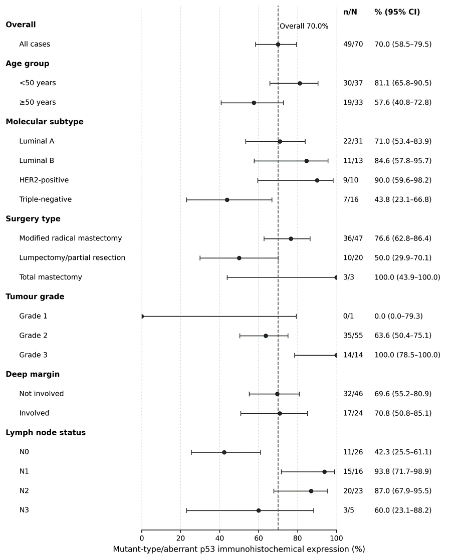
Figure 3. Exploratory subgroup analysis of mutant-type/aberrant p53 immunohistochemical expression.
Footnote. Points represent subgroup proportions and error bars show 95% Wilson confidence intervals. The dashed vertical line marks the overall proportion (70.0%). Estimates from small subgroups are imprecise and should be interpreted descriptively. p53 immunohistochemistry does not establish TP53 mutation status.
DISCUSSION:
This analytical cross-sectional study evaluated p53 immunohistochemical expression in 70 breast carcinoma cases from a tertiary care histopathology setting in Khyber Pakhtunkhwa (KPK), Pakistan. The main finding was the predominance of mutant-type/aberrant p53 immunohistochemical expression in the study cohort. This suggests that abnormal p53 expression may be common among breast carcinoma cases presenting in this local setting. However, because this study was cross-sectional and p53 status was assessed by immunohistochemistry rather than molecular sequencing, the results should be interpreted as clinicopathological associations rather than evidence of causality or confirmed TP53 mutation. The findings are relevant because breast carcinoma remains a major global health burden, while Pakistan-specific literature continues to highlight gaps in awareness, timely diagnosis, screening access, and stage at presentation [1-4]. Breast carcinoma prognosis is influenced by anatomical, histological, and biological factors. Conventional parameters such as tumour size, lymph node status, histological grade, and stage remain central to risk assessment, while receptor status and molecular subtype are increasingly used to refine prognosis and guide management [5,6]. Molecular classification into luminal A, luminal B, HER2-enriched, and basal-like/triple-negative groups have improved understanding of breast cancer heterogeneity [7,8]. In routine pathology practice, especially in resource-limited settings, immunohistochemical surrogates based on ER, PR, HER2, and Ki-67 are commonly used to approximate intrinsic molecular subtypes [9]. Large-scale molecular profiling has also shown that breast cancer subtypes differ in genetic alterations, signalling pathways, and clinical outcomes, supporting biomarker-based stratification [10].
The biological role of p53 provides a plausible basis for its association with aggressive tumour characteristics. TP53 encodes a tumour-suppressor protein that maintains genomic stability through cell-cycle arrest, DNA repair, senescence, apoptosis, and cellular stress responses [11]. Loss of normal p53 function or acquisition of mutant p53 activity may contribute to genomic instability, tumour progression, treatment resistance, and adverse clinical behaviour. Recent Asian breast cancer genomic data suggest that TP53-related effects may vary according to molecular subtype and population background [12].
Moreover, the prognostic impact of TP53 is complex because published studies indicate that TP53 status may interact with treatment type, subtype, and the method used for mutation detection [13]. Therefore, the high frequency of aberrant p53 expression in this cohort should be interpreted in relation to local case selection, tumour grade, stage distribution, and available laboratory methods.
Age group was significantly associated with p53 expression, with mutant-type/aberrant expression more frequent among women younger than 50 years. This is clinically relevant because Pakistani studies have reported that breast carcinoma is often diagnosed at a relatively younger age compared with many Western cohorts [4]. Nevertheless, age should not be interpreted as an independent determinant of p53 expression in this study because multivariable analysis was not performed. Younger age may overlap with other high-risk characteristics, including higher grade, receptor-negative disease, or aggressive molecular phenotype. Pakistani studies have also reported associations between p53 overexpression, hormone receptor negativity, and triple-negative breast carcinoma, supporting the need to interpret p53 together with ER, PR, HER2, grade, and subtype rather than as an isolated marker [14,15].
Molecular subtype was significantly associated with p53 expression in the present cohort. International literature generally reports higher TP53 mutation or aberrant p53 expression in triple-negative and HER2-enriched tumours compared with luminal tumours. However, this study showed a considerable number of mutant-type/aberrant p53 cases among luminal A tumours. This finding should be interpreted cautiously because molecular subtype classification was based on routine immunohistochemical surrogates, and Ki-67 was not available. Pakistani molecular work has also reported abnormal p53 expression across different grades and subtypes of breast carcinoma, suggesting that p53-related abnormalities may not be restricted to a single subgroup [16]. Population-based p53 reclassification studies have shown that p53 expression may vary by breast cancer subtype and may refine clinicopathological interpretation, but it cannot replace complete molecular profiling [17].
Tumour grade showed a significant association with p53 expression. All Grade 3 tumours in the present study demonstrated mutant type/aberrant p53 expression. This finding is biologically plausible because abnormal p53 function is associated with increased genomic instability and aggressive tumour morphology. Recent studies in luminal-like/HER2-negative breast cancer and long-term breast cancer cohorts have reported that p53 or TP53 abnormalities may be associated with adverse outcomes, although the magnitude and independence of this effect vary across studies [18,19]. Meta-analytic evidence in triple-negative breast cancer also suggests that p53 expression may be associated with poorer prognosis, but heterogeneity in staining thresholds, scoring systems, and study populations limits direct comparison [20]. Therefore, in the present study, p53 should be described as associated with higher grade rather than as an independent prognostic marker.
Lymph node status was also significantly associated with p53 expression. Node-positive disease was common in this cohort, and mutant-type/aberrant expression was frequent across node-positive categories. Lymph node involvement remains one of the strongest conventional prognostic indicators in breast carcinoma. The association observed in this study may indicate that aberrant p53 expression is more frequent in tumours with advanced clinicopathological features. However, because survival, recurrence, treatment response, and multivariable outcome data were not available, this study cannot determine whether p53 expression independently predicts nodal metastasis or patient outcome. Evidence linking TP53 mutation with higher genomic risk scores in ER-positive/HER2-negative breast cancer supports the biological relevance of TP53, but such conclusions require molecular confirmation and outcome-based analysis [21].
The exploratory subgroup forest plot further demonstrated variation in mutant-type/aberrant p53 expression across clinicopathological categories. Higher proportions were seen in Grade 3 tumours, HER2-positive tumours, total mastectomy cases, and N1/N2 nodal categories. However, these findings should be interpreted cautiously because several subgroups contained small numbers of cases, producing wide confidence intervals. Therefore, this analysis should be considered descriptive and hypothesis-generating rather than evidence of diagnostic sensitivity, specificity, or independent prognostic effect.
Surgery type was significantly associated with p53 expression, with mutant-type/aberrant expression more frequent among mastectomy cases. This association should be interpreted clinically rather than biologically. Surgical procedure is influenced by tumour size, stage, multifocality, breast-to-tumour ratio, nodal status, patient preference, resource availability, and local surgical practice. In tertiary care settings, mastectomy may be more common when patients present with larger or locally advanced tumours. Therefore, the association between surgery type and p53 expression is more likely to reflect tumour burden and treatment decision-making rather than a direct relationship between p53 biology and surgical procedure.
Deep margin involvement was not significantly associated with p53 expression. This negative finding is important because margin status is mainly determined by tumour extent, anatomical location, surgical clearance, and specimen handling rather than by one molecular marker alone. Although p53 abnormalities may be linked with aggressive tumour behaviour in some studies, the present data do not support using p53 expression as a marker of margin involvement. Larger studies with standardized surgical pathology reporting, tumour size data, and recurrence follow-up are needed to determine whether p53 expression has any meaningful relationship with local recurrence risk in this population.
The interpretation of these findings should also consider the limitations of histopathological and immunohistochemical classification. Histological grading by the modified Bloom-Richardson/Nottingham system remains clinically valuable, but it should be interpreted together with receptor status and other tumour features [22]. ER, PR, and HER2 testing require standardized interpretation because treatment decisions depend heavily on these markers [23,24]. Ki-67 can improve assessment of proliferation and luminal subtype distinction, but reproducibility and cut-off issues remain important [25]. Studies comparing gene-expression molecular subtyping with immunohistochemical surrogate classification have shown that surrogate categories do not always perfectly match intrinsic molecular subtypes [26,27]. Therefore, the absence of Ki-67 and gene-expression profiling may have affected the distinction between luminal A and luminal B tumours in this study.
A key methodological issue is the use of p53 immunohistochemistry as a surrogate for TP53 abnormality. Recent studies have shown that p53 IHC patterns can correlate with TP53 mutation status, particularly when pattern-based interpretation is used, but p53 IHC is not a perfect substitute for sequencing [28-30]. Anderson et al. reported that abnormal p53 expression patterns, including overexpression and null patterns, were strongly associated with underlying TP53 alterations [28].
Armbruster et al. similarly found that aberrant p53 immunostaining patterns in breast carcinoma of no special type strongly correlated with the presence and type of TP53 mutation [29]. In triple-negative breast cancer, Kim et al. showed that refined p53 immunostaining patterns may help correlate IHC findings with somatic TP53 mutation status and functional properties of mutant p53 [30]. These findings support the practical value of p53 IHC in routine pathology, while reinforcing that definitive TP53 mutation status requires molecular testing.
Limitations
This study has several limitations. First, it was conducted in a single tertiary care setting, which may limit generalizability to the wider KPK or Pakistani population. Second, the sample size was modest, particularly for subgroup analyses by molecular subtype, grade, surgery type, nodal category, and stage. Third, the study used p53 immunohistochemistry as a surrogate marker; therefore, mutant-type/aberrant expression should not be considered definitive evidence of TP53 gene mutation. Fourth, molecular subtype classification was based on routine immunohistochemical markers, and Ki-67 was not available, which may have affected subtype assignment, particularly the distinction between luminal A and luminal B tumours.
Fifth, survival, recurrence, treatment response, family history, and detailed tumour size data were not available for outcome-based analysis. The exploratory subgroup analysis was limited by small numbers in some categories, resulting in wide confidence intervals. Finally, because of the cross-sectional design, the study can identify associations but cannot establish temporal, causal, prognostic, or predictive relationships.
CONCLUSION:
This study showed a high frequency of mutant-type/aberrant p53 immunohistochemical expression in breast carcinoma cases from a tertiary care cohort in Khyber Pakhtunkhwa, Pakistan. p53 expression was significantly associated with age group, molecular subtype, surgery type, tumour grade, and lymph node category, while no significant association was observed with deep margin involvement. These findings suggest that p53 immunohistochemistry may serve as a useful adjunct marker when interpreted with established clinicopathological and receptor-based parameters. However, as this was a cross-sectional study and p53 was assessed only by immunohistochemistry, the findings should be interpreted as associations rather than as confirmation of a TP53 mutation or an independent prognostic impact.
Recommendations
Multicentre studies should be conducted across Khyber Pakhtunkhwa and other provinces of Pakistan to improve representativeness and external validity. Future studies should include larger sample sizes to enable reliable subtype-, grade-, and stage-specific analyses. Standardized p53 immunohistochemical scoring criteria should be used, including clear reporting of staining intensity, distribution, and aberrant expression patterns. Molecular testing should be incorporated to distinguish true TP53 gene mutations from surrogate immunohistochemical expression patterns. Future research should also correlate p53 status with disease-free survival, overall survival, recurrence, treatment response, and metastatic progression. Additional clinically relevant markers, including Ki-67, p16, E-cadherin, and PD-L1, may be included to strengthen prognostic and predictive assessment.
REFERENCES:
1. Bray F, Laversanne M, Sung H, Ferlay J, Siegel RL, Soerjomataram I, et al. Global cancer statistics 2022: GLOBOCAN estimates of incidence and mortality worldwide for 36 cancers in 185 countries. CA Cancer J Clin. 2024;74(3):229-263. doi:10.3322/caac.21834
2. Rehman MA, Tahir E, Hussain HG, Khalid A, Taqi SM, Meenai EA. Awareness regarding breast cancer amongst women in Pakistan: a systematic review and meta-analysis. PLoS One. 2024;19(3):e0298275. doi:10.1371/journal.pone.0298275
3. Rehman MA, Taqi SM, Ikram A, Tahir E, Khalid A. Barriers to breast cancer-related care among women in Pakistan: a systematic review. BMJ Public Health. 2025;3:e002559. doi:10.1136/bmjph-2025-002559
4. Soomro R, Faridi S, Khurshaidi N, Zahid N, Mamshad I. Age and stage of breast cancer in Pakistan: an experience at a tertiary care center. J Pak Med Assoc. 2018;68(11):1682-1685.
5. Lee AHS, Ellis IO. The Nottingham Prognostic Index for invasive carcinoma of the breast. Pathol Oncol Res. 2008;14(2):113-115. doi:10.1007/s12253-008-9067-3
6. Tan PH, Ellis I, Allison K, Brogi E, Fox SB, Lakhani S, et al. The 2019 World Health Organization classification of tumours of the breast. Histopathology. 2020;77(2):181-185. doi:10.1111/his.14091
7. Perou CM, Sørlie T, Eisen MB, van de Rijn M, Jeffrey SS, Rees CA, et al. Molecular portraits of human breast tumours. Nature. 2000;406(6797):747-752. doi:10.1038/35021093
8. Sørlie T, Perou CM, Tibshirani R, Aas T, Geisler S, Johnsen H, et al. Gene expression patterns of breast carcinomas distinguish tumour subclasses with clinical implications. Proc Natl Acad Sci U S A. 2001;98(19):10869-10874. doi:10.1073/pnas.191367098
9. Maisonneuve P, Disalvatore D, Rotmensz N, Curigliano G, Colleoni M, Dellapasqua S, et al. Proposed new clinicopathological surrogate definitions of luminal A and luminal B HER2-negative intrinsic breast cancer subtypes. Breast Cancer Res. 2014;16(3):R65. doi:10.1186/bcr3679
10. Cancer Genome Atlas Network. Comprehensive molecular portraits of human breast tumours. Nature. 2012;490(7418):61-70. doi:10.1038/nature11412
11. Marvalim C, Datta A, Lee SC. Role of p53 in breast cancer progression: an insight into p53 targeted therapy. Theranostics. 2023;13(4):1421-1442. doi:10.7150/thno.81847
12. Ragu ME, Lim JMC, Ng PS, Yip CH, Rajadurai P, Teo SH, et al. TP53 somatic mutations in Asian breast cancer are associated with subtype-specific effects. Breast Cancer Res. 2023;25(1):48. doi:10.1186/s13058-023-01635-2
13. Shahbandi A, Nguyen HD, Jackson JG. TP53 mutations and outcomes in breast cancer: reading beyond the headlines. Trends Cancer. 2020;6(2):98-110. doi:10.1016/j.trecan.2020.01.007
14. Qamar S, Khokhar MA, Farooq S, Ashraf S, Humayon WA, Rehman A. Association of p53 overexpression with hormone receptor status and triple negative breast carcinoma. J Coll Physicians Surg Pak. 2019;29(2):164-167. doi:10.29271/jcpsp.2019.02.164
15. Hashmi AA, Naz S, Hashmi SK, Hussain ZF, Irfan M, Khan EY, et al. Prognostic significance of p16 and p53 immunohistochemical expression in triple negative breast cancer. BMC Clin Pathol. 2018;18:9. doi:10.1186/s12907-018-0077-0
16. Sadia H, Bhinder MA, Irshad A, Zahid B, Ahmed R, Ashiq S, et al. Determination of expression profile of p53 gene in different grades of breast cancer tissues by real-time PCR. Afr Health Sci. 2020;20(3):1273-1282. doi:10.4314/ahs.v20i3.32
17. Abubakar M, Guo C, Koka H, Sung H, Shao N, Guida J, et al. Clinicopathological and epidemiological significance of breast cancer subtype reclassification based on p53 immunohistochemical expression. NPJ Breast Cancer. 2019;5:20. doi:10.1038/s41523-019-0117-7
18. Dajti G, Serra M, Cisternino G, Ceccarelli C, Pellegrini A, Melina M, et al. Prognostic role of p53 immunohistochemical status in invasive breast cancer: a retrospective review of 1387 cases with luminal-like/HER2-negative breast tumours. Oncologist. 2024;29(5):384-391. doi:10.1093/oncolo/oyad309
19. Hwang SH, Baek SH, Lee MJ, Kook Y, Bae SJ, Ahn SG, et al. Clinical relevance of TP53 mutation and its characteristics in breast cancer with long-term follow-up data. Cancers (Basel). 2024;16(23):3899. doi:10.3390/cancers16233899
20. Li J, Yang L, Geng J, et al. Association of p53 expression with poor prognosis in patients with triple-negative breast invasive ductal carcinoma: a meta-analysis. Medicine (Baltimore). 2019;98(18):e15449. doi:10.1097/MD.0000000000015449
21. Ji JH, Bae SJ, Kim K, Chu C, Lee KH, Kim Y, et al. Association between TP53 mutation and high 21-gene recurrence score in estrogen receptor-positive/HER2-negative breast cancer. NPJ Breast Cancer. 2022;8(1):19. doi:10.1038/s41523-022-00384-3
22. Elston CW, Ellis IO. Pathological prognostic factors in breast cancer. I. The value of histological grade in breast cancer. Histopathology. 1991;19(5):403-410. doi:10.1111/j.1365-2559.1991.tb00229.x
23. Allison KH, Hammond MEH, Dowsett M, McKernin SE, Carey LA, Fitzgibbons PL, et al. Estrogen and progesterone receptor testing in breast cancer: ASCO/CAP guideline update. J Clin Oncol. 2020;38(12):1346-1366. doi:10.1200/JCO.19.02309
24. Wolff AC, Somerfield MR, Dowsett M, Hammond MEH, Hayes DF, McShane LM, et al. Human epidermal growth factor receptor 2 testing in breast cancer: ASCO-College of American Pathologists guideline update. J Clin Oncol. 2023;41(22):3867-3872. doi:10.1200/JCO.22.02864
25. Nielsen TO, Leung SCY, Rimm DL, Dodson A, Acs B, Badve S, et al. Assessment of Ki67 in breast cancer: updated recommendations from the International Ki67 in Breast Cancer Working Group. J Natl Cancer Inst. 2021;113(7):808-819. doi:10.1093/jnci/djaa201
26. Lundgren C, Bendahl PO, Borg A, Ehinger A, Hegardt C, Larsson C, et al. Agreement between molecular subtyping and surrogate subtype classification: a contemporary population-based study of ER-positive/HER2-negative primary breast cancer. Breast Cancer Res Treat. 2019;178(2):459-467. doi:10.1007/s10549-019-05378-7
27. Holm J, Eriksson L, Ploner A, Eriksson M, Rantalainen M, Li J, et al. Concordance of immunohistochemistry-based and gene expression-based molecular subtyping in breast cancer. J Natl Cancer Inst. 2020;112(10):1046-1053. doi:10.1093/jnci/djaa006
28. Anderson SA, Bartow BB, Harada S, Siegal GP, Wei S, Dal Zotto VL, et al. p53 protein expression patterns associated with TP53 mutations in breast carcinoma. Breast Cancer Res Treat. 2024;207(1):213-222. doi:10.1007/s10549-024-07357-z
29. Armbruster H, Schotte T, Götting I, et al. Aberrant p53 immunostaining patterns in breast carcinoma of no special type strongly correlate with presence and type of TP53 mutations. Virchows Arch. 2024;485(4):631-642. doi:10.1007/s00428-024-03897-3
30. Kim M, Lee M, Lee A, Choi BO, Park WC, Kim SH, et al. Correlating p53 immunostaining patterns with somatic TP53 mutation and functional properties of mutant p53 in triple-negative breast cancer. Histopathology. 2025;87(2):299-309. doi:10.1111/his.15453.