Clinical Profile, Severity Grading and Outcome of Organophosphorus Poisoning in Relation to Serum Pseudocholinesterase Levels: A Tertiary Care Hospital Experience.
- N Bhavana , Postgraduate, Department of Pharmacology, Shri Atal Bihari Vajpayee Medical College and Research Institute, Bengaluru, Karnataka, India.
- Nagesh H N , Associate Professor, Department of Pharmacology, Shri Atal Bihari Vajpayee Medical College and Research Institute, Bengaluru, Karnataka, India.
- S B Nishitha , Assistant Professor, Department of Pharmacology, Shri Atal Bihari Vajpayee Medical College and Research Institute, Bengaluru, Karnataka, India.
- Nalini G K , Professor and Head, Department of Pharmacology, Shri Atal Bihari Vajpayee Medical College and Research Institute, Bengaluru, Karnataka, India.
Article Information:
Abstract:
Background: Organophosphorus (OP) compound poisoning remains one of the most common causes of hospital admission for acute intoxication in India, especially among the rural agricultural workforce. Serum pseudocholinesterase (PChE) is a widely available laboratory marker that reflects the biochemical impact of OP exposure and has been proposed as an objective tool for severity grading and prognostication. Objectives: To describe the demographic and clinical profile of patients with acute OP poisoning admitted to a tertiary care centre, to grade severity on the basis of admission serum PChE and to examine its relationship with clinical outcome. Methods: A hospital-based observational analysis was conducted on 114 consecutive patients admitted with a clinical diagnosis of acute OP poisoning between November 2023 and June 2024. Baseline demographic, clinical, biochemical and treatment data were extracted from a structured case record. Severity was categorised on admission PChE as mild (>2000 U/L), moderate (500–2000 U/L) or severe (<500 U/L). Chi-square, Fisher exact, Pearson correlation and one-way ANOVA were used as appropriate. Results: Mean age was 40.2 ± 12.8 years; 66 patients (57.9%) were male and 99 (86.8%) had suicidal intent. Cypermethrin was the most frequently reported agent (n=42), followed by chlorpyrifos (16), parathion (14), malathion (12) and diazinon (12). Mean admission PChE was 1267 ± 767 U/L, rising to 6109 ± 1730 U/L at discharge. Of 87 patients with a valid admission value, 22 (25.3%) were mild, 53 (60.9%) moderate and 12 (13.8%) severe. Respiratory failure occurred in 24 (21.1%) and intermediate syndrome in 7 (6.1%). Mean hospital stay was 10.0 ± 5.2 days and was longer in the severe subgroup (12.4 ± 9.7 days). All 114 patients survived. Conclusion: Acute OP poisoning at our centre predominantly affected young to middle-aged adults with suicidal intent. Serum PChE was uniformly depressed at admission and recovered by discharge. PChE-based severity grading provided a reproducible framework, but admission PChE alone did not linearly predict complications, suggesting that it should be interpreted alongside clinical scoring rather than in isolation. Protocolised care with early atropinisation achieved excellent short-term survival.
Keywords:
Article :
INTRODUCTION:
Acute poisoning by organophosphorus (OP) compounds is one of the most important preventable causes of morbidity and mortality in the developing world, particularly across South and Southeast Asia [1,2]. A systematic review by Gunnell and colleagues estimated that pesticide self-poisoning accounts for approximately one-third of the world’s suicides, with the majority occurring in the agrarian regions of low- and middle-income countries [3]. India, with its large agricultural workforce and easy over-the-counter availability of OP formulations, continues to contribute a substantial share of this global burden [4,5].
Organophosphorus compounds exert their toxicity by irreversibly phosphorylating the active serine site of acetylcholinesterase (AChE) at cholinergic synapses and neuromuscular junctions, leading to accumulation of acetylcholine and unopposed muscarinic, nicotinic and central nervous system stimulation [6,7]. The classical picture comprises miosis, salivation, lacrimation, bronchorrhoea, bronchospasm, bradycardia, muscle fasciculations, weakness, altered sensorium and, in severe cases, seizures, coma and respiratory failure [8,9]. The clinical spectrum is however not uniform: it is modulated by the specific compound, the quantity ingested, the co-ingestion of alcohol or other substances, and the interval between exposure and initiation of treatment [10,11].
Severity of OP poisoning has traditionally been graded using clinical scores such as the Peradeniya Organophosphorus Poisoning (POP) scale and the Poisoning Severity Score (PSS) proposed by the International Programme on Chemical Safety [12,13]. In parallel, laboratory-based assessment of cholinesterase activity provides an objective and quantitative reflection of the acute cholinergic insult. Erythrocyte or "true" acetylcholinesterase most accurately mirrors synaptic events, but its assay is technically demanding and is not widely available in resource-limited settings [14,15]. In contrast, serum pseudocholinesterase (PChE), also termed butyrylcholinesterase or plasma cholinesterase, is a soluble enzyme synthesised in the liver whose activity can be measured on standard automated chemistry analysers with minimal turnaround time [16,17]. Although PChE depletion is a less specific and slightly less rapid marker than red-cell AChE, its accessibility and low cost make it the practical biomarker of choice in most Indian hospitals [18,19].
The clinical utility of PChE, however, is still debated. Several authors have reported a strong inverse correlation between admission PChE and clinical severity, need for ventilatory support and mortality [10,20,21], while others have observed only modest or no such correlation once patients receive standardised care with atropine and oximes [22,23]. Similarly, the incidence of intermediate syndrome — a distinct proximal muscle weakness syndrome occurring 24–96 hours after apparent recovery from the acute cholinergic crisis, first described by Senanayake and Karalliedde in 1987 — has been variably linked to the depth and duration of PChE inhibition [24,25]. These heterogeneous observations underscore the need for continued hospital-based studies that document the local pattern of exposure, the biochemical response and the clinical outcome.
Against this background, the present study was undertaken at a tertiary care hospital serving the greater Bengaluru region and its surrounding rural belt. We sought to describe the demographic and clinical profile of patients admitted with acute OP poisoning; to grade the severity of the exposure using admission serum PChE; and to examine the association between PChE, complications such as respiratory failure and intermediate syndrome, and the length of hospitalisation. The findings are placed in the context of the published Indian and international literature and the practical implications for bedside triage and resource allocation are discussed.
MATERIALS AND METHODS:
Study design and setting: This was a hospital-based, retrospective observational study conducted in the Department of General Medicine of a tertiary care teaching hospital in Bengaluru, India. The centre receives medical emergencies from urban Bengaluru and adjacent rural taluks, and functions as a referral hub for acute poisoning within its catchment. The study was performed after obtaining institutional ethics committee approval and in accordance with the ethical principles of the Declaration of Helsinki [26]. Written informed consent for the use of anonymised clinical data for research was obtained from each patient or attendant at admission.
Study population: All consecutive patients aged 16 years or older, admitted with a clinical diagnosis of acute organophosphorus compound poisoning between November 2023 and June 2024, were considered for inclusion. Diagnosis was made on the basis of a history of exposure (as reported by the patient, attendants or accompanying containers), clinical features consistent with cholinergic excess (miosis, salivation, bronchorrhoea, vomiting, altered sensorium, muscle fasciculations, bradycardia) and a serum pseudocholinesterase measurement below the laboratory reference range [15,27]. Patients with poisoning attributable to non-OP substances, those with incomplete clinical records, and those who died within six hours of admission before biochemical evaluation was possible, were excluded. A total of 114 patients fulfilled the inclusion criteria and formed the study cohort.
Data collection: A structured proforma was used to extract data from case records. Variables collected included: (i) demographic details — age, sex, address and occupation; (ii) exposure characteristics — name of the compound as declared by patient or attendant, route of exposure, intent (suicidal or accidental), co-ingestion of alcohol and interval to hospital presentation; (iii) presenting clinical features — level of consciousness, pupillary size, oxygen saturation, respiratory rate, pulse rate, blood pressure, cardiovascular, respiratory and abdominal findings; (iv) laboratory investigations — arterial blood gas, complete blood count, serum electrolytes, renal and liver function tests, and serum PChE at admission and again prior to discharge; (v) treatment received — gastric lavage, intravenous atropine dose and regimen, pralidoxime chloride, intravenous fluids and adjunct medications; and (vi) outcomes — occurrence of respiratory failure, development of intermediate syndrome, adverse drug reactions, duration of hospitalisation and status at discharge.
Pseudocholinesterase assay: Serum PChE activity was measured on the hospital’s automated clinical chemistry analyser using the butyrylthiocholine kinetic method, in which the enzyme hydrolyses butyrylthiocholine to butyrate and thiocholine; the latter reacts with 5,5′-dithio-bis-(2-nitrobenzoate) to yield 5-thio-2-nitrobenzoate whose absorbance is measured at 405 nm [28]. The adult laboratory reference interval was 5320–12920 U/L. The first sample was drawn at the time of admission, prior to pharmacological intervention where feasible, and a repeat sample was obtained prior to discharge to document biochemical recovery.
Severity grading: Patients were categorised into three severity grades based on admission PChE, following a modification of the classification used by earlier Indian and Sri Lankan investigators [10,29,30]: mild (PChE >2000 U/L; approximately 40–60% of the lower reference limit), moderate (500–2000 U/L; approximately 10–40%) and severe (<500 U/L; less than 10%). This system was preferred over the POP clinical score because the primary aim of the study was to characterise the utility of PChE as a stand-alone biochemical grading tool.
Treatment protocol: All patients received standardised management following institutional protocols adapted from published guidelines [2,31]. Airway, breathing and circulation were secured, and orogastric lavage was performed within four hours of ingestion in patients presenting early with a protected airway. Intravenous atropine was administered as a 2–4 mg bolus followed by titrated infusion to achieve atropinisation, judged by drying of secretions, resolution of bronchospasm and a heart rate above 80 beats per minute [2,32]. Pralidoxime chloride 1 g intravenously twice daily was administered where clinically indicated and available. Adjunct therapy included intravenous fluids, pantoprazole, ondansetron, thiamine, multivitamin and, where appropriate, insulin for hyperglycaemia and antihypertensive management for underlying comorbidities.
Statistical analysis: Data were entered into a Microsoft Excel spreadsheet and analysed using standard statistical procedures. Categorical variables are expressed as frequencies and percentages, and continuous variables as mean ± standard deviation. Comparisons between severity groups were performed using the chi-square test or Fisher’s exact test for categorical variables and one-way ANOVA for continuous variables. Pearson correlation was used to assess the association between admission PChE and duration of hospitalisation. A two-sided p value of <0.05 was considered statistically significant.
RESULTS:
Demographic profile: A total of 114 patients were included in the study. The mean age was 40.2 ± 12.8 years, with a range from 16 to 65 years. Males comprised 57.9% (n=66) and females 42.1% (n=48), giving a male-to-female ratio of approximately 1.4:1. The largest single age-group was 41–50 years (29.8%), followed by 31–40 years (21.1%) and 51–60 years (21.1%); together, these three decades accounted for more than 70% of admissions (Figure 1). Only four patients (3.5%) were older than 60 years, and 11 (9.6%) were adolescents aged 16–20 years. The majority resided in urban Bengaluru (78.1%), with the remainder drawn from rural taluks and neighbouring peri-urban settlements. Farming was the most frequently recorded occupation (n=19), followed by manual labour (n=12); occupation was not documented in 56 records.
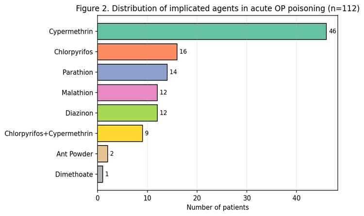
Figure 1. Age and sex distribution of the study population (n=114).
Exposure characteristics: Suicidal intent was the dominant mode of exposure, accounting for 99 patients (86.8%); accidental exposure occurred in 15 (13.2%). Thirty patients (26.3%) had consumed alcohol prior to the poisoning episode. The compound implicated was documented in 112 of the 114 records. Cypermethrin — either alone or as the pyrethroid component of a combination formulation — was the most commonly reported agent (n=42), followed by chlorpyrifos (n=16), parathion (n=14), malathion (n=12), diazinon (n=12) and the popular combination formulation chlorpyrifos 50% + cypermethrin 5% (n=9) (Figure 2). Two records identified only "ant powder" as the ingested agent. The high representation of pyrethroid-containing formulations reflects the ubiquitous household and agricultural use of these products in the study area and the frequent inability to distinguish the pyrethroid component from the co-formulated organophosphate on the basis of clinical presentation alone.
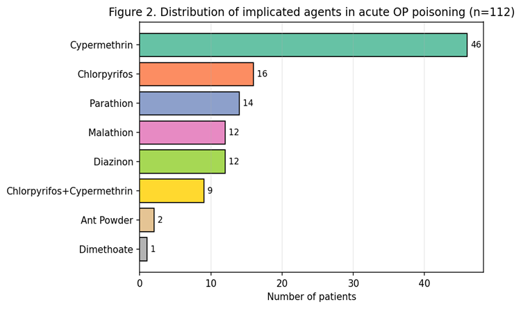
Figure 2. Distribution of implicated agents (n=112).
Clinical features at presentation: At the time of admission, altered sensorium was documented in 37 patients (32.5%), while the remaining 77 (67.5%) were conscious and oriented. Miosis (pupil diameter ≤2 mm) was present in 21 patients (18.4%); a further 39 (34.2%) had a pupil size of 3 mm. The mean oxygen saturation on room air was 95.1 ± 2.7%, mean respiratory rate 17.8 ± 3.7 cycles/min and mean pulse rate 80.4 ± 12.3 beats/min. Cardiac auscultation revealed normal first and second heart sounds in all patients, and bilateral vesicular breath sounds were present at initial presentation in the entire cohort. Table 1 summarises the demographic and baseline clinical characteristics.
Table 1. Demographic, exposure and baseline clinical characteristics (n=114).
|
Characteristic |
Value |
|
Total patients, n |
114 |
|
Age (years), mean ± SD |
40.2 ± 12.8 |
|
Age range |
16–65 |
|
Male, n (%) |
66 (57.9) |
|
Female, n (%) |
48 (42.1) |
|
Suicidal exposure, n (%) |
99 (86.8) |
|
Accidental exposure, n (%) |
15 (13.2) |
|
Alcohol co-ingestion, n (%) |
30 (26.3) |
|
Farmer/agricultural worker, n (%) |
19 (16.7) |
|
Altered sensorium at admission, n (%) |
37 (32.5) |
|
Miosis (pupil ≤2 mm), n (%) |
21 (18.4) |
|
Mean SpO₂ on room air (%) |
95.1 ± 2.7 |
|
Mean respiratory rate (cycles/min) |
17.8 ± 3.7 |
|
Mean pulse rate (beats/min) |
80.4 ± 12.3 |
|
Gastric lavage performed, n (%) |
54 (47.4) |
Serum pseudocholinesterase and severity grading: A valid admission serum PChE estimation was available for 87 patients. The mean admission PChE was 1267.3 ± 767.0 U/L (median 986.5 U/L; range 235.7–3678.9 U/L), which corresponds to marked enzyme depletion when compared with the laboratory lower reference limit of 5320 U/L. A repeat estimation at discharge, available for 102 patients, showed a mean value of 6108.6 ± 1729.8 U/L (median 6222.7 U/L; range 3024.6–8969.3 U/L), documenting substantial biochemical recovery by the time of discharge (Figure 3). Applying the pre-specified cut-offs to the 87 patients with admission PChE data, 22 patients (25.3%) were classified as mild, 53 (60.9%) as moderate and 12 (13.8%) as severe. The mean admission PChE by category was 2402.9 U/L in the mild group, 1001.7 U/L in the moderate group and 358.5 U/L in the severe group (Table 2).
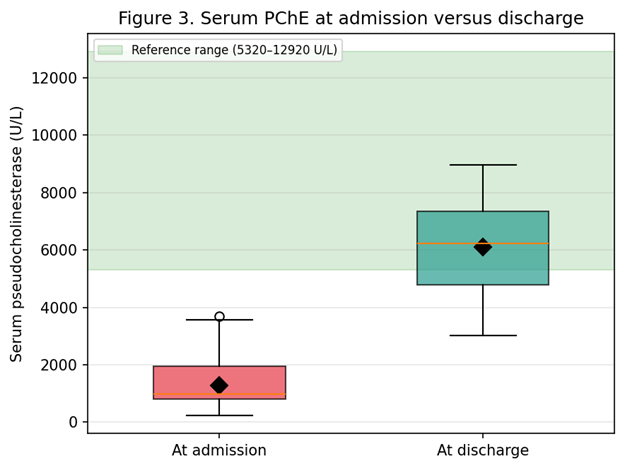
Figure 3. Serum pseudocholinesterase at admission versus discharge. Shaded band = laboratory reference range.
Table 2. Severity classification based on admission serum pseudocholinesterase (n=87).
|
Severity grade |
PChE cut-off (U/L) |
n (%) |
Mean admission PChE (U/L) |
|
Mild |
>2000 |
22 (25.3) |
2402.9 |
|
Moderate |
500–2000 |
53 (60.9) |
1001.7 |
|
Severe |
<500 |
12 (13.8) |
358.5 |
|
Total |
— |
87 (100.0) |
1267.3 |
Treatment: Gastric lavage with normal saline was performed in 54 patients (47.4%) who presented early. Intravenous atropine was administered to every patient, most commonly as a 2–4 mg bolus followed by titrated infusion until atropinisation was achieved; the maintenance dose of 4 mg was used in 89 patients and 2 mg in 25 patients. Pralidoxime chloride 1 g twice daily was administered to seven patients in whom the treating team judged that oxime therapy was likely to be beneficial on the basis of clinical severity and presumed early presentation. Almost all patients received intravenous fluids, intravenous pantoprazole, ondansetron, thiamine and multivitamin support, in keeping with standard institutional protocols. Additional interventions in selected patients included insulin for hyperglycaemia and empirical antibiotics for suspected aspiration pneumonitis.
Complications and outcomes: Twenty-four patients (21.1%) developed acute respiratory failure requiring escalation of respiratory support, and seven (6.1%) developed the classic clinical picture of intermediate syndrome with proximal muscle weakness 24–96 hours after presentation. All 114 patients survived to discharge, resulting in a case-fatality rate of 0% in this cohort. Mean duration of hospitalisation was 10.0 ± 5.2 days (median 8.5; range 5–40). When stratified by severity, the mean stay was 8.9 ± 3.1 days in the mild group, 9.5 ± 3.7 days in the moderate group and 12.4 ± 9.7 days in the severe group (Table 3, Figure 5). The proportion developing respiratory failure was 27.3% (6/22) in the mild group, 24.5% (13/53) in the moderate group and 16.7% (2/12) in the severe group (Figure 4). Although the raw incidence of respiratory failure did not increase in a stepwise fashion with severity, the chi-square test showed no statistically significant difference between groups (χ² = 0.49, p = 0.78), likely reflecting the modest sample sizes in the mild and severe strata and the fact that severe patients received aggressive early management. The Pearson correlation between admission PChE and duration of hospitalisation was weakly negative and non-significant (r = −0.13, p = 0.22). One-way ANOVA of hospital stay across severity groups yielded F = 2.21 (p = 0.12), reflecting the wider dispersion of stay in the severe group.
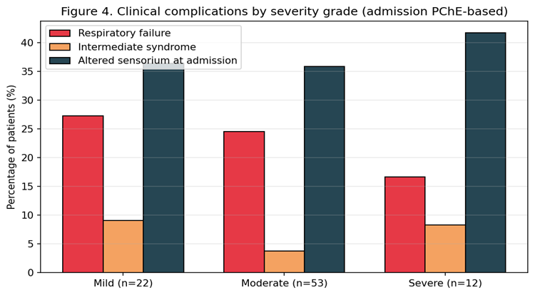
Figure 4. Clinical complications by severity grade (admission-PChE based).
Table 3. Clinical outcomes stratified by severity grade based on admission serum pseudocholinesterase.
|
Outcome |
Mild (n=22) |
Moderate (n=53) |
Severe (n=12) |
|
Respiratory failure, n (%) |
6 (27.3) |
13 (24.5) |
2 (16.7) |
|
Intermediate syndrome, n (%) |
2 (9.1) |
2 (3.8) |
1 (8.3) |
|
Altered sensorium at admission, n |
8 |
19 |
5 |
|
Hospital stay (days), mean ± SD |
8.9 ± 3.1 |
9.5 ± 3.7 |
12.4 ± 9.7 |
|
Mortality, n |
0 |
0 |
0 |
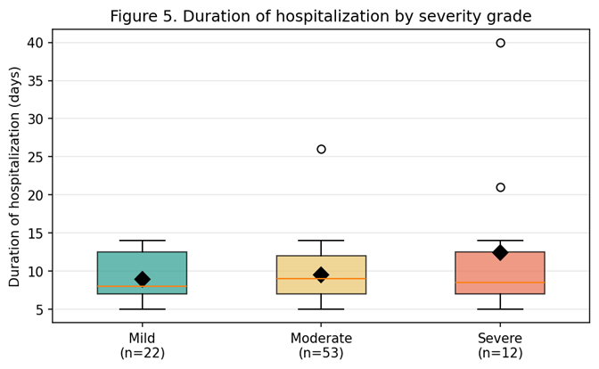
Figure 5. Duration of hospitalisation by severity grade. Black diamond = mean.
DISCUSSION:
The present analysis of 114 consecutive patients admitted with acute organophosphorus poisoning to a tertiary care centre in Bengaluru provides an updated snapshot of the demographic, clinical and biochemical profile of this condition in a mixed urban–rural south Indian population. The findings both reaffirm several long-standing observations and highlight areas in which the utility of a single admission biomarker must be interpreted with caution.
The demographic profile of our cohort — a mean age of 40 years, a male preponderance of approximately 1.4:1, and suicidal exposure in nearly nine of every ten cases — closely mirrors that reported in several other Indian series [4,17,19,33]. Batra and colleagues, in a rural Maharashtrian series of unnatural poisoning, similarly found that OP compounds affected predominantly young adults with self-harm intent [34]. Srinivas Rao et al., in a large multi-centre study from south India, also documented that suicidal exposure dominated the presenting pattern and that agricultural workers and their families were disproportionately represented [35]. Our observation that farmers constituted the largest single occupational category, and that a substantial minority came from the rural Bengaluru belt, is consistent with this pattern. The high proportion of suicidal exposures reinforces the case for population-level interventions such as safe storage of pesticides, restriction of the most toxic World Health Organization Class I compounds and community-based mental health support, all of which have been shown to reduce self-poisoning mortality in South Asia [36,37].
A distinctive feature of the compound distribution in our cohort was the substantial contribution of cypermethrin, either as a pure pyrethroid ingestion or as the co-formulated pyrethroid in the popular chlorpyrifos + cypermethrin agricultural formulation. Combination formulations of an organophosphate with a synthetic pyrethroid are widely marketed across India and are frequently the only labelled product available in local retail outlets. Their inclusion in a cohort presenting with cholinergic features is a familiar clinical reality: patients may be unable to identify the compound with precision, container labels may be missing or damaged, and the initial clinical picture is often indistinguishable from that of a pure OP exposure until enzyme assays return [15,38]. This heterogeneity of compound identification is an important caveat when interpreting hospital-based OP series and has been noted by other Indian authors [19,35].
The mean admission serum pseudocholinesterase in our cohort was 1267 U/L, corresponding to less than one-quarter of the lower reference limit, and rose to 6109 U/L by discharge. This biochemical footprint — profound early depletion followed by near-normalisation over the hospital stay — is entirely consistent with the natural history of the disease. Serum PChE is synthesised de novo in the liver with a half-life of approximately 8–14 days, and functional recovery of the plasma enzyme therefore lags behind clinical recovery of the neuromuscular junction, where new AChE synthesis is required unless enzyme reactivation is achieved with oximes before ageing occurs [6,14]. That a substantial proportion of our patients achieved discharge-level PChE values approaching the lower reference limit reflects both the interval between exposure and discharge and, in some cases, ongoing biochemical recovery beyond the point at which clinical stability was attained.
When patients were categorised on the basis of admission PChE, one-quarter were mild, three-fifths moderate and one in seven severe. The dominance of the moderate category is in keeping with several previous Indian and Sri Lankan studies [10,29,30,33]. Rehiman et al., in a Nepalese hospital-based series, similarly reported that most of their patients fell in the moderate band and that clinical grading by the POP score largely paralleled the enzyme-based grading [10]. Rajpal and colleagues, in an Indian series of 50 patients, likewise found that most fell into intermediate categories of PChE depression and that the concordance between biochemical and clinical severity was strong but not perfect [19]. In our cohort, admission PChE captured the overall biochemical burden of exposure well, but the incidence of respiratory failure did not increase monotonically with severity grade and the correlation between admission PChE and duration of hospitalisation was weak. Several plausible reasons for this observation deserve consideration.
First, admission PChE captures a single time-point and is highly dependent on the interval between ingestion and blood sampling. Two patients with an identical ingested dose may present with very different admission enzyme values if one delays presentation. Serial measurements, ideally at admission, 24 hours and 72 hours, would better capture the true depth and dynamics of enzyme inhibition [21,39]. Second, PChE assays reflect plasma butyrylcholinesterase, which is not the enzyme whose activity determines the acute cholinergic crisis; that role belongs to red-cell acetylcholinesterase and, more directly, to synaptic AChE [14,15]. Third, and perhaps most importantly in a well-resourced tertiary centre, patients graded severe on admission tend to receive intensified monitoring, earlier atropine escalation, earlier oxime therapy and lower thresholds for airway support. Aggressive early management can mitigate what would otherwise be a poor prognosis in the most heavily poisoned patients, thereby weakening the observed correlation between admission PChE and downstream complications. This "treatment effect" has been described in randomised and observational studies of pralidoxime, in which favourable outcomes in intervention groups partly obscured biomarker–outcome relationships [22,23]. A fourth explanation is the mixed compound cohort discussed earlier: pure pyrethroid exposures may produce a less profound PChE fall while still causing significant respiratory symptoms through non-cholinergic mechanisms.
Respiratory failure occurred in 21.1% of our patients, which lies within the range reported in comparable Indian and Sri Lankan cohorts, where figures between 15% and 35% are typical [20,32,33]. Intermediate syndrome, first characterised by Senanayake and Karalliedde as a distinct proximal muscle weakness syndrome appearing after the resolution of the acute cholinergic phase, was identified in 6.1% of our patients [24]. This is at the lower end of the published range of 8–40% [25,26]. Under-recognition, aggressive early atropinisation and prompt weaning from ventilation may all contribute to the lower observed incidence in our series. A notable finding was that all 114 patients survived to discharge. While tertiary-centre studies from India and abroad have reported case-fatality rates as low as 2–5% with modern intensive care [20,29,30], a zero-mortality outcome in a cohort of this size is at the favourable end of the published spectrum. It should be interpreted with caution: it may reflect a genuine effect of protocol-driven care, but it may also reflect referral bias, with patients dying en route or shortly after admission being excluded from the retrievable records.
The average length of hospital stay in our patients was 10 days, with a longer stay of 12.4 days in the severe-PChE group. This is consistent with the observations of Munidasa and colleagues, who reported prolonged ICU and hospital stay in patients with the greatest enzyme depression [20], and of Sam and colleagues, who used the PSS and APACHE II scores to demonstrate a comparable prolonged stay in severely poisoned patients [40]. The wide standard deviation in the severe group (±9.7 days) reflects the influence of a small number of patients with prolonged stays for intermediate syndrome or nosocomial complications, and highlights the ceiling effect of small subgroup sizes.
Several limitations of this study warrant explicit mention. It is a single-centre retrospective analysis and is subject to the recognised limitations of that design, including incomplete documentation of the ingested compound, absence of serial cholinesterase assays and lack of a parallel clinical severity score such as POP or PSS with which to compare the biochemical grading. Occupation was not recorded in about half the case notes, limiting inferences about occupational exposure. Erythrocyte AChE, which better reflects the synaptic enzyme, was not measured. Finally, the modest size of the severe subgroup (n=12) limits the statistical power to detect associations between PChE grade and less common outcomes. Notwithstanding these caveats, the study provides a systematic description of the current pattern of OP poisoning at a large south Indian tertiary centre, documents excellent short-term survival with protocol-driven care and cautions against reliance on any single laboratory value in isolation for prognostication.
CONCLUSION:
Acute organophosphorus poisoning at our tertiary care centre in Bengaluru predominantly affected young to middle-aged adults, with a male preponderance and an overwhelming preponderance of suicidal intent. Cypermethrin — often as a pyrethroid co-formulated with chlorpyrifos — chlorpyrifos, parathion, malathion and diazinon were the most commonly implicated compounds. Serum pseudocholinesterase was markedly depressed at admission and returned to near-normal values by the time of discharge, providing a clear biochemical footprint of the exposure. Severity grading based on admission PChE identified a moderate depression in the majority of patients, but the incidence of respiratory failure and the duration of hospital stay were not linearly determined by admission PChE alone. All patients in this cohort survived, suggesting that a protocolised approach with early atropinisation, judicious use of oximes and aggressive supportive care can achieve very favourable short-term outcomes. Serum pseudocholinesterase is a useful, accessible and inexpensive biomarker for the initial biochemical characterisation of OP poisoning, but is best used in conjunction with clinical scoring systems and serial measurement, rather than as an isolated prognostic tool. Broader public health measures aimed at safe pesticide storage, restriction of the most toxic compounds and community mental health support remain essential complements to hospital-level care.
REFERENCES:
1. Eddleston M. Patterns and problems of deliberate self-poisoning in the developing world. QJM. 2000;93(11):715–31.
2. Eddleston M, Buckley NA, Eyer P, Dawson AH. Management of acute organophosphorus pesticide poisoning. Lancet. 2008;371(9612):597–607.
3. Gunnell D, Eddleston M, Phillips MR, Konradsen F. The global distribution of fatal pesticide self-poisoning: systematic review. BMC Public Health. 2007;7:357.
4. Peter JV, Sudarsan TI, Moran JL. Clinical features of organophosphate poisoning: a review of different classification systems and approaches. Indian J Crit Care Med. 2014;18(11):735–45.
5. Goel A, Aggarwal P. Pesticide poisoning. Natl Med J India. 2007;20(4):182–91.
6. Bardin PG, van Eeden SF, Moolman JA, Foden AP, Joubert JR. Organophosphate and carbamate poisoning. Arch Intern Med. 1994;154(13):1433–41.
7. Aardema H, Meertens JHJM, Ligtenberg JJM, Peters-Polman OM, Tulleken JE, Zijlstra JG. Organophosphorus pesticide poisoning: cases and developments. Neth J Med. 2008;66(4):149–53.
8. Karalliedde L, Senanayake N. Organophosphorus insecticide poisoning. Br J Anaesth. 1989;63(6):736–50.
9. Sungur M, Guven M. Intensive care management of organophosphate insecticide poisoning. Crit Care. 2001;5(4):211–5.
10. Rehiman S, Lohani SP, Bhattarai MD. Correlation of serum cholinesterase level, clinical score at presentation and severity of organophosphorus poisoning. J Nepal Med Assoc. 2008;47(170):47–52.
11. Peter JV, Cherian AM. Organic insecticides. Anaesth Intensive Care. 2000;28(1):11–21.
12. Senanayake N, de Silva HJ, Karalliedde L. A scale to assess severity in organophosphorus intoxication: POP scale. Hum Exp Toxicol. 1993;12(4):297–9.
13. Persson HE, Sjöberg GK, Haines JA, Pronczuk de Garbino J. Poisoning severity score: grading of acute poisoning. J Toxicol Clin Toxicol. 1998;36(3):205–13.
14. Coye MJ, Barnett PG, Midtling JE, Velasco AR, Romero P, Clements CL, et al. Clinical confirmation of organophosphate poisoning by serial cholinesterase analyses. Arch Intern Med. 1987;147(3):438–42.
15. Vale JA. Toxicokinetic and toxicodynamic aspects of organophosphorus (OP) insecticide poisoning. Toxicol Lett. 1998;102–103:649–52.
16. Chowdhary S, Bhattacharyya R, Banerjee D. Acute organophosphorus poisoning. Clin Chim Acta. 2014;431:66–76.
17. Kar SM, Timsinha S, Agrawal P. An epidemiological study of organophosphorus poisoning at Manipal Teaching Hospital, Pokhara, Nepal. J Indian Acad Forensic Med. 2010;32(2):108–9.
18. Dandapani M, Zachariah A, Kavitha MR, Jeyaseelan L, Oommen A. Oxidative damage in intermediate syndrome of acute organophosphorus poisoning. Indian J Med Res. 2003;117:253–9.
19. Rajpal S, Ali R, Bhatnagar A, Bhandari SK, Mittal G. Clinical and bio-chemical profile of acute organophosphorus insecticide poisoning cases. Toxicol Int. 2010;17(1):17–21.
20. Munidasa UM, Gawarammana IB, Kularatne SAM, Kumarasiri PVR, Goonasekera CDA. Survival pattern in patients with acute organophosphate poisoning receiving intensive care. J Toxicol Clin Toxicol. 2004;42(4):343–7.
21. Nouira S, Abroug F, Elatrous S, Boujdaria R, Bouchoucha S. Prognostic value of serum cholinesterase in organophosphate poisoning. Chest. 1994;106(6):1811–4.
22. Buckley NA, Eddleston M, Li Y, Bevan M, Robertson J. Oximes for acute organophosphate pesticide poisoning. Cochrane Database Syst Rev. 2011;(2):CD005085.
23. Pawar KS, Bhoite RR, Pillay CP, Chavan SC, Malshikare DS, Garad SG. Continuous pralidoxime infusion versus repeated bolus injection to treat organophosphorus pesticide poisoning: a randomised controlled trial. Lancet. 2006;368(9553):2136–41.
24. Senanayake N, Karalliedde L. Neurotoxic effects of organophosphorus insecticides: an intermediate syndrome. N Engl J Med. 1987;316(13):761–3.
25. Karki P, Ansari JA, Bhandary S, Koirala S. Cardiac and electrocardiographical manifestations of acute organophosphate poisoning. Singapore Med J. 2004;45(8):385–9.
26. World Medical Association. WMA Declaration of Helsinki: ethical principles for medical research involving human subjects. JAMA. 2013;310(20):2191–4.
27. Namba T, Nolte CT, Jackrel J, Grob D. Poisoning due to organophosphate insecticides: acute and chronic manifestations. Am J Med. 1971;50(4):475–92.
28. Ellman GL, Courtney KD, Andres V Jr, Featherstone RM. A new and rapid colorimetric determination of acetylcholinesterase activity. Biochem Pharmacol. 1961;7:88–95.
29. Sam KG, Kondabolu K, Pati D, Kamath A, Kumar GP, Rao PGM. Poisoning severity score, APACHE II and GCS: effective clinical indices for estimating severity and predicting outcome of acute organophosphorus and carbamate poisoning. J Forensic Leg Med. 2009;16(5):239–47.
30. Sundaray NK, Vinod KV, Dutta TK. Serum cholinesterase in acute organophosphorus poisoning: a study from a tertiary care centre in southern India. Trop Doct. 2015;45(2):85–90.
31. Roberts DM, Aaron CK. Management of acute organophosphorus pesticide poisoning. BMJ. 2007;334(7594):629–34.
32. Jokanović M. Medical treatment of acute poisoning with organophosphorus and carbamate pesticides. Toxicol Lett. 2009;190(2):107–15.
33. Kavya ST, Srinivas V, Chandana, Madhumati R. Clinical profile of patients with organophosphorus poisoning in an intensive care unit in a tertiary hospital. Int J Clin Cases Investig. 2012;4(3):24–31.
34. Batra AK, Keoliya AN, Jadhav GU. Poisoning: an unnatural cause of morbidity and mortality in rural India. J Assoc Physicians India. 2003;51:955–9.
35. Srinivas Rao Ch, Venkateswarlu V, Surender T, Eddleston M, Buckley NA. Pesticide poisoning in south India: opportunities for prevention and improved medical management. Trop Med Int Health. 2005;10(6):581–8.
36. World Health Organization. Preventing suicide: a global imperative. Geneva: World Health Organization; 2014.
37. Gunnell D, Fernando R, Hewagama M, Priyangika WDD, Konradsen F, Eddleston M. The impact of pesticide regulations on suicide in Sri Lanka. Int J Epidemiol. 2007;36(6):1235–42.
38. Jaga K, Dharmani C. Sources of exposure to and public health implications of organophosphate pesticides. Rev Panam Salud Publica. 2003;14(3):171–85.
39. Eyer P. The role of oximes in the management of organophosphorus pesticide poisoning. Toxicol Rev. 2003;22(3):165–90.
40. Grmec S, Mally S, Klemen P. Glasgow coma scale score and QTc interval in the prognosis of organophosphate poisoning. Acad Emerg Med. 2004;11(9):925–30.