Dosimetric Evaluation of Bladder, Rectal, and Sigmoid Colon Doses Using CT-Based 3D Planning for Vaginal Cuff High-Dose-Rate Brachytherapy in Endometrial Cancer: A Prospective Observational Study.
- S B Nikhitha , Radiation Oncologist, India.
- Vinay M Ural , Senior Consultant, Department of Radiation Oncology, Apollo Hospitals Bengaluru, Bengaluru, Karnataka, India.
- S B Nishitha , Assistant Professor, Department of Pharmacology, Shri Atal Bihari Vajpayee Medical College and Research Institute, Bengaluru, Karnataka, India.
Article Information:
Abstract:
Background: Vaginal cuff high-dose-rate (HDR) brachytherapy is a cornerstone of adjuvant management of endometrial cancer. Conventional two-dimensional (2D) planning inadequately quantifies dose to organs at risk (OARs) — the bladder, rectum, sigmoid colon and small bowel — because it depends on point-dose surrogates rather than volumetric estimates. Computed tomography (CT)-based three-dimensional (3D) planning permits individualised OAR dosimetry and offers a rational route to reducing toxicity. Objective: To quantify the doses received by the bladder, rectum and sigmoid colon during CT-based 3D-planned vaginal cuff HDR brachytherapy and to correlate these doses, expressed as EQD2, with acute genitourinary, gastrointestinal and vaginal toxicity graded by CTCAE v5.0. Methods: Fifty-eight post-hysterectomy patients with histologically confirmed endometrial carcinoma treated at a tertiary care centre between February 2022 and July 2023 were enrolled in this prospective observational study. Twenty-eight received vaginal brachytherapy (VBT) alone and 30 received external-beam radiotherapy followed by VBT (EBRT + VBT). Planning CT was acquired with a single-channel vaginal cylinder in situ; the CTV, bladder, rectum and sigmoid colon were contoured per RTOG guidelines and D0.1cc, D1cc, D2cc and D5cc were recorded per GEC-ESTRO recommendations. Acute toxicity was graded at two weeks. Results: Mean per-fraction D2cc was 2.89 ± 0.71 Gy (bladder), 3.59 ± 0.56 Gy (rectum) and 1.74 ± 0.65 Gy (sigmoid) in the EBRT + VBT group and 2.88 ± 0.64 Gy, 3.66 ± 0.56 Gy and 1.90 ± 0.66 Gy respectively in the VBT-alone group. Acute grade ≥ 2 events were uncommon: proctitis 6.7 % and diarrhoea 13.3 % at EQD2 ≈ 32 Gy in the EBRT + VBT arm, and cystitis 3.6 % at EQD2 ≈ 70 Gy in the VBT arm. A stepwise increase in any-grade toxicity was observed with rising EQD2. Conclusion: CT-based 3D planning for vaginal cuff HDR brachytherapy delivers OAR D2cc values well within accepted tolerance thresholds and is associated with a low incidence of grade ≥ 2 acute toxicity. Volumetric planning should be preferred over library-based 2D plans wherever the technology is available.
Keywords:
Article :
INTRODUCTION:
Endometrial carcinoma is the sixth most common cancer diagnosed in women and, in 2020, accounted for an estimated 417 367 new cases and 97 370 deaths worldwide.[1] India recorded 16 413 new cases and 6 385 deaths from the disease in the same year, making it the fourth most common gynaecological malignancy nationally, with the highest incidence in South Asia observed among Indian women.[2,3] Although the age-standardised incidence in India (2.6 per 100 000) is lower than in high-income countries, an ageing population, rising obesity and delayed menopause are expected to increase disease burden substantially over the next two decades.[3]
The primary treatment of localised endometrial cancer is total abdominal hysterectomy with bilateral salpingo-oophorectomy, with or without pelvic and para-aortic lymphadenectomy. Adjuvant radiotherapy is tailored to stage, grade, depth of myometrial invasion and lymphovascular space invasion.[4] Following the 2023 revision of the FIGO staging system — which now incorporates histological type, patterns of invasion and molecular classification — risk stratification and treatment decisions have become increasingly individualised.[4] The PORTEC-2 trial established that vaginal brachytherapy (VBT) alone is non-inferior to pelvic external-beam radiotherapy (EBRT) for local control in high-intermediate risk disease and is associated with significantly less acute gastrointestinal toxicity (12.6 % vs 53.8 % grade 1–2 events) and better long-term quality of life.[5,6] VBT has consequently become the adjuvant treatment of choice for this population, and is used as a boost after pelvic EBRT in higher-risk patients.[7,8]
Historically, vaginal cuff HDR brachytherapy was planned from orthogonal radiographs using the International Commission on Radiation Units and Measurements (ICRU) point-dose formalism, which quantifies rectal and bladder dose at single reference points and ignores dose to the sigmoid and small bowel.[9,10] After hysterectomy, however, small bowel loops and the sigmoid colon frequently descend into the pelvis and lie in close apposition to the vaginal vault, exposing them to steep dose gradients.[11] Recent surveys of American practice patterns confirm that dose to OARs is often inadequately documented when 2D planning is used.[12]
The advent of CT-based 3D planning has enabled volumetric contouring of the target volume and adjacent OARs and reporting of the minimum dose to the most exposed 0.1, 1, 2 and 5 cm³ (D0.1cc, D1cc, D2cc, D5cc), as recommended by the Groupe Européen de Curiethérapie–European Society for Radiotherapy and Oncology (GEC-ESTRO) working group.[13,14] Comparative studies have shown that library-based 2D plans significantly overestimate coverage while under-reporting OAR dose, and that individualised 3D planning meaningfully reduces D2cc to the rectum and bladder without compromising target coverage.[15–17] Nevertheless, prospective Indian data on OAR dose distributions and correlations with acute toxicity remain limited.
This prospective observational study was undertaken to quantify the doses delivered to the bladder, rectum and sigmoid colon during CT-based 3D-planned vaginal cuff HDR brachytherapy in patients with endometrial carcinoma and to correlate these dose–volume parameters, expressed as equivalent dose in 2 Gy fractions (EQD.
MATERIALS AND METHODS:
Study design and setting
This prospective observational study was conducted at the Department of Radiation Oncology, Apollo Hospitals, Bannerghatta Road, Bengaluru, between February 2022 and July 2023. The protocol was reviewed and approved by the institutional scientific review board and ethics committee, and written informed consent was obtained from every participant before enrolment.
Sample size
The sample size was calculated using the mean bladder maximum dose reported by Russo et al.[15] Assuming a standard deviation of 32.7, a 5 % type I error, 95 % confidence and 10 % absolute precision, the formula n = (Z2₁₋α/₂ × SD²) / d² yielded a minimum of 58 patients. Fifty-eight patients were therefore recruited.
Eligibility criteria
Women who had undergone total abdominal hysterectomy with bilateral salpingo-oophorectomy (with or without pelvic and para-aortic nodal dissection) for histologically confirmed endometrial carcinoma and were referred for adjuvant vaginal cuff brachytherapy, either alone or after pelvic external-beam radiotherapy, were eligible. Patients with vaginal recurrence, previous pelvic irradiation, distant metastatic disease or pregnancy were excluded.
Applicator selection and planning CT
Every patient underwent gynaecological examination to determine vaginal vault length and the widest cylinder that could be comfortably accommodated, in accordance with American Brachytherapy Society (ABS) consensus recommendations.[7] Single-channel stackable vaginal cylinders (diameters 2.0–3.5 cm) were used. A planning CT extending from the L4 vertebral level to mid-thigh was acquired with the applicator in situ using a 2 mm slice thickness, with a bladder-filling protocol (indwelling catheter clamped after 100 mL saline instillation) to standardise anatomy between fractions.
Contouring, plan generation and dose reporting
The CT dataset was transferred to a Varian Eclipse™ Brachytherapy treatment planning system. The bladder, rectum and sigmoid colon were contoured on every slice by a single radiation oncologist in accordance with Radiation Therapy Oncology Group (RTOG) female pelvic normal-tissue delineation guidelines.[19] The clinical target volume to depth (CTVdepth) was defined by an isotropic 5 mm expansion of the treatment length around the applicator; the CTV to surface (CTVsurface) was the volume encompassing the treatment length at the applicator surface. Single-channel applicator reconstruction, dwell-position loading and inverse graphical optimisation were performed to conform the prescription isodose to the CTV while minimising exposure of the OARs.
Dose to the bladder, rectum and sigmoid was reported as the minimum dose to the most exposed 0.1, 1, 2 and 5 cm³ (D0.1cc, D1cc, D2cc, D5cc), following the GEC-ESTRO consensus that D2cc is the most clinically relevant volumetric determinant of OAR dose in gynaecological brachytherapy.[13,14] Volumes of each OAR receiving 3 Gy, 4 Gy and 4.2 Gy were also recorded. Treatment was delivered using an Iridium-192 source on a GammaMed™ HDR remote afterloader in accordance with standard institutional practice. Representative planning images from the CT-based 3D planning workflow used in this study are shown in Figures 1–3.
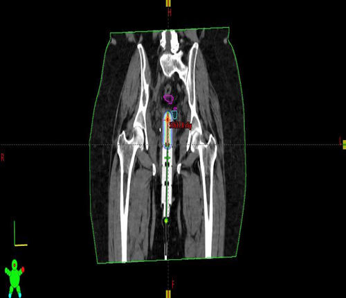
Figure 1. Coronal reconstruction of the planning CT with the single-channel vaginal cylinder in situ, illustrating the prescription isodose conforming to the cylinder surface.
Figure 2. Sagittal CT section showing the isodose distribution with the rectum contoured in blue and the bladder in yellow, demonstrating the steep dose fall-off away from the applicator.
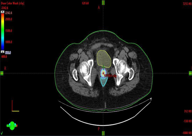
Figure 3. Axial CT section with dose colour-wash overlay for a 6 Gy prescription to the applicator surface; the rectum (blue) and bladder (yellow) are contoured for D2cc dose-volume histogram analysis.
Fractionation and total dose
Patients receiving VBT alone were prescribed 5.5–6 Gy per fraction to a depth of 5 mm from the applicator surface for 3–5 fractions delivered twice weekly, in line with ABS and ASTRO evidence-based recommendations.[7,8] Patients receiving EBRT (46–50 Gy in 23–25 fractions) followed by VBT were boosted with 5–6 Gy per fraction for 3 fractions. Cumulative biologically equivalent dose was expressed as EQD2 assuming α/β = 3 Gy for late-responding normal tissues, in accordance with GEC-ESTRO recommendations.[13,14]
Toxicity assessment
Patients were reviewed two weeks after completion of brachytherapy. Acute proctitis, cystitis, vaginitis and diarrhoea were graded prospectively using the CTCAE version 5.0.[18]
Statistical analysis
Data were entered in Microsoft Excel and analysed using SPSS version 22.0 (IBM Corp., Armonk, NY, USA). Continuous variables are summarised as mean ± standard deviation and categorical variables as frequencies and percentages. The chi-square test was used to test associations between EQD2 tiers and toxicity grades. A two-sided p-value < 0.05 was considered statistically significant.
RESULTS:
Patient characteristics
Fifty-eight women were enrolled; 28 received VBT alone and 30 received EBRT followed by VBT. The median age was 62 years (range 42–78 years), and the majority (58.6 %) were older than 60. The FIGO stage distribution mirrored expected age dependence: patients under 50 predominantly presented with stage I disease, whereas stage III accounted for the largest share of tumours in patients over 60 (Table 1). Histological grade 2 tumours were most frequent overall (51.7 %).
Table 1. Age and FIGO stage distribution by treatment group
|
Age (yr) |
VBT alone (n = 28) |
EBRT + VBT (n = 30) |
FIGO I / II / III |
Grade 1 / 2 / 3 |
|
< 50 |
3 |
3 |
3 / 0 / 0 |
0 / 3 / 0 |
|
50–60 |
8 |
8 |
8 / 0 / 0 |
2 / 6 / 0 |
|
> 60 |
17 |
19 |
16 / 1 / 0 |
20 / 16 / 0 |
|
Total |
28 |
30 |
27 / 1 / 0 |
22 / 25 / 0 |
FIGO = International Federation of Gynaecology and Obstetrics.
Fractionation delivered
In the VBT-alone group, 18 patients received 6 Gy × 5 fractions, 8 received 5.5 Gy × 5 fractions and 2 received 4 Gy × 6 fractions. In the EBRT + VBT group, 15 patients received 6 Gy × 3 fractions, 10 received 5.5 Gy × 3 fractions and 5 received 5 Gy × 3 fractions after 46 Gy pelvic EBRT. Older patients were more likely to be prescribed higher per-fraction doses in both groups, reflecting clinical preference for shorter overall treatment times when comorbidities preclude prolonged attendance.
Dose to organs at risk
Per-fraction OAR doses at each volumetric interval are summarised in Table 2. Mean D2cc values remained well below published tolerance thresholds for the bladder (< 90 Gy EQD2 cumulative), rectum (< 75 Gy) and sigmoid (< 75 Gy).[14,20] The rectum received the highest mean per-fraction D2cc in both groups (3.59 Gy in EBRT + VBT and 3.66 Gy in VBT-alone), reflecting its proximity to the posterior surface of the cylinder. Bladder and sigmoid doses were consistently lower, with the sigmoid receiving the smallest mean dose across all volume intervals.
Table 2. Mean per-fraction OAR doses (Gy) by volume interval and treatment group
|
Organ / Volume |
EBRT + VBT mean ± SD (Gy) |
EBRT + VBT range (Gy) |
VBT alone mean ± SD (Gy) |
VBT alone range (Gy) |
|
Bladder D0.1cc |
3.69 ± 0.86 |
2.21 – 5.78 |
3.60 ± 0.71 |
2.60 – 5.08 |
|
Bladder D1cc |
3.09 ± 0.77 |
1.83 – 4.70 |
3.07 ± 0.67 |
1.99 – 4.25 |
|
Bladder D2cc |
2.89 ± 0.71 |
1.67 – 4.31 |
2.88 ± 0.64 |
1.95 – 3.94 |
|
Bladder D5cc |
2.47 ± 0.62 |
1.37 – 3.70 |
2.47 ± 0.54 |
1.68 – 3.48 |
|
Rectum D0.1cc |
4.38 ± 0.55 |
3.38 – 5.16 |
4.44 ± 0.49 |
3.40 – 5.10 |
|
Rectum D1cc |
3.83 ± 0.60 |
2.58 – 4.68 |
3.90 ± 0.57 |
2.55 – 4.69 |
|
Rectum D2cc |
3.59 ± 0.56 |
2.57 – 4.58 |
3.66 ± 0.56 |
2.57 – 4.59 |
|
Rectum D5cc |
3.18 ± 0.67 |
2.35 – 4.92 |
3.30 ± 0.78 |
2.00 – 4.94 |
|
Sigmoid D0.1cc |
2.31 ± 0.95 |
1.06 – 4.16 |
2.55 ± 0.96 |
1.42 – 4.17 |
|
Sigmoid D1cc |
1.90 ± 0.74 |
1.04 – 3.32 |
2.10 ± 0.75 |
1.06 – 3.32 |
|
Sigmoid D2cc |
1.74 ± 0.65 |
0.90 – 3.06 |
1.90 ± 0.66 |
0.95 – 3.08 |
|
Sigmoid D5cc |
1.52 ± 0.55 |
0.80 – 2.62 |
1.66 ± 0.57 |
0.79 – 2.64 |
OAR = organ at risk; SD = standard deviation.
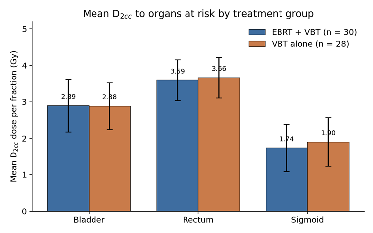
Figure 4. Mean per-fraction D2cc for the bladder, rectum and sigmoid colon in the EBRT + VBT and VBT-alone groups. Error bars indicate one standard deviation.
Volume of OAR receiving high doses
In the EBRT + VBT group, the mean bladder volume receiving 3 Gy per fraction (V3Gy) was 2.53 ± 2.63 cm³ and the mean rectal V3Gy was 3.80 ± 4.22 cm³. Very small volumes of any OAR received ≥ 4 Gy per fraction (mean bladder V4Gy 0.85 cm³, rectum V4Gy 1.57 cm³, sigmoid V4Gy 0.08 cm³). In the VBT-alone group, mean bladder V3Gy was 3.12 ± 2.96 cm³ and rectal V3Gy 2.89 ± 3.14 cm³; no patient in this group received ≥ 4 Gy to any measurable sigmoid volume.
Acute toxicity by treatment group
Acute toxicity was overwhelmingly mild. In the VBT-alone group, grade 1 proctitis occurred in 25.0 % of patients (7/28), grade 1 cystitis in 10.7 % (3/28), grade 1 vaginitis in 32.1 % (9/28) and grade 1 diarrhoea in 7.1 % (2/28). Grade 2 events were rare: cystitis 3.6 % (1/28) and diarrhoea 7.1 % (2/28). No grade ≥ 3 events were observed. In the EBRT + VBT group, grade 1 proctitis occurred in 20.0 %, grade 1 cystitis in 26.7 % and grade 1 diarrhoea in 33.3 %; grade 2 events were limited to proctitis (6.7 %) and diarrhoea (13.3 %). Table 3 summarises the toxicity distribution.
Table 3. Distribution of acute toxicity (CTCAE v5.0) by treatment group
|
Toxicity |
VBT alone Gr 0 (%) |
VBT alone Gr 1 (%) |
VBT alone Gr 2 (%) |
EBRT+VBT Gr 0 (%) |
EBRT+VBT Gr 1 (%) |
EBRT+VBT Gr 2 (%) |
|
Proctitis |
75.0 |
25.0 |
0.0 |
73.3 |
20.0 |
6.7 |
|
Cystitis |
85.7 |
10.7 |
3.6 |
73.3 |
26.7 |
0.0 |
|
Vaginitis |
67.9 |
32.1 |
0.0 |
96.7 |
3.3 |
0.0 |
|
Diarrhoea |
85.7 |
7.1 |
7.1 |
53.3 |
33.3 |
13.3 |
Gr = grade; VBT = vaginal brachytherapy; EBRT = external beam radiotherapy.
Correlation between EQD2 and toxicity
A stepwise relationship between rising EQD2 and acute toxicity was evident in both groups. In the EBRT + VBT arm, EQD2 of 24 Gy produced grade 1 proctitis in 3.3 % of patients and no diarrhoea; at 28 Gy, grade 1 diarrhoea appeared in 16.7 %, grade 2 in 3.3 % and grade 1 cystitis in 6.7 %. At 32.4 Gy, grade 1 and 2 proctitis (13.3 % and 6.7 %), grade 1 and 2 diarrhoea (16.7 % and 10.0 %) and grade 1 cystitis (20.0 %) were recorded (Figure 5).
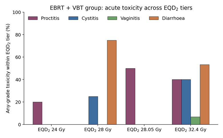
Figure 5. Any-grade acute toxicity within each EQD2 tier in the EBRT + VBT group.
A similar gradient was observed in the VBT-alone group: EQD2 ≤ 46 Gy was associated with no cystitis, whereas at EQD2 ≈ 70 Gy, grade 1 cystitis rose to 10.7 % and grade 1 vaginitis to 21.4 % (Figure 6). The chi-square test showed a significant positive association between EQD2 tier and any-grade diarrhoea in the EBRT + VBT group (p = 0.02) and between EQD2 and vaginitis in the VBT group (p = 0.04).
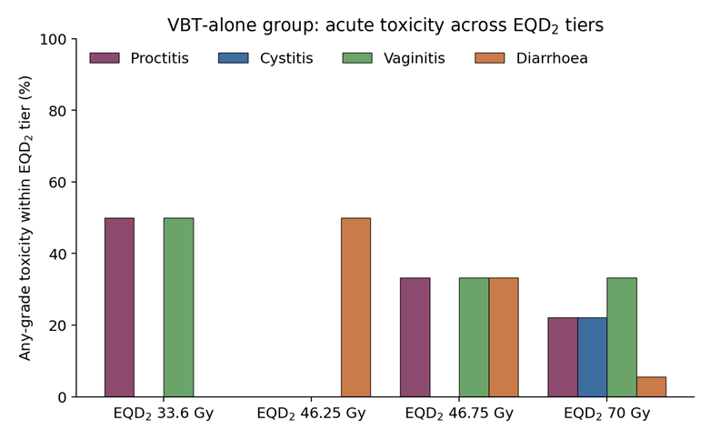
Figure 6. Any-grade acute toxicity within each EQD2 tier in the VBT-alone group.
DISCUSSION:
This prospective study demonstrates that CT-based 3D planning for vaginal cuff HDR brachytherapy in endometrial cancer can consistently deliver per-fraction D2cc values to the bladder, rectum and sigmoid colon that lie well within contemporary tolerance thresholds, and is associated with a low incidence of acute grade ≥ 2 toxicity in both VBT-alone and EBRT + VBT settings. These findings extend the growing evidence base supporting a routine shift from 2D library-based planning to volumetric image-guided planning in the post-hysterectomy setting.[13–17]
Our mean per-fraction bladder D2cc (2.89 Gy) and rectum D2cc (3.59 Gy) in the EBRT + VBT arm compare favourably with the values reported by Kim and Beriwal, whose 3D CT-based plans achieved rectum and bladder D2cc doses significantly below those derived from parallel 2D library plans (p ≤ 0.001).[16] Russo et al., in a comparison of 91 patients planned with 2D and 3D techniques, similarly documented that 2D planning both overestimates coverage and can underestimate OAR dose, with the rectum D2cc exceeding the prescription dose in a subset of 2D plans — a phenomenon that would go undetected without volumetric imaging.[15] Yaghoobi Notash and colleagues, in a cobalt-60 series of 37 patients, also observed that D0.1cc, D1cc and D2cc for the rectum, sigmoid and bladder were consistently lower with 3D planning than with 2D planning while V100 for the target was preserved.[21] Our data, obtained with an iridium-192 source and inverse graphical optimisation, corroborate these observations in a South Asian cohort.
The rectum received the highest per-fraction dose across all volume intervals in both treatment groups. This anatomical reality — the rectum lies immediately posterior to the vaginal apex, separated only by fascial planes — has been a consistent theme in dosimetric studies and underpins the current GEC-ESTRO recommendation that D2cc rather than a single point dose be used for reporting.[13,14]
Damast and colleagues, in a series of 177 endometrial cancer patients treated with 21 Gy in 3 fractions, reported mean rectum D2cc of 5.7 Gy per course, sigmoid D2cc 3.3 Gy, small bowel D2cc 3.8 Gy and bladder D2cc 5.4 Gy — values broadly consistent with those observed here.[22] Small volumes of the OARs received doses ≥ 4 Gy per fraction in our series, and cumulative EQD2 remained under the accepted thresholds of 90 Gy (bladder) and 75 Gy (rectum, sigmoid).[14,20]
The clear stepwise relationship between rising EQD2 and acute toxicity mirrors findings from the biological planning study of DE Sanctis et al., in which acute urinary toxicity was significantly higher with mean bladder dose per fraction > 2.5 Gy or total bladder dose > 7.5 Gy.[23] Delishaj et al., in a 20-year single-institution series of endovaginal HDR brachytherapy, similarly demonstrated a dose-dependent rise in vaginal toxicity but noted that severe (grade 3) events remained rare when GEC-ESTRO constraints were respected.[24] Arden and colleagues, using 30 Gy in 6 fractions of adjuvant VBT, reported grade 2 or higher genitourinary toxicity in only 1 % of patients at two-year follow-up, again consistent with our observation that grade ≥ 2 events are uncommon when volumetric planning is used.[25]
Two additional observations merit comment. First, the age-dependent distribution of stage — with FIGO stage I predominating in patients under 50 and stage III becoming more common in those over 60 — matches national tertiary-care data reported by Indian cancer registries and by the recent survival analysis of a large South Asian institutional series, in which the median age at diagnosis was 58 years.[3,26] Second, the finding that older patients were more frequently prescribed higher per-fraction doses reflects clinical trade-offs between shorter overall treatment time and increased biological dose per fraction. The favourable toxicity profile suggests that a modestly hypofractionated schedule remains safe when 3D planning is used to constrain OAR dose.
The PORTEC-2 trial established the equivalence of VBT alone to pelvic EBRT for vaginal control in high-intermediate risk disease, with significantly less acute gastrointestinal toxicity.[5] Our EBRT + VBT and VBT-alone cohorts, drawn from a heterogeneous risk group that included stage I–III disease, showed grade 1–2 gastrointestinal toxicity rates (46.6 % in EBRT + VBT; 14.2 % in VBT-alone) that are of a similar order to those reported by Nout and colleagues (53.8 % vs 12.6 %), supporting the external validity of the PORTEC-2 findings when 3D planning is applied.[5,6] The long-term quality-of-life data from the PORTEC-2 trial further reinforce the rationale for minimising OAR dose in the acute setting.[6]
Several limitations should be acknowledged. This was a single-centre study with a modest sample size, and follow-up was restricted to acute toxicity assessed at two weeks. Late toxicities — vaginal stenosis, chronic proctitis and urinary dysfunction — may emerge years after treatment and were not captured. Only D0.1cc–D5cc and V3–V4.2Gy were reported; small-bowel dose, although anatomically relevant, was not systematically contoured owing to inconsistent inclusion within the CT field of view.
The prospective observational design precludes a direct within-patient comparison with 2D plans, though the study was not designed as a comparative dosimetric analysis and its findings should be interpreted against the substantial body of already-published 2D-versus-3D data.[15–17,21] Multicentre studies with long-term follow-up, incorporating patient-reported outcomes and prospective quality-of-life measures, are needed to further characterise the therapeutic ratio of CT-based 3D planned vaginal cuff HDR brachytherapy in Indian populations.
CONCLUSION:
CT-based three-dimensional planning for adjuvant vaginal cuff HDR brachytherapy in endometrial cancer delivers per-fraction D0.1cc, D1cc, D2cc and D5cc doses to the bladder, rectum and sigmoid colon that lie well within GEC-ESTRO and QUANTEC tolerance thresholds. Acute grade ≥ 2 toxicity is uncommon and shows a clear stepwise relationship with cumulative EQD2, supporting the routine use of volumetric image-based planning as a means of individualising treatment, sparing organs at risk and rationally selecting fractionation. Volumetric planning should be preferred to point-dose 2D planning wherever technology permits.
REFERENCES:
1. Sung H, Ferlay J, Siegel RL, Laversanne M, Soerjomataram I, Jemal A, et al. Global cancer statistics 2020: GLOBOCAN estimates of incidence and mortality worldwide for 36 cancers in 185 countries. CA Cancer J Clin. 2021;71(3):209–49.
2. Nandakumar A, Rath GK, Kataki AC, Poonamalle BV, Parkin DM, Sebastian P, et al. Decadal trends of endometrial cancer in India: analysis from the National Cancer Registry Programme. Indian J Cancer. 2020;57(1):86–94.
3. Kamal J, Basumatary G, Kandagatla P, Bhalavat R. Endometrial carcinoma: survival analysis at a tertiary care centre in India. Indian J Gynecol Oncol. 2022;20(4):85.
4. Berek JS, Matias-Guiu X, Creutzberg C, Fotopoulou C, Gaffney D, Kehoe S, et al. FIGO staging of endometrial cancer: 2023. Int J Gynaecol Obstet. 2023;162(2):383–94.
5. Nout RA, Smit VT, Putter H, Jürgenliemk-Schulz IM, Jobsen JJ, Lutgens LC, et al. Vaginal brachytherapy versus pelvic external beam radiotherapy for patients with endometrial cancer of high-intermediate risk (PORTEC-2): an open-label, non-inferiority, randomised trial. Lancet. 2010;375(9717):816–23.
6. Nout RA, Putter H, Jürgenliemk-Schulz IM, Jobsen JJ, Lutgens LC, van der Steen-Banasik EM, et al. Quality of life after pelvic radiotherapy or vaginal brachytherapy for endometrial cancer: first results of the randomized PORTEC-2 trial. J Clin Oncol. 2009;27(21):3547–56.
7. Small W Jr, Beriwal S, Demanes DJ, Dusenbery KE, Eifel P, Erickson B, et al. American Brachytherapy Society consensus guidelines for adjuvant vaginal cuff brachytherapy after hysterectomy. Brachytherapy. 2012;11(1):58–67.
8. Harkenrider MM, Block AM, Alektiar KM, Gaffney DK, Jones E, Klopp A, et al. American Brachytherapy Task Group Report: adjuvant vaginal brachytherapy for early-stage endometrial cancer: a comprehensive review. Brachytherapy. 2017;16(1):95–108.
9. International Commission on Radiation Units and Measurements. ICRU Report 38: dose and volume specification for reporting intracavitary therapy in gynecology. Bethesda: ICRU; 1985.
10. Nag S, Erickson B, Parikh S, Gupta N, Varia M, Glasgow G. The American Brachytherapy Society recommendations for high-dose-rate brachytherapy for carcinoma of the endometrium. Int J Radiat Oncol Biol Phys. 2000;48(3):779–90.
11. Schoeppel SL, Lavigne ML, Martel MK, McShan DL, Fraass BA, Roberts JA. Three-dimensional treatment planning of intracavitary gynaecologic implants: analysis of ten cases and implications for dose specification. Int J Radiat Oncol Biol Phys. 1994;28(1):277–83.
12. Harkenrider MM, Grover S, Erickson BA, Viswanathan AN, Small C, Kliethermes S, et al. Vaginal brachytherapy for postoperative endometrial cancer: 2014 survey of the American Brachytherapy Society. Brachytherapy. 2016;15(1):23–9.
13. Haie-Meder C, Pötter R, Van Limbergen E, Briot E, De Brabandere M, Dimopoulos J, et al. Recommendations from Gynaecological (GYN) GEC-ESTRO Working Group (I): concepts and terms in 3D image based 3D treatment planning in cervix cancer brachytherapy with emphasis on MRI assessment of GTV and CTV. Radiother Oncol. 2005;74(3):235–45.
14. Pötter R, Haie-Meder C, Van Limbergen E, Barillot I, De Brabandere M, Dimopoulos J, et al. Recommendations from Gynaecological (GYN) GEC-ESTRO Working Group (II): concepts and terms in 3D image-based treatment planning in cervix cancer brachytherapy—3D dose volume parameters and aspects of 3D image-based anatomy, radiation physics, radiobiology. Radiother Oncol. 2006;78(1):67–77.
15. Russo JK, Armeson KE, Richardson S. Comparison of 2D and 3D imaging and treatment planning for postoperative vaginal apex high-dose rate brachytherapy for endometrial cancer. Int J Radiat Oncol Biol Phys. 2012;83(1):e75–80.
16. Kim H, Rajagopalan MS, Beriwal S, Huq MS, Smith KJ. Is there any advantage to three-dimensional planning for vaginal cuff brachytherapy? Brachytherapy. 2012;11(5):398–401.
17. Hellebust TP, Kirisits C, Berger D, Pérez-Calatayud J, De Brabandere M, De Leeuw A, et al. Recommendations from Gynaecological (GYN) GEC-ESTRO Working Group: considerations and pitfalls in commissioning and applicator reconstruction in 3D image-based treatment planning of cervix cancer brachytherapy. Radiother Oncol. 2010;96(2):153–60.
18. US National Cancer Institute. Common Terminology Criteria for Adverse Events (CTCAE) version 5.0. Bethesda: NCI; 2017.
19. Small W Jr, Mell LK, Anderson P, Creutzberg C, De Los Santos J, Gaffney D, et al. Consensus guidelines for delineation of clinical target volume for intensity-modulated pelvic radiotherapy in postoperative treatment of endometrial and cervical cancer. Int J Radiat Oncol Biol Phys. 2008;71(2):428–34.
20. Marks LB, Yorke ED, Jackson A, Ten Haken RK, Constine LS, Eisbruch A, et al. Use of normal tissue complication probability models in the clinic. Int J Radiat Oncol Biol Phys. 2010;76(3 Suppl):S10–9.
21. Yaghoobi Notash A, Ranjbar M, Mahdavi SR, Nikoofar AR, Bakhshandeh M, Fatemi A, et al. A comparison between 2D and 3D planning of high-dose-rate vaginal cuff brachytherapy in patients with stage I–II endometrial cancer using cobalt-60. J Contemp Brachytherapy. 2021;13(5):525–32.
22. Damast S, Alektiar KM, Goldfarb S, Eaton A, Patil S, Mosenkis J, et al. Sexual functioning among endometrial cancer patients treated with adjuvant high-dose-rate intra-vaginal radiation therapy. Int J Radiat Oncol Biol Phys. 2012;84(2):e187–93.
23. De Sanctis V, Agolli L, Visco V, Monaco F, Muni R, Spagnoletti I, et al. Cystitis and rectitis after in vivo dosimetry-based post-operative vaginal-cuff HDR brachytherapy in endometrial cancer. Cancers (Basel). 2021;13(21):5405.
24. Delishaj D, Barcellini A, D'Amico R, Ursino S, Pasqualetti F, Fumagalli IC, et al. Vaginal toxicity after high-dose-rate endovaginal brachytherapy: 20 years of results. J Contemp Brachytherapy. 2018;10(6):559–66.
25. Arden JD, Dokter J, Almahariq MF, Marvin K, Nandalur SR, Al-Wahab Z, et al. Toxicity and efficacy after adjuvant vaginal brachytherapy using 30 Gy in 6 fractions for stages I and II endometrial cancer. Adv Radiat Oncol. 2021;6(6):100773.
26. Bagga N, Chopra S, Engineer R, Mahantshetty U, Deodhar K, Shrivastava SK. Endometrial cancer at a tertiary care centre in India: patterns of care and clinical outcomes. South Asian J Cancer. 2021;10(1):41–5.
27. Creutzberg CL, Nout RA, Lybeert ML, Wárlám-Rodenhuis CC, Jobsen JJ, Mens JW, et al. Fifteen-year radiotherapy outcomes of the randomized PORTEC-1 trial for endometrial carcinoma. Int J Radiat Oncol Biol Phys. 2011;81(4):e631–8.
28. Anderson JM, Stea B, Hallum AV, Rogoff E, Childers J. High-dose-rate postoperative vaginal cuff irradiation alone for stage IB and IC endometrial cancer. Int J Radiat Oncol Biol Phys. 2000;46(2):417–21.
29. Alektiar KM, Venkatraman E, Chi DS, Barakat RR. Intravaginal brachytherapy alone for intermediate-risk endometrial cancer. Int J Radiat Oncol Biol Phys. 2005;62(1):111–7.
30. Miller DA, Richardson S, Grigsby PW. A new method of anatomically conformal vaginal cuff HDR brachytherapy. Gynecol Oncol. 2010;116(3):413–8.
31. Sabater S, Andres I, Lopez-Honrubia V, Berenguer R, Sevillano M, Jimenez-Jimenez E, et al. Vaginal cuff brachytherapy in endometrial cancer: a review of major clinical trials with a focus on fractionation. Rep Pract Oncol Radiother. 2020;25(3):378–84.
32. Guy CL, Fields EC, Quinn BA, Song J, Chen KS, Ban T, et al. Vaginal cuff brachytherapy: do we need to treat to more than a two-centimeter active length? J Contemp Brachytherapy. 2021;13(3):249–56.
33. Randall ME, Filiaci VL, Muss H, Spirtos NM, Mannel RS, Fowler J, et al. Randomized phase III trial of whole-abdominal irradiation versus doxorubicin and cisplatin chemotherapy in advanced endometrial carcinoma: a Gynecologic Oncology Group study. J Clin Oncol. 2006;24(1):36–44.
34. Keys HM, Roberts JA, Brunetto VL, Zaino RJ, Spirtos NM, Bloss JD, et al. A phase III trial of surgery with or without adjunctive external pelvic radiation therapy in intermediate risk endometrial adenocarcinoma: a Gynecologic Oncology Group study. Gynecol Oncol. 2004;92(3):744–51.
35. Wortman BG, Creutzberg CL, Putter H, Jürgenliemk-Schulz IM, Jobsen JJ, Lutgens LC, et al. Ten-year results of the PORTEC-2 trial for high-intermediate risk endometrial carcinoma: improving patient selection for adjuvant therapy. Br J Cancer. 2018;119(9):1067–74.