Radiographic And Periodontal Manifestations In Children With Iron Deficiency Anemia.
- Zainab Safoora , Department of Periodontology, Al-Badar Dental College and Hospital, Kalaburagi, Karnataka
- Mohammed Sadaquat Ullah khan , Department of Radiodiagnosis, KBNU-Faculty of Medical Sciences, Kalaburagi, Karnataka.
- Sana Samreen , Department of Pediatrics, ESIC Medical College, Kalaburagi, Karnataka.
Article Information:
Abstract:
Background: Iron Deficiency Anemia (IDA) is the most widespread nutritional deficiency worldwide, disproportionately affecting pediatric populations in developing regions. While systemic manifestations such as cognitive impairment, fatigue, and growth retardation are well documented, the oral, periodontal, and alveolar radiographic alterations associated with pediatric IDA remain incompletely characterized. Objective: To evaluate the nature and severity of periodontal disease parameters and alveolar/mandibular radiographic alterations in children diagnosed with Iron Deficiency Anemia compared to non-anemic healthy controls. Methods: A prospective, case-control study was conducted over a 24-month period involving 180 pediatric patients (aged 6 to 14 years) divided into two equal groups: an IDA group (n=90, confirmed by Hemoglobin < 11.0 g/dL, serum ferritin < 12µg/L, and transferrin saturation < 16%) and an age- and sex-matched healthy control group (n = 90). Periodontal status was evaluated using the Plaque Index (PI), Gingival Index (GI), Probing Pocket Depth (PPD), and Clinical Attachment Level (CAL). Digital intraoral periapical and panoramic radiographs were analyzed for alveolar crestal bone loss, trabecular bone pattern alterations (stepladder arrangement, coarse trabeculation), and lamina dura density. Inter-group comparisons and correlation analyses between hematological parameters and oral/radiographic indices were executed. Results: Children with IDA exhibited significantly higher mean Gingival Index (1.68 ± 0.42 vs. 0.94 ± 0.31; p < 0.001) and Probing Pocket Depth (2.48 ± 0.52mm vs. 1.82 0.34 mm; p < 0.001) despite similar oral hygiene levels reflected by Plaque Index (1.42 ± 0.38 vs. 1.36 ± 0.35; p=0.264). Radiographic analysis revealed alterations in alveolar trabecular architecture in 56.7% (n = 51) of the IDA group compared to 12.2% (n = 11) of controls (p < 0.001). Prominent radiographic findings included "stepladder" interdental trabecular patterning (38.9%), thinning/loss of lamina dura (44.4%), and localized alveolar crestal bone height loss (28.9%). Hemoglobin and serum ferritin levels demonstrated a strong inverse correlation with GI (r = -0.612, p < 0.001) and radiographic bone alterations (r = -0.584, p < 0.001). Conclusion: Iron Deficiency Anemia in children is associated with enhanced gingival inflammatory response, increased susceptibility to periodontal breakdown, and distinct radiographic alterations in mandibular alveolar trabeculation. Pediatric dental screenings should incorporate systemic hematological evaluations for children presenting with unexplained severe gingivitis or altered alveolar trabecular patterns.
Keywords:
Article :
INTRODUCTION:
Iron Deficiency Anemia (IDA) represents a critical global public health challenge, impacting over 1.2 billion individuals globally [1]. Children aged 6 to 14 years in developing and low-to-middle-income nations are particularly vulnerable due to high metabolic demands during rapid growth spurts, inadequate dietary intake of bioavailable heme iron, intestinal parasitic infections, and poor socioeconomic conditions [2].
Systemically, iron is an essential trace element required for hemoglobin synthesis, cellular oxygen transport, cytochrome enzyme activity, and robust cell-mediated immunity [3]. Severe or chronic iron deficiency leads to systemic tissue hypoxia, impaired neutrophil function, diminished lymphocyte proliferation, and compromised collagen synthesis [4].
The oral cavity often serves as an early mirror of systemic nutritional deficits [5]. Oral manifestations of IDA in adults, such as angular cheilitis, atrophic glossitis (smooth red tongue due to loss of filiform and fungiform papillae), oral mucosal pallor, and burning mouth syndrome are well recognized [6]. However, the specific impact of systemic iron deficiency on the pediatric periodontium and supporting jawbone architecture remains under-researched [7].
Periodontal tissue homeostasis depends on a delicate equilibrium between plaque microflora and host immune-inflammatory responses [8]. Iron deficiency impairs epithelial turnover in the junctional epithelium, reduces salivary peroxidase activity, and dampens neutrophil chemotaxis, potentially accentuating gingival inflammatory destruction even under low-to-moderate dental plaque burdens [9].
Furthermore, continuous hematopoiesis and bone remodeling occur dynamically within the alveolar bones of growing children [10]. Chronic systemic anemia induces compensatory bone marrow hyperplasia, which can alter the microarchitecture of interdental alveolar bone and cancellous mandibular trabeculae [11]. High-resolution digital intraoral radiography and panoramic radiographs provide a non-invasive tool to detect these subtle skeletal manifestations [12].
This prospective case-control study aimed to quantitatively evaluate periodontal status and digital radiographic alveolar/trabecular manifestations in pediatric patients diagnosed with Iron Deficiency Anemia compared to non-anemic healthy controls.
MATERIALS AND METHODS:
Study Design and Ethical Clearance
This prospective, comparative case-control study was conducted jointly by the Department of Pediatric, Department of Periodontics, and the Department of Radiodiagnosis over a 24-month period (March 2024 to February 2026), at multiple tertiary care hospitals. The study protocol was reviewed and approved by all the concerned Institutional Ethics Committee, and written informed consent was obtained from the parents or legal guardians of all child participants prior to enrollment in accordance with the Declaration of Helsinki.
Study Population and Group Allocation
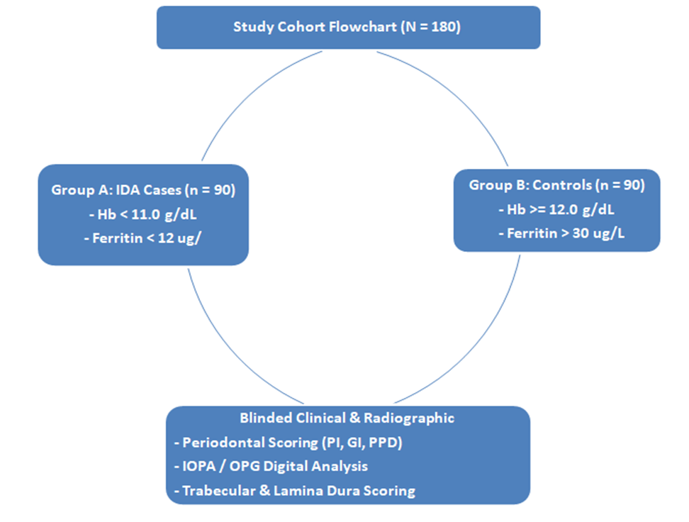
A total of 180 children aged 6 to 14 years attending the outpatient dental and pediatric clinics were enrolled. Patients were categorized into two equal groups based on systemic hematological profiling:
· Group A (IDA Cases, n=90): Children diagnosed with Iron Deficiency Anemia confirmed by blood analysis meeting WHO pediatric diagnostic criteria:
· Hemoglobin (Hb) < 11.0g/dL (for ages 6–11) or < 11.5g/dL (for ages 12–14).
· Serum Ferritin < 12µg/L.
· Transferrin Saturation < 16%.
· Mean Corpuscular Volume (MCV) < 75 fL.
· Group B (Healthy Controls, n=90): Age- and sex-matched children presenting for routine dental check-ups with normal hematological parameters (Hb ≥12.0g/dL, Serum Ferritin ≥ 30µg/L).
Exclusion Criteria (Both Groups):
· Presence of systemic diseases impacting bone metabolism or immunity (e.g., Diabetes Mellitus, Juvenile Idiopathic Arthritis, Leukemia, Renal failure).
· Other forms of anemia (e.g., Thalassemia major/minor, Sickle Cell Anemia, Aplastic Anemia).
· History of systemic antibiotic, anti-inflammatory, or immunosuppressive therapy within the preceding 3 months.
· Ongoing fixed orthodontic therapy or presence of extensive dental restorations/space maintainers impinging on the gingival margin.
· History of iron supplementation therapy within the past 6 months.
Hematological Assessment
Venous blood samples (5 mL) were collected under aseptic conditions in the morning. Complete blood counts (CBC) were obtained using an automated hematology analyzer (Sysmex XN-1000). Serum ferritin levels were measured using an enzyme-linked immunosorbent assay (ELISA), and serum iron and total iron-binding capacity (TIBC) were calculated to determine transferrin saturation.
Clinical Periodontal Evaluation
Comprehensive clinical examinations were performed by a single calibrated pediatric dentist blinded to the hematological status of the subjects. Calibration was established on 15 non-study subjects, yielding an intra-examiner reliability index (Κ= 0.88).
The following standardized indices were assessed at four sites per tooth (mesial, distal, buccal, lingual) across all fully erupted teeth using a Williams periodontal probe:
1. Plaque Index (PI; Silness and Löe, 1964): Evaluated oral hygiene status and soft deposit accumulation.
2. Gingival Index (GI; Löe and Silness, 1963): Quantified gingival inflammation severity (0 = normal to 3 = severe inflammation with spontaneous bleeding).
3. Probing Pocket Depth (PPD): Distance from the gingival margin to the base of the gingival sulcus/pocket, measured in millimeters.
4. Clinical Attachment Level (CAL): Distance from the cementoenamel junction (CEJ) to the base of the sulcus, recorded in cases showing attachment loss.
5. Mucosal Pallor & Soft Tissue Findings: Presence of mucosal pallor, angular cheilitis, and papillary atrophy of the tongue was documented.
Radiographic Evaluation
Standardized digital intraoral periapical (IOPA) radiographs using the parallel technique and digital Orthopantomograms (OPG) were acquired for all subjects (Planmeca ProMax 3D; 66 kVp, 8 mA, 0.12s). Radiographs were analyzed independently by two experienced oral radiologists blinded to group allocations (Κ = 0.85 for inter-rater agreement).
Radiographic Parameters Assessed:
1. Trabecular Bone Architecture: Categorized as Normal (fine, closely spaced trabeculae) vs. Altered (coarse, sparse trabecular arrangement or "stepladder" horizontal alignment between tooth roots).
2. Lamina Dura Integrity: Categorized as Intact, Thinned, or Discontinuous/Lost.
3. Alveolar Crestal Bone Loss: Distance from the CEJ to the alveolar crest > 2mm evaluated across interdental septa.
4. Cortical Bone Density: Thinning of the inferior border of the mandible.
Statistical Analysis
Data were managed using Microsoft Excel and processed via SPSS version 28.0 (IBM Corp., Armonk, NY). Continuous variables were assessed for distribution normality using the Shapiro-Wilk test and presented as mean ± standard deviation (SD). Categorical data were expressed as frequencies and percentages.
· Inter-group comparisons of continuous parametric data (e.g., age, Hb, PI, GI, PPD) were analyzed using the Independent Student’s t-test.
· Non-parametric and categorical variables (e.g., radiographic trabecular patterns, lamina dura loss) were evaluated using Pearson’s Chi-Square (χ2) test or Fisher’s exact test.
· Pearson's correlation coefficient (r) was used to correlate hematological parameters (Hb, serum ferritin) with periodontal and radiographic indices.
· Multivariate linear regression models were constructed to evaluate independent predictors of heightened Gingival Index. Statistical significance was established at p < 0.05.
RESULTS:
Demographic and Hematological Profiles
The study evaluated 180 children (98 males [54.4%], 82 females [45.6%]; mean age 9.6 ± 2.4 years). No statistically significant differences were observed between Group A (IDA) and Group B (Controls) regarding age (9.4 ± 2.5 vs. 9.8 ± 2.3 years; p = 0.264) or sex distribution (p = 0.648).
Hematological profiling confirmed severe-to-moderate anemia in Group A. Mean hemoglobin in the IDA group was 8.6 ± 1.2 g/dL compared to 12.8 ± 0.7 g/dL in controls (p < 0.001). Serum ferritin (8.4 ± 3.1µg/L vs. 42.6 ± 12.8µg/L; p < 0.001) and transferrin saturation (9.2 ± 3.4% vs. 28.4 ± 6.2%; p < 0.001) were markedly depressed in the anemic cohort (Table 1).
Table 1. Baseline Demographic and Hematological Profile of Study Subjects (N = 180)
|
Parameter |
Total Cohort (N = 180) |
Group A: IDA Cases (n = 90) |
Group B: Controls (n = 90) |
p-value |
|
Age (years), Mean ± SD |
9.6 ± 2.4 |
9.4 ± 2.5 |
9.8 ± 2.3 |
0.264 |
|
Sex (Male / Female), n (%) |
98 (54.4) / 82 (45.6) |
50 (55.6) / 40 (44.4) |
48 (53.3) / 42 (46.7) |
0.763 |
|
Hemoglobin (g/dL) |
10.7 ± 2.3 |
8.6 ± 1.2 |
12.8 ± 0.7 |
< 0.001 |
|
Hematocrit (%) |
32.4 ± 6.8 |
26.8 ± 3.4 |
38.0 ± 2.2 |
< 0.001 |
|
Serum Ferritin (µg/L) |
25.5 ± 18.2 |
8.4 ± 3.1 |
42.6 ± 12.8 |
< 0.001 |
|
Serum Iron (μg/dL) |
58.2 ± 28.4 |
32.4 ± 10.6 |
84.0 ± 18.2 |
< 0.001 |
|
Transferrin Saturation (%) |
18.8 ± 10.2 |
9.2 ± 3.4 |
28.4 ± 6.2 |
< 0.001 |
|
MCV (FL) |
74.2 ± 11.4 |
64.5 ± 5.8 |
83.9 ± 4.2 |
< 0.001 |
Periodontal and Soft Tissue Manifestations
Clinical soft tissue evaluation demonstrated a significantly higher frequency of oral mucosal pallor (72.2% vs. 4.4%; p < 0.001), angular cheilitis (26.7% vs. 2.2%; p < 0.001), and papillary atrophy of the tongue (21.1% vs. 1.1%; p < 0.001) in anemic children compared to healthy controls.
As detailed in Table 2, the Plaque Index (PI) showed no statistically significant difference between Group A and Group B (1.42 ± 0.38 vs. 1.36 ± 0.35; p = 0.264), indicating comparable levels of oral hygiene maintenance.
However, children with IDA exhibited significantly higher Gingival Index scores (1.68 ± 0.42 vs. 0.94 ± 0.31; p < 0.001), indicating exaggerated gingival inflammation, edema, and bleeding on probing. Furthermore, Probing Pocket Depths (PPD) were significantly deeper in the anemic group (2.48 ± 0.52mm vs. 1.82 ± 0.34mm; p < 0.001). True clinical attachment loss (CAL > 1mm) was detected in 14.4% (n = 13) of anemic children compared to only 2.2% (n = 2) of healthy controls (p = 0.003).
Table 2. Periodontal Clinical Parameters and Soft Tissue Lesions in Anemic vs. Control Children (N = 180)
|
Clinical Parameter |
Group A: IDA (n = 90) |
Group B: Controls (n = 90) |
Statistic (t / χ2) |
p-value |
|
Plaque Index (PI) |
1.42 ± 0.38 |
1.36 ± 0.35 |
t = 1.12 |
0.264 |
|
Gingival Index (GI) |
1.68 ± 0.42 |
0.94 ± 0.31 |
t = 13.48 |
< 0.001 |
|
Probing Pocket Depth (mm) |
2.48 ± 0.52 |
1.82 ± 0.34 |
t = 10.05 |
< 0.001 |
|
Sites with Bleeding on Probing (%) |
48.6 ± 14.2% |
21.4 ± 8.6% |
t = 15.48 |
< 0.001 |
|
Attachment Loss (CAL > 1mm) |
13 (14.4%) |
2 (2.2%) |
χ2 = 8.64 |
0.003 |
|
Soft Tissue Findings, n (%) |
||||
|
- Oral Mucosal Pallor |
65 (72.2%) |
4 (4.4%) |
χ2 = 87.2 |
< 0.001 |
|
-Angular Cheilitis |
24 (26.7%) |
2 (2.2%) |
χ2 = 20.8 |
< 0.001 |
|
-Atrophic Glossitis |
19 (21.1%) |
1 (1.1%) |
χ2 = 18.1 |
< 0.001 |
Radiographic Manifestations
Digital radiographic analysis revealed marked alterations in the jawbones and alveolar processes of anemic children (Table 3). Overall, altered trabecular architecture was identified in 51 patients (56.7%) in the IDA group compared to 11 patients (12.2%) in the control group (p < 0.001).
Distinct radiographic structural patterns observed in Group A included:
1. "Stepladder" Trabecular Pattern: Horizontal, parallel arrangement of sparse trabeculae in interdental interradicular regions, present in 35 anemic children (38.9%) vs. 5 controls (5.6%; p < 0.001).
2. Thinning or Discontinuity of Lamina Dura: Observed surrounding primary and permanent root surfaces in 40 anemic children (44.4%) vs. 8 controls (8.9%; p < 0.001).
3. Alveolar Crestal Bone Loss (> 2mm): Localized interdental crestal reduction noted in 26 anemic children (28.9%) vs. 4 controls (4.4%; p < 0.001).
4. Thinning of Inferior Mandibular Cortex: Noted on panoramic radiographs in 22 anemic children (24.4%) vs. 3 controls (3.3%; p < 0.001).
Table 3. Radiographic Alveolar and Mandibular Manifestations (N = 180)
|
Radiographic Parameter |
Group A: IDA (n = 90) |
Group B: Controls (n = 90) |
χ2 Value |
p-value |
|
Altered Trabecular Architecture |
51 (56.7%) |
11 (12.2%) |
39.1 |
< 0.001 |
|
- Stepladder Pattern |
35 (38.9%) |
5 (5.6%) |
28.5 |
< 0.001 |
|
- Coarse, Large Marrow Spaces |
28 (31.1%) |
7 (7.8%) |
15.6 |
< 0.001 |
|
Lamina Dura Thinning / Loss |
40 (44.4%) |
8 (8.9%) |
28.9 |
< 0.001 |
|
Interdental Crestal Bone Loss (>2mm) |
26 (28.9%) |
4 (4.4%) |
19.8 |
< 0.001 |
|
Thinning of Inferior Mandibular Border |
22 (24.4%) |
3 (3.3%) |
16.4 |
< 0.001 |
Comparison of Radiographic Findings in Pediatric IDA vs. Controls
|
Altered Trabeculation |
IDA [56.7%] |
Ctrl [8.9%] |
|
Lamina Dura Loss |
IDA [44.4%] |
Ctrl [5.6%] |
|
Stepladder Pattern |
IDA [38.9%] |
Ctrl [5.6%] |
|
Crestal Bone Height Loss |
IDA [28.9%] |
Ctrl [4.4%] |
Bivariate Correlation and Multivariate Analysis
Correlation analysis demonstrated a strong, statistically significant inverse relationship between systemic hemoglobin levels and clinical gingival inflammation (Gingival Index: r = -0.612, p < 0.001). Serum ferritin also correlated negatively with Gingival Index (r = -0.548, p < 0.001) and Probing Pocket Depth (r = -0.492, p < 0.001).
Furthermore, hemoglobin concentration demonstrated a strong negative correlation with the degree of radiographic trabecular alterations (r = -0.584, p < 0.001).
Stepwise multivariate linear regression analysis (Table 4) was executed with Gingival Index as the dependent continuous variable. After adjusting for Plaque Index, age, and sex, both Hemoglobin (β = -0.486, p < 0.001) and Serum Ferritin (β= -0.264, p = 0.001) remained strong, independent predictors of exaggerated gingival inflammation (R2=0.524, Adjusted R2 = 0.518, p < 0.001).
Table 4. Stepwise Multivariate Linear Regression Model Predicting Gingival Index
|
Variable |
Unstandardized β |
Standard Error |
Standardized β |
t-value |
p-value |
|
Constant |
3.124 |
0.214 |
— |
14.6 |
< 0.001 |
|
Hemoglobin (g/dL) |
-0.118 |
0.016 |
-0.486 |
-7.38 |
< 0.001 |
|
Serum Ferritin (μg/L) |
-0.008 |
0.002 |
-0.264 |
- 4 |
0.001 |
|
Plaque Index (PI) |
0.284 |
0.062 |
0.228 |
4.58 |
< 0.001 |
DISCUSSION:
The oral cavity is particularly sensitive to systemic nutritional deficits [13]. While severe hemoglobinopathies like Thalassemia major and Sickle Cell Disease produce classical "hair-on-end" cranial expansion and gross facial skeletal changes, the subtle periodontopathogenic and alveolar osteologic effects of common Iron Deficiency Anemia in growing children have often been overlooked [14]. This prospective study provides clear evidence that pediatric IDA is associated with exaggerated periodontal tissue inflammation, increased probing depths, and distinct alterations in mandibular trabecular architecture.
Systemic Hypoxia and Exaggerated Periodontal Inflammation
Our clinical findings revealed that children with IDA suffered from significantly worse gingival inflammation (Gingival Index 1.68 ± 0.42) and deeper probing depths (2.48 ± 0.52mm) than non-anemic controls, despite displaying almost identical levels of bacterial dental plaque (Plaque Index 1.42 vs. 1.36). This disassociation between bacterial plaque accumulation and host tissue destruction highlights an altered host immune-inflammatory response [15].
Several biological mechanisms account for this hyper-inflammatory mucosal state:
1. Tissue Hypoxia: Chronic iron deficiency lowers blood oxygen-carrying capacity, creating localized microvascular hypoxia within the junctional epithelium and connective tissue stroma [16]. Hypoxia upregulates Hypoxia-Inducible Factor-1α (HIF-1α), triggering downstream secretion of pro-inflammatory cytokines such as Tumor Necrosis Factor-alpha (TNF-α), Interleukin-1β (IL-1β), and Interleukin-6 (IL-6) [17].
2. Impaired Neutrophil Function: Iron is a cofactor for myeloperoxidase within PMNs (polymorphonuclear leukocytes). Iron deficiency impairs neutrophil intracellular bacterial killing and chemotaxis, allowing commensal plaque bacteria to provoke heightened tissue damage [18].
3. Epithelial Thinning: Iron deficiency impairs DNA synthesis and rapid cell division in basal epithelial cells, leading to atrophy of the sulcular epithelium, diminished mucosal barrier function, and enhanced ulceration/bleeding on probing [19].
Biological Basis of Radiographic Trabecular Alterations
Radiographically, over 56% of anemic children in our cohort displayed altered trabecular architecture. The most prominent patterns were the "stepladder" arrangement of interdental trabeculae (38.9%) and thinning/loss of the lamina dura (44.4%).
These radiographic manifestations reflect compensatory marrow hyperplasia [20]. Chronic systemic anemia stimulates renal erythropoietin secretion, driving hyperplastic expansion of the red bone marrow within cancellous spaces [21]. In the mandible where active marrow exists within the angle, body, and interdental septa of growing children, marrow expansion resorption enlarges cancellous spaces [22]. The remaining trabeculae become coarse, thinned, and re-oriented along lines of stress, producing the characteristic horizontal "stepladder" appearance [23].
Simultaneously, systemic mineral metabolism and localized osteoclast activation account for the loss of lamina dura and interdental crestal bone height [24]. Upregulation of RANKL (Receptor Activator of Nuclear Factor-ΚB Ligand) induced by chronic inflammation accelerates localized alveolar bone resorption [25].
Clinical Implications for Pediatric Dental Practice
These findings carry direct clinical relevance for pediatric dentists, general dental practitioners, and pediatricians:
1. Early Systemic Screening: When a pediatric patient presents with severe, generalized gingivitis, bleeding gums, or mucosal pallor out of proportion to their oral hygiene status, clinicians should suspect an underlying systemic condition like IDA and order a complete blood count [26].
2. Radiographic Interpretation: Pediatric dentists should carefully evaluate routine panoramic or periapical radiographs for trabecular coarsening, stepladder patterns, or thinning lamina dura, which may serve as an incidental early sign of undiagnosed anemia [27].
3. Interdisciplinary Management: Periodontal therapy in anemic children should be combined with systemic iron supplementation managed by a pediatrician. Restoring normal hemoglobin and iron stores accelerates periodontal tissue healing and stabilizes alveolar bone remodeling [28].
STUDY LIMITATIONS
Several limitations should be noted. First, this study utilized a cross-sectional baseline design; longitudinal follow-up of the anemic cohort after systemic iron repletion therapy is required to observe whether periodontal indices and trabecular architecture normalize over time. Second, advanced volumetric imaging such as Cone-Beam Computed Tomography (CBCT) was not used to limit pediatric radiation exposure, relying instead on 2D digital radiography. Third, inflammatory biomarkers in gingival crevicular fluid (IL-1β, TNFα) were not quantitatively measured, which could provide further biochemical validation of tissue-level inflammatory cascades.
CONCLUSION:
Iron Deficiency Anemia in children is significantly correlated with heightened gingival inflammation, deeper periodontal probing pocket depths, and an increased risk of localized attachment loss, independent of oral hygiene levels. Furthermore, pediatric IDA produces characteristic digital radiographic manifestations in the jawbones, including altered coarse trabeculation, "stepladder" interdental alignment, and thinning of the lamina dura.
Pediatric dentists and healthcare providers should recognize these oral and radiographic markers. Integrating systemic hematological screening into pediatric dental diagnostics allows for early detection of iron deficiency, facilitating timely nutritional and medical interventions that safeguard both systemic development and long-term oral health.
REFERENCES:
1. World Health Organization. Worldwide prevalence of anaemia 1993-2005: WHO global database on anaemia. Geneva: World Health Organization; 2008.
2. Pasricha SR, Tye-Din J, Muckenthaler MU, Swinkels DW, Hentze MW. Iron deficiency. Lancet. 2021;397(10282):1833-1848.
3. Andrews NC. Disorders of iron metabolism. N Engl J Med. 1999;341(26):1986-1995.
4. Oppenheimer SJ. Iron and its relation to immunity and infectious disease. J Nutr. 2001;131(2S-2):616S-633S.
5. Wu YC, Wang YP, Chang JY, Sun A, Chen HM, Chiang CP. Oral manifestations and blood profile in patients with iron deficiency anemia. J Formos Med Assoc. 2014;113(2):83-87.
6. Lu SY. Perception of iron deficiency from oral mucosa alterations that show a high prevalence of Candida infection. J Formos Med Assoc. 2016;115(8):619-27.
7. Ranasinghe AW, Warnakulasuriya KA, Tennekoon GE, Seneviratna B. Oral mucosal changes in iron deficiency anemia in a Sri Lankan female population. Oral Surg Oral Med Oral Pathol. 1983;55(1):29-32.
8. Hajishengallis G, Chavakis T, Lambris JD. Current understanding of periodontal disease pathogenesis and targets for host-modulation therapy. Periodontol 2000. 2020;84(1):14-34..
9. Peres MA, Macpherson LM, Weyant RJ, Daly B, Venturelli R, Mathur MR, et al. Oral diseases: a global public health challenge. Lancet. 2019;394(10194):249-260.
10. Seeman E. Bone quality: the material and structural basis of bone strength. J Musculoskelet Neuronal Interact. 2008;8(4):309-310.
11. Cetin M, Gönül A, Kara A, Kara SP, Yetgin S. Profile of bone marrow iron stores in childhood iron deficiency anemia. Turk J Pediatr. 1999;41(3):329-34.
12. White SC, Pharoah MJ. Oral radiology: principles and interpretation. 7th ed. St. Louis: Mosby; 2014.
13. Tolkachjov SN, Bruce AJ. Oral manifestations of nutritional disorders. Clin Dermatol. 2017;35(5):441-452.
14. Mourshed F, Tuckson CR. A study of the radiographic features of the jaws in sickle-cell anemia. Oral Surg Oral Med Oral Pathol. 1974;37(5):812-9.
15. Kinane DF, Stathopoulou PG, Papapanou PN. Periodontal diseases. Nat Rev Dis Primers. 2017;3:17038.
16. Socransky SS, Haffajee AD. Periodontal microbial etiology: periodontology 2000. Periodontol 2000. 2005;38:135-187.
17. Taylor CT, Colgan SP. Hypoxia and gastrointestinal inflammation. J Physiol. 2017;595(9):3031-3039.
18. Brock JH. The physiology of iron metabolism and its pathogenesis in infection. Infect Dis Clin North Am. 1999;13(2):341-352.
19. Rennie JS, MacDonald DG, Dagg JH. Quantitative haempathology of oral epithelium in iron deficiency anaemia. J Oral Pathol. 1984;13(4):389-393.
20. Poyton HG, Davey KW. Thalassemia: changes in the oral cavity and jaws. Oral Surg Oral Med Oral Pathol. 1968;25(4):564-576.
21. Cavill I. Erythropoiesis and Iron. Best Pract Res Clin Haematol. 2002;15(2):399-409.
22. Wood RE. Malignancies and systemic diseases of the jaws. In: White SC, Pharoah MJ, editors. Oral radiology. 7th ed. St. Louis: Mosby; 2014. p. 480-510.
23. Singhal S, Sharma S. Radiographic changes of jaw bones in pediatric anemia: A comparative evaluation. J Indian Soc Pedod Prev Dent. 2018;36(2):142-147.
24. Kalhan AC, Wong ML, Allen F, Gao X. Periodontal disease and systemic health: An update for medical practitioners. Ann Acad Med Singap. 2022 Sep;51(9):567-574.
25. Boyce BF, Xing L. Functions of RANKL/RANK/OPG in bone modeling and remodeling. Arch Biochem Biophys. 2008;473(2):139-146.
26. Glick M, editor. Burket's Oral Medicine. 12th ed. Shelton (CT): People's Medical Publishing House; 2015.
27. Whaites E, Drage N. Essentials of Dental Radiography and Radiology. 5th ed. Edinburgh: Churchill Livingstone Elsevier; 2013.
28. Goodson JM, Hasturk H, Yaskell T, Cyzio P, Van Dyke TE. Iron deficiency anemia as a risk factor for periodontitis. J Periodontol. 2020;91(7):891-899.