A STUDY OF ECHOCARDIOGRAPY IN PATIENTS OF TYPE 2 DIABETES MELLITUS.
- Pokuru Mohan Krishna , DNB (General Medicine), Department of Medicine, ESIC Hospital and Dental College, Rohini Sector -15, Delhi-85.
- Kapil Kumar Verma , Professor and HOD Department of Medicine, ESIC Medical College and Hospital, Bapunagar Ahmedabad.
- Vijay Kumar Malviya , Professor and HOD, Department of Medicine, ESIC Medical College, Indore (M.P).
- Meher Shikha Verma , Associate Professor, Department of Anaesthesia, Sri Aurobindo Medical College and Postgraduate institute, Indore (M.P).
Article Information:
Abstract:
Background: Type 2 diabetes mellitus (T2DM) is associated with an increased risk of cardiovascular complications due to structural and functional myocardial changes. Echocardiography is a reliable, noninvasive imaging modality for detecting early cardiac abnormalities before the onset of symptomatic heart disease. This study evaluated echocardiographic changes and their association with demographic and clinical characteristics in patients with T2DM. Methods: A hospital-based cross-sectional observational study was conducted among 220 adult patients with T2DM attending the inpatient and outpatient departments of ESIC Model Hospital and Occupational Disease Centre, Indore, India, between May 2023 and April 2024. All participants underwent clinical evaluation, laboratory investigations including glycated hemoglobin (HbA1c), and comprehensive two-dimensional transthoracic echocardiography. Echocardiographic findings were analyzed in relation to age, gender, duration of diabetes, and glycemic control. Statistical analysis was performed using SPSS version 24.0, with p<0.05 considered statistically significant. Results: The mean age of the participants was 53.53±9.41 years, and 50.9% were males. Diastolic dysfunction was the most common echocardiographic abnormality (26.0%), followed by left ventricular hypertrophy (19.5%), reduced left ventricular ejection fraction (13.6%), diabetic cardiomyopathy (13.0%), left atrial enlargement (10.5%), and dilated left ventricle (4.5%). Increasing age was significantly associated with left atrial enlargement, left ventricular hypertrophy, and diabetic cardiomyopathy (p<0.001). Longer duration of diabetes was significantly associated with left atrial enlargement and left ventricular hypertrophy (p<0.001). Poor glycemic control (HbA1c >8.5%) showed significant associations with left atrial enlargement, left ventricular hypertrophy, dilated left ventricle, and diabetic cardiomyopathy (p<0.001), whereas gender showed no significant association with most echocardiographic abnormalities. Conclusions: Echocardiographic abnormalities are common in patients with T2DM, particularly diastolic dysfunction and left ventricular hypertrophy. Advancing age, longer duration of diabetes, and poor glycemic control are important determinants of cardiac structural and functional changes. Routine echocardiographic screening may facilitate early detection and timely management of diabetic cardiac complications, thereby improving long-term cardiovascular outcomes.
Keywords:
Article :
INTRODUCTION:
Diabetes mellitus (DM) is a chronic metabolic disorder characterized by impaired insulin secretion, insulin action, or both, resulting in persistent hyperglycemia and abnormalities in carbohydrate, fat, and protein metabolism [1]. Type 2 diabetes mellitus (T2DM) is the most common form of diabetes and has become a major global public health concern due to its rapidly increasing prevalence. India is among the countries with the highest burden of diabetes, with an estimated 77 million affected individuals, making it the second most affected country after China [2]. The rising prevalence of T2DM has been accompanied by a marked increase in chronic complications, particularly cardiovascular disease (CVD), which remains the leading cause of morbidity and mortality in this population [3].
Patients with T2DM have a two- to four-fold higher risk of developing cardiovascular complications, including coronary artery disease, diabetic cardiomyopathy, heart failure, and stroke. Diabetic cardiomyopathy is characterized by structural and functional myocardial abnormalities that occur independently of hypertension or coronary artery disease. Moreover, coronary artery disease in diabetic patients is frequently asymptomatic because of autonomic neuropathy, leading to delayed diagnosis and poor clinical outcomes [4]. Early detection of cardiac involvement is therefore essential to reduce cardiovascular morbidity and improve long-term prognosis.
Echocardiography is a safe, noninvasive, readily available, and cost-effective imaging modality that provides real-time assessment of cardiac anatomy and function. It plays a central role in evaluating ventricular dimensions, chamber size, myocardial thickness, systolic and diastolic function, and other structural abnormalities [5]. Echocardiography is particularly useful for identifying subclinical cardiac changes before the onset of overt heart failure or symptomatic cardiovascular disease, thereby facilitating early therapeutic intervention and risk stratification.
Several echocardiographic parameters, including left ventricular ejection fraction, interventricular septal thickness, left ventricular posterior wall thickness, left ventricular internal diameter, left atrial size, and left ventricular mass, are important indicators of cardiac remodeling in patients with diabetes. Assessment of these parameters enables the identification of early myocardial involvement and may help predict future cardiovascular events.
Therefore, the present study was undertaken to evaluate echocardiographic changes in patients with type 2 diabetes mellitus attending the inpatient and outpatient departments of ESIC Model Hospital and Occupational Disease Centre, Nanda Nagar, Indore, with the objective of identifying structural and functional cardiac abnormalities associated with T2DM and emphasizing the importance of early cardiovascular evaluation in this high-risk population.
MATERIALS AND METHODS:
This hospital-based cross-sectional observational study was conducted in the Department of Medicine at ESIC Model Hospital and Occupational Disease Centre, Nanda Nagar, Indore, Madhya Pradesh, India, between May 2023 and April 2024. The study was approved by the Institutional Ethics Committee, and written informed consent was obtained from all participants prior to enrollment.
A total of 220 consecutive adult patients with type 2 diabetes mellitus (T2DM) attending the inpatient and outpatient departments during the study period were included in the study. The diagnosis of T2DM was established according to the American Diabetes Association (ADA) criteria, defined as fasting plasma glucose (FPG) ≥126 mg/dL, 2-hour plasma glucose ≥200 mg/dL during an oral glucose tolerance test, HbA1c ≥6.5%, or documented treatment for diabetes mellitus.
Inclusion and Exclusion Criteria
Patients aged more than 30 years with known or newly diagnosed type 2 diabetes mellitus who provided written informed consent were eligible for inclusion.
Patients with known coronary artery disease, valvular heart disease, chronic obstructive pulmonary disease (COPD), chronic alcoholism, smoking history, pregnancy, age below 30 years or above 75 years, and those unwilling to participate were excluded from the study.
Clinical Assessment and Data Collection
A detailed clinical history was obtained from all participants, including demographic characteristics, duration of diabetes, treatment history, associated comorbidities, and cardiovascular risk factors. A comprehensive physical examination was performed, and vital parameters were recorded at the time of evaluation. Laboratory investigations included fasting blood sugar (FBS), postprandial blood sugar (PPBS), glycated hemoglobin (HbA1c), renal function tests, urine routine and microscopy, electrocardiography (ECG), and chest radiography wherever clinically indicated.
Echocardiographic Assessment
All enrolled participants underwent comprehensive two-dimensional transthoracic echocardiography performed by experienced cardiologists using standard imaging protocols. Echocardiographic measurements were obtained primarily in the parasternal long-axis (PLAX) view.
The following echocardiographic parameters were evaluated:
• Left atrial (LA) volume
• Peak early mitral inflow velocity (E wave)
• Peak late mitral inflow velocity (A wave)
• Interventricular septal thickness at end-diastole (IVSd)
• Left ventricular posterior wall thickness at end-diastole (LVPWd)
• Left ventricular internal diameter at end-diastole (LVIDd)
• Left ventricular end-diastolic volume (EDV)
• Left ventricular end-systolic volume (ESV)
• Left ventricular ejection fraction (LVEF)
• Left ventricular mass (LVM)
Echocardiographic abnormalities including left ventricular hypertrophy, left atrial enlargement, reduced ejection fraction, ventricular dilatation, and diastolic dysfunction were documented according to standard echocardiographic reference values.
Definition of Echocardiographic Abnormalities
• Diabetes mellitus was defined as fasting plasma glucose ≥126 mg/dL, 2-hour plasma glucose ≥200 mg/dL, HbA1c ≥6.5%, or current treatment for diabetes.
• Hypertension was classified according to the Eighth Joint National Committee (JNC-8) recommendations.
• Left ventricular hypertrophy was defined as interventricular septal thickness or left ventricular posterior wall thickness >1.0 cm or an increased left ventricular mass index according to standard guideline recommendations.
• Left atrial enlargement was defined as a left atrial volume index (LAVI) ≥34 mL/m².
• Reduced left ventricular systolic function was defined as a left ventricular ejection fraction (LVEF) <55%.
• Left ventricular dilatation was defined as an increased left ventricular internal diameter at end-diastole beyond normal sex-specific reference values.
Study Outcome Measures
The primary objective of the study was to evaluate echocardiographic changes among patients with type 2 diabetes mellitus. The prevalence of structural and functional cardiac abnormalities, including left ventricular hypertrophy, left atrial enlargement, ventricular dilatation, reduced left ventricular ejection fraction, and diastolic dysfunction, was assessed. The association of these echocardiographic findings with clinical and biochemical characteristics of diabetes was also evaluated.
Statistical Analysis
Data were entered into Microsoft Excel and analyzed using Statistical Package for the Social Sciences (SPSS) software version 24.0 (IBM Corp., Armonk, NY, USA). Continuous variables were expressed as mean ± standard deviation (SD), whereas categorical variables were presented as frequencies and percentages. Comparisons between categorical variables were performed using the chi-square test or Fisher's exact test, as appropriate. Continuous variables were compared using the independent Student's t-test or one-way analysis of variance (ANOVA), wherever applicable. A p-value <0.05 was considered statistically significant.
RESULTS:
A total of 220 patients with type 2 diabetes mellitus (T2DM) were included in the study. The mean age of the study population was 53.53 ± 9.41 years, with the majority of patients (53.6%) belonging to the 51–65 years age group. The gender distribution was nearly equal, with 112 (50.9%) males and 108 (49.1%) females. Most patients had a duration of diabetes of 6–10 years (36.8%), followed by <5 years (35.0%). Regarding glycemic control, 113 (51.4%) patients had HbA1c levels between 7.0% and 8.5%, while 32 (14.5%) had HbA1c levels >8.5%. Hypertension was present in 100 (45.0%) patients (Table 1, Figure 1).
Table 1. Baseline demographic and clinical characteristics of the study participants
|
Variable |
Category |
n |
% |
|
Age (years) |
<40 |
15 |
6.8 |
|
41–50 |
69 |
31.4 |
|
|
51–65 |
118 |
53.6 |
|
|
>65 |
15 |
6.8 |
|
|
Gender |
Male |
112 |
50.9 |
|
Female |
108 |
49.1 |
|
|
Duration of T2DM (years) |
<5 |
77 |
35.0 |
|
6–10 |
81 |
36.8 |
|
|
11–15 |
37 |
16.8 |
|
|
>15 |
25 |
11.4 |
|
|
HbA1c (%) |
<7.0 |
75 |
34.1 |
|
7.0–8.5 |
113 |
51.4 |
|
|
>8.5 |
32 |
14.5 |
|
|
Blood Pressure |
Normal |
120 |
55.0 |
|
Hypertension |
100 |
45.0 |
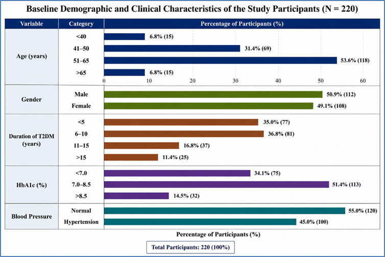
Figure 1: Baseline demographic and clinical characteristics of the study participants
Overall echocardiographic evaluation demonstrated that 57 (26.0%) patients had diastolic dysfunction, making it the most common abnormality observed. Left ventricular hypertrophy was present in 43 (19.5%) patients, followed by reduced left ventricular ejection fraction in 30 (13.6%), diabetic cardiomyopathy in 28 (13.0%), left atrial enlargement in 23 (10.5%), and dilated left ventricle in 10 (4.5%) patients (Table 2, Figure 2).
Table 2. Distribution of echocardiographic abnormalities among study participants
|
Variable |
Frequency (%) |
|
Left atrial enlargement |
23 (10.5) |
|
Diastolic dysfunction |
57 (26.0) |
|
Left ventricular hypertrophy |
43 (19.5) |
|
Dilated left ventricle |
10 (4.5) |
|
Reduced ejection fraction (<55%) |
30 (13.6) |
|
Diabetic cardiomyopathy |
28 (13.0) |
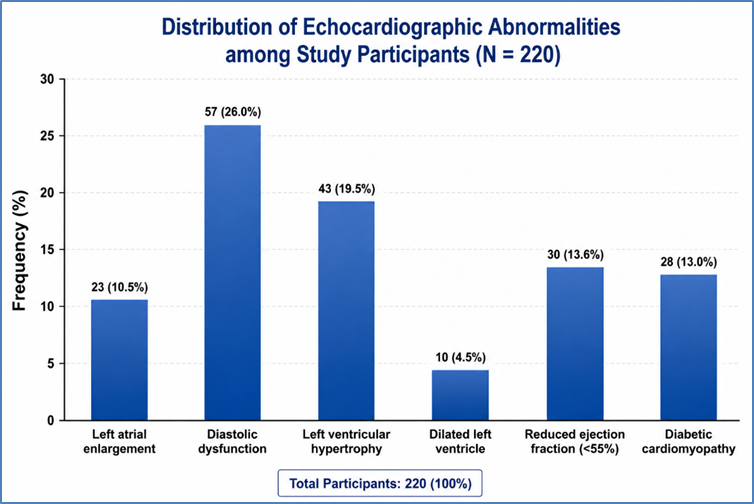
Figure 2: Distribution of echocardiographic abnormalities among study participants
Among patients with diastolic dysfunction, Grade I dysfunction was the most common finding, observed in 40 (18.2%) patients, followed by Grade II dysfunction in 12 (5.5%) and Grade III dysfunction in 5 (2.3%) patients. Valvular abnormalities were uncommon. Aortic stenosis was identified in 7 (3.2%) patients, aortic regurgitation in 1 (0.5%), and mitral regurgitation in 6 (2.7%), while the majority of patients had structurally normal aortic and mitral valves (Table 3, Figure 3).
Table 3. Distribution of diastolic dysfunction grades and valvular abnormalities
|
Variable |
Frequency (%) |
|
Diastolic dysfunction grade |
|
|
Normal |
163 (74.0) |
|
Grade I |
40 (18.2) |
|
Grade II |
12 (5.5) |
|
Grade III |
5 (2.3) |
|
Aortic valve |
|
|
Normal |
212 (96.4) |
|
Aortic stenosis |
7 (3.2) |
|
Aortic regurgitation |
1 (0.5) |
|
Mitral valve |
|
|
Normal |
214 (97.3) |
|
Mitral regurgitation |
6 (2.7) |
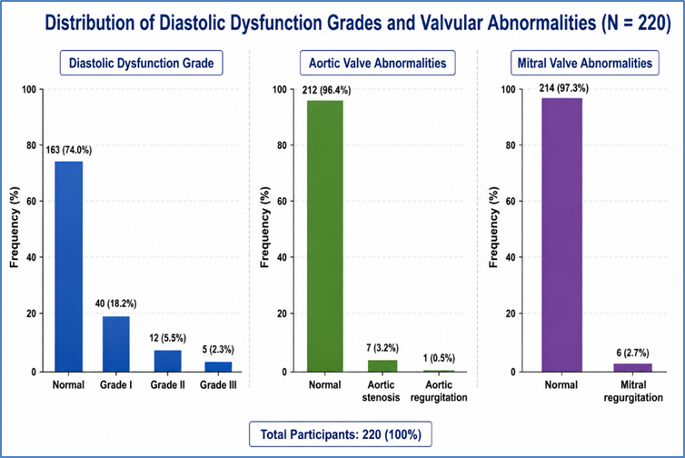
Distribution of diastolic dysfunction grades and valvular abnormalities
Comparative analysis showed that increasing age was significantly associated with left atrial enlargement, left ventricular hypertrophy, and diabetic cardiomyopathy (p<0.001). The prevalence of left atrial enlargement increased from 6.7% in patients aged <40 years to 50.0% in those aged >65 years, while left ventricular hypertrophy increased from 6.7% to 61.1% across the same age groups. Diabetic cardiomyopathy was also more common among older patients (27.8% vs. 6.7%). Grade II and III diastolic dysfunction were more frequent with advancing age, whereas reduced ejection fraction showed no significant association with age (χ²=5.246, p=0.342) (Table 4).
Table 4: Comparison of echocardiographic change in study population according to Age
|
Parameters |
Age |
Chi Square |
P Value |
|||
|
<40 (N=15) |
41 to 50 (N=69) |
51 to 65 (N=118) |
>65 (N=18) |
|||
|
Left Atrial Enlargement (+) |
1 (6.67%) |
2 (2.90%) |
11 (9.32%) |
9 (50.0%) |
82.28 |
0 |
|
GRADES OF DIASTOLIC DYSFUNCTION |
||||||
|
Grade 1 |
4(26.67%) |
13(18.84%) |
20(16.96%) |
3(16.67%) |
* |
*** |
|
Grade 2 |
0 |
1(1.45%) |
9(7.63%) |
2(11.11%) |
||
|
Grade 3 |
0 |
2(2.90%) |
1(0.85%) |
2 (11.11%) |
||
|
LEFT VENTRICULAR HYPERTROPHY(LVH) |
||||||
|
Normal |
14 (93.33%) |
62 (89.60%) |
94 (79.66%) |
7 (38.89%) |
* |
*** |
|
LVH |
1 (6.67%) |
7 (10.14%) |
24 (20.34%) |
11 (61.11%) |
||
|
DILATED LEFT VENTRICLE |
||||||
|
Dilated Left Ventricle |
0 |
3(4.35%) |
5(4.24%) |
2(11.11%) |
* |
*** |
|
Normal |
15(100.0%) |
66(95.65%) |
113(95.76%) |
16(88.89%) |
||
|
EJECTION FRACTION |
||||||
|
EF% (Reduced) |
0 |
5 (7.25%) |
22 (18.64%) |
3 (16.6%) |
5.246 |
0.342 |
|
DIABETIC CARDIOMYOPATHY |
||||||
|
Diabetic Cardiomyopathy |
1 (6.67%) |
6 (8.69%) |
16 (13.56%) |
5 (27.78%) |
88.64 |
0 |
A longer duration of type 2 diabetes mellitus was significantly associated with left atrial enlargement and left ventricular hypertrophy (p<0.001), whereas no significant association was observed with reduced ejection fraction or diabetic cardiomyopathy. Poor glycemic control (HbA1c >8.5%) was significantly associated with left atrial enlargement, left ventricular hypertrophy, dilated left ventricle, and diabetic cardiomyopathy (p<0.001). (Table 5).
Table 5: Comparison of echocardiographic change in study Duration of DM2
|
Variable |
Duration of T2DM(Yrs) |
χ² |
P-value |
|||
|
<5 years (n=77) |
6–10 years (n=81) |
11–15 years (n=37) |
>15 years (n=25) |
|||
|
Left atrial enlargement, n (%) |
1 (1.3) |
5 (6.2) |
10 (27.0) |
7 (28.0) |
155.11 |
<0.001* |
|
GRADES OF DIASTOLIC DYSFUNCTION |
||||||
|
Grade I |
14 (18.2) |
16 (19.8) |
7 (18.9) |
3 (12.0) |
* |
*** |
|
Grade II |
4 (5.2) |
2 (2.5) |
4 (10.8) |
2 (8.0) |
||
|
Grade III |
0 |
2 (2.5) |
1 (2.7) |
2 (8.0) |
||
|
LEFT VENTRICULAR HYPERTROPHY (LVH) |
||||||
|
Normal |
70 (90.91%) |
71 (87.65%) |
24 (64.86%) |
12 (48.0%) |
* |
*** |
|
Left ventricular hypertrophy, n (%) |
7 (9.1) |
10 (12.4) |
13 (35.1) |
13 (52.0) |
||
|
DILATED LEFT VENTRICLE |
||||||
|
Dilated left ventricle, n (%) |
3 (3.9) |
4 (4.9) |
2 (5.4) |
1 (4.0) |
* |
*** |
|
Normal |
74 (96.10%) |
77 (95.06%) |
35 (94.59%) |
24 (96.0%) |
||
|
EJECTION FRACTION |
||||||
|
Reduced ejection fraction (<55%), n (%) |
5 (6.5) |
12 (14.8) |
6 (16.2) |
7 (28.0) |
6.211 |
0.411 |
|
DIABETIC CARDIOMYOPATHY |
||||||
|
Diabetic cardiomyopathy, n (%) |
5 (6.5) |
10 (12.3) |
8 (21.6) |
5 (20.0) |
8.244 |
0.370 |
However, HbA1c was not significantly associated with reduced ejection fraction (p=0.774). Gender was not significantly associated with the majority of echocardiographic abnormalities, although females showed slightly higher frequencies of left atrial enlargement, left ventricular hypertrophy, reduced ejection fraction, dilated left ventricle, and diabetic cardiomyopathy than males.
DISCUSSION:
Type 2 diabetes mellitus (T2DM) is associated with a substantially increased risk of cardiovascular disease because chronic hyperglycemia promotes structural and functional myocardial remodeling. Echocardiography provides a noninvasive method for detecting these changes at an early stage, even before the development of overt cardiovascular symptoms. The present study evaluated echocardiographic abnormalities in 220 patients with T2DM and examined their association with age, gender, duration of diabetes, and glycemic control. Overall, diastolic dysfunction was the most common echocardiographic abnormality, followed by left ventricular hypertrophy (LVH), reduced ejection fraction, diabetic cardiomyopathy, and left atrial enlargement. These abnormalities were significantly associated with advancing age, longer duration of diabetes, and poor glycemic control, emphasizing the importance of early cardiovascular assessment in patients with T2DM. The study population predominantly consisted of middle-aged adults, with more than half of the participants belonging to the 51-65 years age group and a mean age of 53.53±9.41 years. The gender distribution was almost equal, indicating that T2DM affects both sexes similarly in the study population. These findings are consistent with previous studies by Almigbal et al. (2023) [6] and Jaiswal et al. (2022) [7], who also reported that most patients with T2DM were middle-aged with a nearly equal distribution between males and females. Similar observations have been reported in South Asian populations, where increasing age remains one of the strongest determinants of diabetes prevalence [8,9].
Most participants had diabetes for less than 10 years, while only a small proportion had disease duration exceeding 15 years. Nevertheless, echocardiographic abnormalities increased progressively with longer duration of diabetes. Left atrial enlargement increased from 1.3% in patients with diabetes duration of less than five years to 28.0% in those with disease duration greater than 15 years, while LVH increased from 9.9% to 52.0% across the same groups. These findings indicate that prolonged exposure to hyperglycemia contributes to adverse cardiac remodeling. Similar observations were reported by Hodgar (2019) [10] and Liao et al. (2022) [11], who demonstrated significant deterioration in diastolic function with increasing duration of diabetes. Assessment of glycemic control revealed that approximately half of the patients had HbA1c values between 7.0% and 8.5%, whereas 14.5% had poor glycemic control (HbA1c >8.5%). Poor glycemic control showed a strong association with adverse echocardiographic findings, particularly left atrial enlargement, LVH, dilated left ventricle, and diabetic cardiomyopathy. Patients with HbA1c levels greater than 8.5% demonstrated markedly higher frequencies of these abnormalities compared with patients having better glycemic control. These observations are in agreement with studies by Liao et al. (2022) [11], Zhao et al. (2020) [12], and Guria et al. (2022) [13], which reported that higher HbA1c levels were independently associated with impaired ventricular relaxation, increased ventricular mass, and left ventricular diastolic dysfunction. Similar findings have also been reported by previous investigators demonstrating progressively increasing rates of diastolic dysfunction with worsening glycemic control [14].
Diastolic dysfunction was the most common echocardiographic abnormality observed in the present study, affecting 26.0% of patients, with Grade I dysfunction accounting for the majority of cases. This finding supports the concept that impaired ventricular relaxation represents one of the earliest manifestations of diabetic cardiomyopathy. LVH was observed in 19.5% of patients, whereas diabetic cardiomyopathy and reduced ejection fraction were identified in 13.0% and 13.6% of patients, respectively. Left atrial enlargement was present in 10.5% of patients, while dilated left ventricle and valvular abnormalities were relatively uncommon. Comparable findings have been reported by Jørgensen et al. (2016) [15], who observed a high prevalence of diastolic dysfunction, LVH, and left atrial enlargement among patients with T2DM. Similar echocardiographic changes were also described by Zhao et al. (2020) [7], Sardesai et al. (2022) [16], Jaiswal et al. (2022) [17], and other studies evaluating diabetic cardiac remodeling [18-20]. Advancing age demonstrated a significant association with left atrial enlargement, LVH, and diabetic cardiomyopathy. The prevalence of LVH increased substantially among patients older than 65 years, while severe grades of diastolic dysfunction were more frequently observed in older age groups. These findings indicate that ageing, together with prolonged metabolic stress, accelerates myocardial structural changes in patients with T2DM. Similar observations have been reported by Jørgensen et al. (2016) [15], who demonstrated that echocardiographic abnormalities become increasingly prevalent with advancing age. Previous studies have also identified LVH as an established predictor of cardiovascular morbidity and mortality in elderly diabetic patients [21].
Gender-based differences were relatively modest in the present study. Although female patients demonstrated slightly higher frequencies of left atrial enlargement, LVH, reduced ejection fraction, and diabetic cardiomyopathy, these differences were not statistically significant. Similar trends have been reported by Jørgensen et al. (2016) [15], who found greater LVH and diastolic dysfunction among women, whereas men more frequently exhibited reduced systolic function. However, findings from the Framingham Study [22] and the Strong Heart Study [23] suggest that the influence of gender on cardiac remodeling in diabetes remains inconsistent and is likely modified by other cardiovascular risk factors. Overall, the findings of the present study reinforce the established relationship between T2DM and subclinical cardiac dysfunction. Advancing age, prolonged disease duration, and poor glycemic control were the major determinants of echocardiographic abnormalities. These observations support the incorporation of routine echocardiographic evaluation into the clinical assessment of patients with T2DM, particularly those with long-standing disease or inadequate glycemic control, to facilitate early diagnosis and timely intervention aimed at reducing future cardiovascular complications.
CONCLUSION:
Echocardiographic abnormalities are frequently observed in patients with type 2 diabetes mellitus, with diastolic dysfunction being the most common finding, followed by left ventricular hypertrophy, reduced ejection fraction, left atrial enlargement, and diabetic cardiomyopathy. Advancing age, longer duration of diabetes, and poor glycemic control were significantly associated with these cardiac abnormalities, whereas gender showed no significant association. These findings emphasize the importance of routine echocardiographic screening and strict glycemic control for the early identification and management of subclinical cardiac involvement, thereby reducing the risk of cardiovascular complications and improving long-term clinical outcomes.
REFERENCES:
1. Powers AC, Stafford JM, Rickels MR. 20th ed. New York: McGraw Hill Education; 2018. Harrison's Principles of Internal Medicine.
2. Whiting DR, Guariguata L, Weil C, Shaw J. IDF diabetes atlas: global estimates of the prevalence of diabetes for 2011 and 2030. Diabetes research and clinical practice. 2011 Dec 1;94(3):311-21.
3. Ramachandran A. Know the signs and symptoms of diabetes. Indian Journal of Medical Research. 2014 Nov 1;140(5):579-81.
4. Jia G, Hill MA, Sowers JR. Diabetic cardiomyopathy: an update of mechanisms contributing to this clinical entity. Circulation research. 2018 Feb 16;122(4):624-38.
5. Zarich SW, Nesto RW. Diabetic cardiomyopathy. Am Heart J. 2001;35:166-8.
6. Almigbal TH, Almunif DS, Aburisheh KH, Barhoush MM, Aldhahi RA, Anabi MJ, Alotaibi OA. Echocardiographic Changes in Saudi Patients with Type 2 Diabetes Mellitus. Medicina. 2023 Nov 10;59(11):1985.
7. Jaiswal NK, Karki M, Dhakal PR, Dahal M, Kandel S, Shahi A, Chaudhary S. Clinical Profile and Echocardiographic Changes in Patients with Newly Diagnosed Type 2 Diabetes. Journal of Universal College of Medical Sciences. 2022 Dec 31;10(02):27-30.
8. Jayawardena R, Ranasinghe P, Byrne NM, Soares MJ, Katulanda P, Hills AP. Prevalence and trends of the diabetes epidemic in South Asia: a systematic review and meta-analysis. BMC public health. 2012 Dec;12:1-1.
9. Nordström* A, Hadrévi J, Olsson T, Franks PW, Nordström P. Higher prevalence of type 2 diabetes in men than in women is associated with differences in visceral fat mass. The Journal of Clinical Endocrinology & Metabolism. 2016 Oct 1;101(10):3740-6.
10. Hodgar DD. Echocardiographic evaluation of diastolic dysfunction in type 2 diabetes mellitus without cardiac manifestations in rural hospital (Doctoral dissertation).2019.
11. Liao L, Shi B, Ding Z, Chen L, Dong F, Li J, Zhong Y, Xu J. Echocardiographic study of myocardial work in patients with type 2 diabetes mellitus. BMC Cardiovascular Disorders. 2022 Feb 17;22(1):59.
12. Zhao Z, Hou C, Ye X, Cheng J. Echocardiographic changes in newly diagnosed type 2 diabetes mellitus patients with and without hypertension. Medical science monitor: international medical journal of experimental and clinical research. 2020;26:e918972-1.
13. Guria RT, Prasad MK, Mishra B, Marandi S, Kumar A, Dungdung A. Association of glycosylated haemoglobin (HbA1c) level with left ventricular diastolic dysfunction in patients with type 2 diabetes. Cureus. 2022 Nov;14(11).
14. Meena RK, Ravichandran R, Sharma S, Sudharsan S, Kumari P. Subclinical diastolic dysfunction and its correlation with laboratory parameters in type 2 diabetes mellitus in India: a case control study. Romanian Journal of Diabetes Nutrition and Metabolic Diseases. 2018 Jun 16;25(2):157-64.
15. Jørgensen PG, Biering-Sørensen T, Mogelvang R, Fritz-Hansen T, Vilsbøll T, Rossing P, Jensen MT. Predictive value of echocardiography in Type 2 diabetes. European Heart Journal-Cardiovascular Imaging. 2019 Jun 1;20(6):687-93.
16. Sardesai VV, Kokane HT, Mukherjee S, Sangle SA. A study of electrocardiographic and 2D echocardiographic changes in type 2 diabetes mellitus patients without cardiovascular symptoms. Journal of Family Medicine and Primary Care. 2022 Mar 1;11(3):1036-9.
17. Jaiswal NK, Karki M, Dhakal PR, Dahal M, Kandel S, Shahi A, Chaudhary S. Clinical Profile and Echocardiographic Changes in Patients with Newly Diagnosed Type 2 Diabetes. Journal of Universal College of Medical Sciences. 2022 Dec 31;10(02):27-30.
18. Drazner MH, Rame JE, Marino EK, Gottdiener JS, Kitzman DW, Gardin JM, Manolio TA, Dries DL, Siscovick DS. Increased left ventricular mass is a risk factor for the development of a depressed left ventricular ejection fraction within five years: the Cardiovascular Health Study. Journal of the American College of Cardiology. 2004 Jun 16;43(12):2207-15.
19. Levy D, Garrison RJ, Savage DD, Kannel WB, Castelli WP. Prognostic implications of echocardiographically determined left ventricular mass in the Framingham Heart Study. New England Journal of Medicine. 1990 May 31;322(22):1561-6.
20. Levy D, Anderson KM, Savage DD, Kannel WB, Christiansen JC, Castelli WP. Echocardiographically detected left ventricular hypertrophy: prevalence and risk factors: the Framingham Heart Study. Annals of internal medicine. 1988 Jan 1;108(1):7-13.
21. Kuperstein R, Hanly P, Niroumand M, Sasson Z. The importance of age and obesity on the relation between diabetes and left ventricular mass. Journal of the American College of Cardiology. 2001 Jun 1;37(7):1957-62.
22. Galderisi M, Anderson KM, Wilson PW, Levy D. Echocardiographic evidence for the existence of a distinct diabetic cardiomyopathy (the Framingham Heart Study). The American journal of cardiology. 1991 Jul 1;68(1):85-9.
23. Devereux RB, Roman MJ, Paranicas M, O’Grady MJ, Lee ET, Welty TK, Fabsitz RR, Robbins D, Rhoades ER, Howard BV. Impact of diabetes on cardiac structure and function: the strong heart study. Circulation. 2000 May 16;101(19):2271-6.