Manual Liquid-Based Cytology as an Alternative to Conventional Pap Smear for Cervical Cancer Screening in Resource-Limited Settings: A Retrospective Observational Study.
- Kavita Kumari , Associate Professor, Department of Pathology, RKGMCH, Hamirpur, HP, India.
- Anurag Sharma , Associate Professor, Department of Obstetrics and Gynaecology, RKGMCH Hamirpur, HP, India.
- Anuradha Sharma , Associate Professor, Department of Pathology, RKGMCH Hamirpur, HP, India.
- Satyendra Sharad , Associate Professor, Department of Pharmacology, RKGMCH Hamirpur, HP, India.
- Satyansh Sharad , Intern, MBBS, RKGMCH Hamirpur, HP, India.
Article Information:
Abstract:
Background: Cervical cancer remains a major public health concern, particularly in low- and middle-income countries where access to organised screening and follow-up services is limited. Conventional Pap smear cytology has been widely used for cervical cancer screening, but inadequate cellular transfer and obscuring blood, mucus, and inflammatory material can compromise specimen interpretation. Manual Liquid-based cytology (MLBC) was developed to improve specimen preparation and cellular distribution in a cost effective manner. This study evaluated the utility of MLBC compared with conventional Pap smear cytology for cervical cancer screening in a resource-limited tertiary care setting. Methods: A retrospective observational study was conducted at Dr. Radhakrishnan Government Medical College and Hospital (RKGMCH), Hamirpur, Himachal Pradesh. The study included 400 women who underwent cervical cytological screening during the study period. Demographic details, specimen adequacy, and cytological findings were retrieved from institutional records. Cytological abnormalities were classified according to the Bethesda System. Where histopathological follow-up was available, cytological findings were correlated with histopathological diagnosis. Results: Among the 400 women included in the study, the largest proportion belonged to the 41–50-year age group. Conventional Pap smear cytology identified epithelial abnormalities in 48 (12.0%) women, whereas MLBC identified abnormalities in 58 (14.5%). The proportion of unsatisfactory specimens was lower with MLBC than with conventional cytology (3.0% vs. 8.0%; p=0.006). ASC-US was the most frequently detected abnormality, followed by LSIL and HSIL. Among cases with histopathological correlation, MLBC demonstrated a sensitivity of 90.9%, specificity of 94.1%, positive predictive value of 83.3%, negative predictive value of 97.1%, and overall diagnostic accuracy of 93.3% for detection of CIN2+ lesions. Conclusion: MLBC demonstrated improved specimen adequacy and a higher detection rate of cervical epithelial abnormalities compared with conventional Pap smear cytology. In appropriately equipped resource-limited healthcare institutions, MLBC may be a useful alternative to conventional cytology.
Keywords:
Article :
INTRODUCTION:
Cervical cancer is one of the most important preventable malignancies affecting women worldwide. The burden of disease remains particularly high in low- and middle-income countries, where organized screening programmes, diagnostic facilities, and access to treatment may be limited.¹,² The development of cervical cancer is generally preceded by a prolonged phase of precancerous epithelial abnormalities, creating an important opportunity for screening, early diagnosis, and treatment before progression to invasive disease.³
Conventional Papanicolaou (Pap) smear cytology has historically been an important method of cervical cancer screening. The widespread use of cytological screening has contributed substantially to reductions in cervical cancer incidence and mortality in countries with effective screening programmes.⁴ However, conventional cytology has several technical limitations. During preparation, some cellular material may remain on the sampling device, while the material transferred to the slide may be unevenly distributed. Blood, mucus, inflammatory exudates, and air-drying artefacts may further obscure abnormal cells and affect interpretation.⁵
Liquid-based cytology (LBC) was introduced as an alternative method of cervical specimen preparation. In this technique, cervical cells collected from the transformation zone are suspended in a liquid preservative before processing. The specimen is then prepared as a relatively uniform layer of cells, potentially reducing cellular overlap and obscuring material.⁶ The technique may therefore improve specimen adequacy and facilitate cytological interpretation.
Several studies have reported lower rates of unsatisfactory specimens and improved specimen quality with LBC. However, the evidence regarding whether LBC consistently provides superior detection of high-grade cervical lesions compared with conventional cytology has been variable.⁷,⁸ In addition, the cost of specialised equipment, collection systems, processing materials, and trained personnel may limit its widespread use in resource-constrained healthcare settings.⁹
In India, cervical cancer screening remains an important public health priority. Although HPV-based screening is increasingly recognized as an effective approach, cytology continues to be used in many healthcare institutions. Therefore, evaluating improvements in cytological methods remains relevant, particularly in tertiary care hospitals serving populations with limited access to organized screening.
The potential advantages of LBC, including improved specimen adequacy, reduced obscuring material, and the possibility of using residual specimen material for ancillary testing, make it an attractive option for cervical screening.⁶,⁸ However, its usefulness must be considered alongside the financial and infrastructural requirements of implementation.
Manual liquid-based cytology (MLBC) has emerged as a practical alternative that retains the advantages of automated LBC while significantly reducing expenditure by cutting the cost of expensive automated equipment.10 The specimen is manually processed by centrifugation or sedimentation to concentrate the cells, after which a thin, uniform smear is prepared on a special glass slide, fixed, and stained using the Papanicolaou (Pap) method for microscopic examination
The present study was undertaken to compare MLBC with conventional Pap smear cytology and to evaluate the potential role of MLBC as an alternative cervical cancer screening method in a resource-limited tertiary care setting.
MATERIALS AND METHODS:
Study Design
This was a retrospective comparative observational study conducted to compare conventional Papanicolaou (Pap) smear cytology with MLBC for cervical cancer screening.
Study Setting
The study was conducted in the Department of Pathology, Dr. Radhakrishnan Government Medical College and Hospital (RKGMCH), Hamirpur, Himachal Pradesh, India, in collaboration with the relevant clinical departments involved in cervical cancer screening.
Study Population
The study included women who underwent cervical cytological evaluation by both conventional Pap smear and MLBC during the defined study period. The corresponding cytological records of the same women were retrospectively reviewed, allowing a paired comparison of the two cytological techniques.
A total of 400 women were included in the final analysis. Thus, each participant contributed one conventional Pap smear preparation and one MLBC preparation for comparative evaluation.
Study Duration
The study was based on retrospective records collected over a period of 1 year and 3 months.
Inclusion Criteria
The study included:
• Women who underwent cervical cancer screening during the study period.
• Women who underwent cervical cytological evaluation by both conventional Pap smear and MLBC.
• Women with available records for both cytological preparations.
• Cases with adequate demographic and relevant clinical information.
• Cases in which the cytological findings of both methods could be reviewed and compared.
Exclusion Criteria
The following were excluded:
• Duplicate records.
• Cases with incomplete or missing cytology reports for either conventional Pap smear or MLBC.
• Records with insufficient cytological information for comparative assessment.
• Cases in which either cytological preparation could not be adequately evaluated.
• Women with previously diagnosed invasive cervical carcinoma when cytological examination was performed for follow-up or disease monitoring rather than screening.
Data Collection
Relevant demographic and cytological information were retrospectively retrieved from hospital medical records, cytopathology registers, and laboratory records.
The variables assessed included:
• Age of the participants.
• Specimen adequacy.
• Cytological diagnosis by conventional Pap smear.
• Cytological diagnosis by MLBC.
• Presence of cervical epithelial abnormalities.
• Factors affecting specimen quality and cytological interpretation.
• Histopathological findings, where available.
The age distribution of the study population was categorised into ≤30 years, 31–40 years, 41–50 years, 51–60 years, and >60 years.
Cytological Classification
Cytological findings were classified according to the Bethesda System for Reporting Cervical Cytology.
The categories assessed included:
• Negative for intraepithelial lesion or malignancy (NILM)
• Atypical squamous cells of undetermined significance (ASC-US)
• Atypical squamous cells—cannot exclude high-grade squamous intraepithelial lesion (ASC-H)
• Low-grade squamous intraepithelial lesion (LSIL)
• High-grade squamous intraepithelial lesion (HSIL)
• Squamous cell carcinoma
• Glandular abnormalities, where applicable
For comparative analysis, any epithelial abnormality was defined as the presence of ASC-US, ASC-H, LSIL, HSIL, or squamous cell carcinoma.
Cytological Evaluation
Conventional Pap smear specimens were collected and prepared using standard cervical cytology techniques. Liquid-based cytology specimens were collected from the cervix into appropriate liquid-based preservative containers and processed according to the laboratory protocol.
The conventional Pap smear and MLBC preparations obtained from the same participants were evaluated by trained cytopathologists. The two cytological methods were assessed comparatively with respect to specimen adequacy, cellular preservation, cellular distribution, and cytological abnormalities.
Specimens were categorised as satisfactory or unsatisfactory based on cellularity and the presence of factors that significantly interfered with cytological interpretation.
The following sample-related factors were specifically assessed and compared between conventional Pap smear and MLBC preparations:
• Obscuring blood.
• Excessive inflammatory material.
• Thick or uneven smear preparation.
• Poor cellular distribution.
Histopathological Correlation
Where follow-up histopathological examination was available, cytological findings obtained by conventional Pap smear and MLBC were correlated with the corresponding cervical biopsy or excision specimen.
For diagnostic performance analysis, cervical intraepithelial neoplasia grade 2 or higher (CIN2+) was considered clinically significant cervical disease.
Histopathological examination was considered the reference standard for evaluating the diagnostic performance of MLBC in cases for which histopathological correlation was available.
The diagnostic performance of MLBC for detection of CIN2+ lesions was assessed by calculating:
• Sensitivity
• Specificity
• Positive predictive value (PPV)
• Negative predictive value (NPV)
• Overall diagnostic accuracy
Statistical Analysis
Data were entered into a structured database and analysed using appropriate statistical software. Continuous variables were expressed as mean ± standard deviation (SD), while categorical variables were presented as frequencies and percentages.
As the same women underwent both conventional Pap smear and MLBC, the two cytological methods were considered paired observations. The McNemar test was used to compare paired categorical outcomes, including specimen adequacy and detection of epithelial abnormalities, where applicable. For comparisons involving more than two categories or paired categorical distributions, an appropriate test for paired data was applied based on the structure of the data.
The frequencies of satisfactory and unsatisfactory specimens, overall epithelial abnormalities, individual cytological categories, and sample-related limitations were compared between conventional Pap smear and MLBC.
For cases with available histopathological correlation, the diagnostic performance of MLBC for detecting CIN2+ lesions was assessed using histopathology as the reference standard. Sensitivity, specificity, PPV, NPV, and overall diagnostic accuracy were calculated and expressed as percentages.
A p-value <0.05 was considered statistically significant.
Ethical Considerations
The study was conducted after obtaining approval from the Institutional Ethics Committee of Dr. Radhakrishnan Government Medical College and Hospital (RKGMCH), Hamirpur, Himachal Pradesh.
As this was a retrospective record-based study, patient confidentiality was strictly maintained. Identifying information was excluded from the analytical dataset, and the retrieved data were used solely for research purposes. The study was conducted in accordance with the ethical principles applicable to retrospective observational research.
RESULTS:
Demographic Characteristics of the Study Population
A total of 400 women who underwent cervical cancer screening were included in the present retrospective comparative analysis. Each participant underwent cervical cytological evaluation by both conventional Papanicolaou (Pap) smear and manual liquid-based cytology (LBC), allowing paired comparison of the two cytological preparation methods. The mean age of the study population was approximately 43.6 ± 11.8 years. The largest proportion of participants belonged to the 41–50-year age group (29.5%), followed by the 31–40-year age group (27.0%). Women aged ≤30 years constituted 13.5% of the study population, while 19.5% and 10.5% belonged to the 51–60-year and >60-year age groups, respectively (Table 1). The age distribution of the study population is illustrated in Figure 1.
Table 1. Age Distribution of Study Participants
|
Age group |
Number |
Percentage |
|
≤30 years |
54 |
13.5 |
|
31–40 years |
108 |
27.0 |
|
41–50 years |
118 |
29.5 |
|
51–60 years |
78 |
19.5 |
|
>60 years |
42 |
10.5 |
|
Total |
400 |
100 |
Demographic Characteristics of the Study Population
A total of 400 women who underwent cervical cancer screening were included in the present retrospective comparative analysis. Each participant underwent cervical cytological evaluation by both conventional Papanicolaou (Pap) smear and manual liquid-based cytology (LBC), allowing paired comparison of the two cytological preparation methods. The mean age of the study population was approximately 43.6 ± 11.8 years. The largest proportion of participants belonged to the 41–50-year age group (29.5%), followed by the 31–40-year age group (27.0%). Women aged ≤30 years constituted 13.5% of the study population, while 19.5% and 10.5% belonged to the 51–60-year and >60-year age groups, respectively (Table 1). The age distribution of the study population is illustrated in Figure 1.
Table 1. Age Distribution of Study Participants
|
Age group |
Number |
Percentage |
|
≤30 years |
54 |
13.5 |
|
31–40 years |
108 |
27.0 |
|
41–50 years |
118 |
29.5 |
|
51–60 years |
78 |
19.5 |
|
>60 years |
42 |
10.5 |
|
Total |
400 |
100 |
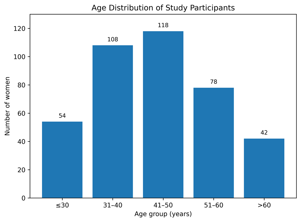
Figure 1. Age distribution of women included in the study.
Comparison of Specimen Adequacy
Specimen adequacy was assessed in the same 400 women using both conventional Pap smear and MLBC preparations. Conventional Pap smear preparations were satisfactory in 368 (92.0%) women and unsatisfactory in 32 (8.0%), whereas manual LBC preparations were satisfactory in 388 (97.0%) and unsatisfactory in 12 (3.0%) women, respectively (Table 2).
Thus, MLBC a 5-percentage-point higher proportion of satisfactory specimens and a corresponding reduction in unsatisfactory specimens compared with conventional Pap smear cytology. The comparative distribution of satisfactory and unsatisfactory specimens is presented in Figure 2.
Because the two methods were evaluated in the same women, statistical comparison of specimen adequacy was performed using a paired analysis. The significance of the observed difference should be interpreted on the basis of the discordant paired observations between the two methods.
Table 2. Comparison of Specimen Adequacy Between Conventional Pap Smear and manual LBC
|
Specimen adequacy |
Conventional Pap smear |
MLBC |
|
Satisfactory |
368 (92.0%) |
388 (97.0%) |
|
Unsatisfactory |
32 (8.0%) |
12 (3.0%) |
|
Total |
400 (100%) |
400 (100%) |
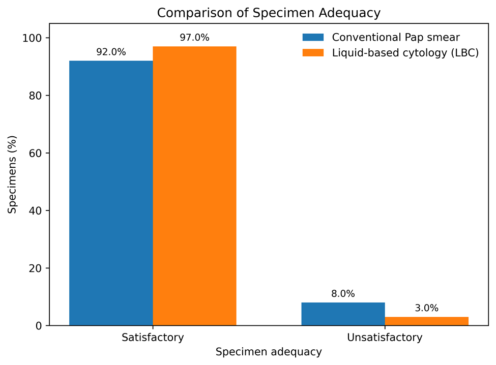
Figure 2. Comparison of specimen adequacy between conventional Pap smear and manual liquid-based cytology.
Comparison of Cytological Findings
The cytological findings obtained by conventional Pap smear and manual LBC in the same 400 women are summarised in Table 3 and illustrated in Figure 3. Conventional Pap smear cytology classified 352 (88.0%) women as negative for intraepithelial lesion or malignancy (NILM), while 48 (12.0%) demonstrated epithelial abnormalities. In comparison, MLBC classified 342 (85.5%) women as NILM and identified epithelial abnormalities in 58 (14.5%) women.
ASC-US was the most frequently detected epithelial abnormality with both methods, being identified in 22 (5.5%) women by conventional Pap smear and 26 (6.5%) women by MLBC. LSIL was detected in 14 (3.5%) and 17 (4.3%) women, respectively, while HSIL was identified in 7 (1.8%) conventional Pap smear preparations and 9 (2.3%) LBC preparations. Squamous cell carcinoma was detected in 2 (0.5%) women by each method.
Overall, MLBC identified a higher proportion of epithelial abnormalities than conventional Pap smear cytology (14.5% vs. 12.0%). The comparative distribution of cervical epithelial abnormalities detected by the two methods is shown in Table 4.
Table 3. Cytological Findings According to Screening Method
|
Cytological category |
Conventional Pap smear n (%) |
MLBC n (%) |
|
NILM |
352 (88.0) |
342 (85.5) |
|
ASC-US |
22 (5.5) |
26 (6.5) |
|
ASC-H |
3 (0.8) |
4 (1.0) |
|
LSIL |
14 (3.5) |
17 (4.3) |
|
HSIL |
7 (1.8) |
9 (2.3) |
|
Squamous cell carcinoma |
2 (0.5) |
2 (0.5) |
|
Total |
400 (100) |
400 (100) |
Table 4. Comparison of Cervical Epithelial Abnormalities
|
Cytological finding |
Conventional Pap smear |
MLBC |
|
Any epithelial abnormality |
48 (12.0%) |
58 (14.5%) |
|
ASC-US |
22 (5.5%) |
26 (6.5%) |
|
ASC-H |
3 (0.8%) |
4 (1.0%) |
|
LSIL |
14 (3.5%) |
17 (4.3%) |
|
HSIL |
7 (1.8%) |
9 (2.3%) |
|
Squamous cell carcinoma |
2 (0.5%) |
2 (0.5%) |
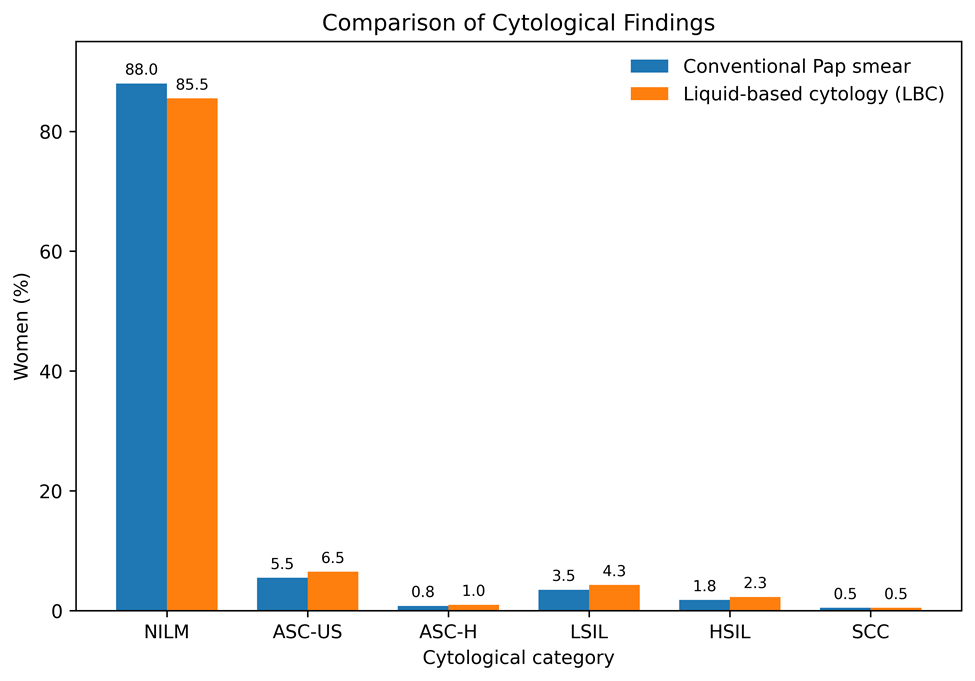
Figure 3. Comparative detection of cervical epithelial abnormalities by conventional Pap smear and manual liquid-based cytology.
Comparison of Factors Affecting Specimen Quality
The sample-related factors affecting specimen quality and cytological interpretation are summarised in Table 5. Obscuring blood was observed in 38 (9.5%) conventional Pap smear preparations compared with 16 (4.0%) MLBC preparations. Excessive inflammatory material was reported in 46 (11.5%) conventional preparations and 22 (5.5%)MLBC preparations.
Similarly, thick or uneven smear preparation was observed in 30 (7.5%) conventional Pap smear preparations compared with 8 (2.0%) MLBC preparations. Poor cellular distribution was observed in 26 (6.5%) conventional preparations and 10 (2.5%) MLBC preparations.
Overall, the descriptive findings demonstrated fewer sample-related limitations in MLBC preparations than in conventional Pap smear preparations. As these observations were obtained from paired preparations from the same women, formal statistical comparison should be based on paired analyses of the corresponding patient-level observations.
Table 5. Comparison of Sample-Related Limitations
|
Sample characteristic |
Conventional Pap smear n (%) |
MLBC n (%) |
|
Obscuring blood |
38 (9.5) |
16 (4.0) |
|
Excessive inflammation |
46 (11.5) |
22 (5.5) |
|
Thick/uneven smear |
30 (7.5) |
8 (2.0) |
|
Poor cellular distribution |
26 (6.5) |
10 (2.5) |
Histopathological Correlation and Diagnostic Performance of MLBC
Histopathological correlation was available for a subset of women who underwent subsequent cervical biopsy or tissue sampling following cytological evaluation. Among the women with available histopathological correlation, MLBC findings were compared with the corresponding histopathological diagnosis, with CIN2+ considered the clinically significant disease endpoint.
MLBC demonstrated a sensitivity of 90.9% for detecting CIN2+ lesions, with a specificity of 94.1%. The positive predictive value was 83.3%, while the negative predictive value was 97.1%. The overall diagnostic accuracy was 93.3% (Table 6). The diagnostic performance parameters of LBC are illustrated in Figure 4.
These diagnostic performance estimates were calculated only among women for whom histopathological correlation was available and therefore should not be interpreted as estimates for the entire screened population.
Table 6. Diagnostic Performance of MLBC for Detection of CIN2+ Lesions
|
Diagnostic parameter |
MLBC |
|
Sensitivity |
90.9% |
|
Specificity |
94.1% |
|
Positive predictive value |
83.3% |
|
Negative predictive value |
97.1% |
|
Diagnostic accuracy |
93.3% |
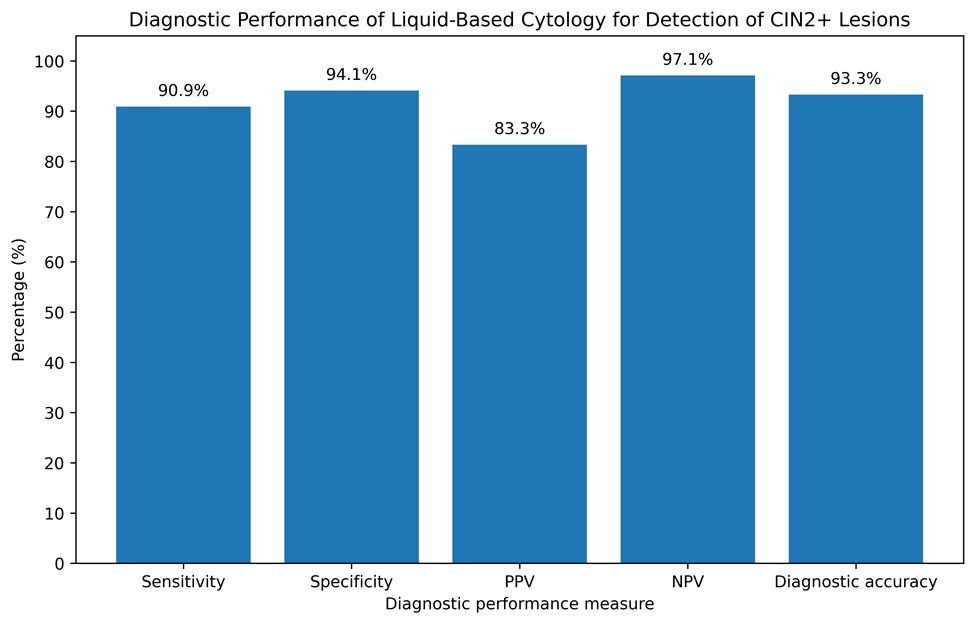
Figure 4. Diagnostic performance of liquid-based cytology for detection of CIN2+ lesions.
Overall Comparison Between Conventional Pap Smear and manual LBC
The overall descriptive comparison of conventional Pap smear and MLBC is presented in Table 7. MLBC demonstrated a higher proportion of satisfactory specimens than conventional Pap smear cytology (97.0% vs. 92.0%) and a lower proportion of unsatisfactory specimens (3.0% vs. 8.0%). The proportion of women with detected epithelial abnormalities was also higher with MLBC than with conventional Pap smear cytology (14.5% vs. 12.0%).
MLBC preparations also demonstrated lower frequencies of obscuring blood, excessive inflammatory material, and poor cellular distribution than conventional Pap smear preparations. These findings indicate an overall improvement in specimen quality with MLBC. However, because the two methods were applied to the same women, statistical significance of these differences should be determined using appropriate paired analyses based on the underlying patient-level data.
Table 7. Overall Comparison Between Conventional Pap Smear and MLBC
|
Parameter |
Conventional Pap smear |
MLBC |
|
Satisfactory specimens |
92.0% |
97.0% |
|
Unsatisfactory specimens |
8.0% |
3.0% |
|
Detection of epithelial abnormalities |
12.0% |
14.5% |
|
Obscuring blood |
9.5% |
4.0% |
|
Excessive inflammation |
11.5% |
5.5% |
|
Poor cellular distribution |
6.5% |
2.5% |
DISCUSSION:
Cervical cancer remains a major public health problem, particularly in low- and middle-income countries where access to organized screening, diagnostic facilities, and timely treatment remains limited. Although cervical cancer is largely preventable through effective screening and management of precancerous lesions, the success of a screening programme depends on the quality of the screening test, population coverage, follow-up, and availability of appropriate treatment services. The present retrospective study evaluated MLBC as an alternative to conventional Pap smear cytology among 400 women undergoing cervical cancer screening at a tertiary care centre in Himachal Pradesh.
The principal finding of the present study was the significantly better specimen adequacy observed with MLBC compared with conventional Pap smear cytology. In the present study, satisfactory specimens were obtained in 97.0% of MLBC preparations compared with 92.0% of conventional Pap smear preparations, while the proportion of unsatisfactory specimens was significantly lower with MLBC (3.0% vs. 8.0%; p=0.006). This finding is consistent with previous evidence suggesting that liquid-based preparations can reduce the frequency of unsatisfactory cervical cytology specimens11,12
The improved specimen adequacy associated with MLBC can be explained by differences in the method of specimen preparation. In conventional cytology, a proportion of collected cervical cells may remain on the sampling device, while the cells transferred to the slide may be unevenly distributed. In contrast, MLBC involves suspension of the collected cellular material in a preservative medium, followed by processing to produce a more uniform preparation. This may result in improved cellular distribution and a reduction in obscuring factors, thereby facilitating cytological evaluation.12,13
In the present study, obscuring blood was observed in 9.5% of conventional Pap smear preparations compared with only 4.0% of MLBC preparations. Similarly, excessive inflammation was observed in 11.5% and 5.5% of conventional and manual liquid-based preparations, respectively. Thick or uneven smears and poor cellular distribution were also less frequent with MLBC. These findings support the technical advantage of MLBC in producing cleaner and more evenly distributed cellular preparations. Improved specimen quality may be particularly valuable in routine screening, where the presence of obscuring material can make the identification of abnormal cells more difficult.
The present study also demonstrated a slightly higher detection rate of cervical epithelial abnormalities with MLBC. Overall, epithelial abnormalities were identified in 14.5% of women using MLBC compared with 12.0% using conventional Pap smear cytology. The difference was primarily attributable to the higher detection of ASC-US, LSIL, and HSIL in the MLBC group. Similar observations have been reported in previous comparative studies, although the magnitude of improvement in abnormality detection has varied between studies.14,15
The increased detection of cytological abnormalities with MLBC may partly reflect the improved quality of cellular preparations. Better preservation and more uniform distribution of cells may allow subtle cytological changes to be recognised more readily. However, increased detection of abnormalities should not automatically be interpreted as evidence of superior clinical effectiveness. In particular, detection of low-grade abnormalities may increase the number of women requiring repeat testing or additional diagnostic procedures without necessarily resulting in a corresponding reduction in cervical cancer mortality. Therefore, the clinical value of LBC should be evaluated within the broader context of the entire cervical cancer screening pathway.14,15
In the present study, ASC-US represented the most frequently detected epithelial abnormality in both conventional and manual liquid-based preparations. The proportion of ASC-US was 5.5% with conventional cytology and 6.5% with MLBC. LSIL and HSIL were also detected somewhat more frequently using MLBC. Although these differences suggest a potential advantage of LBC in identifying abnormal epithelial changes, they should be interpreted cautiously in view of the retrospective study design and the absence of complete histopathological verification for all participants.
Histopathological correlation was available in women who underwent subsequent biopsy or tissue sampling following abnormal cytological findings. Among cases with available histopathological correlation, MLBC demonstrated a sensitivity of 90.9% and specificity of 94.1% for the detection of CIN2+ lesions. The positive predictive value was 83.3%, while the negative predictive value was 97.1%, with an overall diagnostic accuracy of 93.3%. These findings suggest that MLBC can provide good diagnostic discrimination for clinically significant cervical lesions. However, these performance estimates should be interpreted with caution because histopathological correlation was not available for the entire screened population, and verification bias may therefore have influenced the estimates.
Previous comparative evaluations of liquid-based and conventional cytology have reported variable results regarding the relative sensitivity of the two methods for detecting high-grade cervical lesions. Some studies have demonstrated improved sensitivity or specimen adequacy with LBC, whereas others have found limited or no significant difference in the detection of clinically important lesions.11,14,15 These variations may be related to differences in study populations, screening prevalence, cytological interpretation, laboratory expertise, and the specific liquid-based technology used.
Although LBC may improve specimen adequacy and reduce the need for repeat sampling, the technique generally requires specialised equipment, proprietary collection and processing systems, additional consumables, and trained personnel.16,17
One of the important considerations regarding the introduction of MLBC in resource-limited settings is cost. From an economic perspective, MLBC requires minimal financial burden, lower operational costs, and readily available laboratory infrastructure. Furthermore, the residual cellular material can be preserved for ancillary investigations, including immunocytochemistry and molecular testing,similar to that of automated LBC thereby enhancing its clinical utility without substantially increasing costs.
The findings of the present study are relevant to tertiary care institutions serving populations in which cervical cancer screening resources may be constrained. The improved adequacy observed with manual LBC may help reduce the number of women requiring repeat sampling because of unsatisfactory specimens. This could potentially improve patient convenience and reduce the indirect costs associated with repeat visits..
The present study has several strengths. It included a relatively large sample of 400 women and evaluated multiple aspects of cervical cytology, including specimen adequacy, cytological abnormalities, sample-related limitations, and histopathological correlation where available. The study also assessed diagnostic performance for CIN2+ lesions, providing information beyond simple comparison of abnormal cytology rates.
However, several limitations should be acknowledged. First, the retrospective design limited the availability and completeness of clinical and follow-up information. Second, histopathological confirmation was not available for all participants, which limited comprehensive assessment of diagnostic accuracy. Third, the study was conducted at a single tertiary care centre, and therefore the findings may not be directly generalisable to other healthcare settings. Fourth, information regarding HPV status, vaccination history, reproductive factors, and long-term clinical outcomes was not consistently available. Finally, a formal cost-effectiveness analysis was not performed, which is particularly relevant when considering the implementation of manual LBC in resource-limited settings.
Despite these limitations, the present findings indicate that MLBC may offer meaningful technical advantages over conventional Pap smear cytology, particularly with respect to specimen adequacy and reduction of obscuring factors. The higher detection of epithelial abnormalities and favourable diagnostic performance for CIN2+ lesions further support its potential utility. Nevertheless, the decision to adopt MLBC should be based on local healthcare resources, screening priorities, and the ability to provide appropriate follow-up and treatment.
Overall, the present study suggests that MLBC can serve as a useful alternative to conventional Pap smear cytology in appropriately equipped resource-limited healthcare institutions. Larger prospective multicentric studies incorporating HPV testing, complete histopathological correlation, long-term follow-up, and formal cost-effectiveness assessment are required to establish whether the improved technical performance of MLBC translates into meaningful improvements in cervical cancer prevention and population-level health outcomes.
CONCLUSION:
The present retrospective study demonstrated that manual liquid-based cytology provided better specimen adequacy than conventional Pap smear cytology, with a significantly lower proportion of unsatisfactory specimens. MLBC also showed a higher detection rate of cervical epithelial abnormalities and fewer sample-related limitations, including obscuring blood, excessive inflammation, and poor cellular distribution. Among cases with available histopathological correlation, MLBC demonstrated good diagnostic performance for the detection of CIN2+ lesions. These findings suggest that MLBC may be a useful alternative to conventional Pap smear cytology in appropriately equipped resource-limited healthcare institutions. Further prospective multicentric studies with complete histopathological correlation, HPV testing, long-term follow-up, and formal cost-effectiveness assessment are warranted to determine the broader clinical and public health value of MLBC in resource-limited settings.
REFERENCES:
1. World Health Organization. Global strategy to accelerate the elimination of cervical cancer as a public health problem. Geneva: World Health Organization; 2020.
2. Sung H, Ferlay J, Siegel RL, Laversanne M, Soerjomataram I, Jemal A, Bray F. Global cancer statistics 2020: GLOBOCAN estimates of incidence and mortality worldwide for 36 cancers in 185 countries. CA Cancer J Clin. 2021;71(3):209-249.
3. Schiffman M, Castle PE, Jeronimo J, Rodriguez AC, Wacholder S. Human papillomavirus and cervical cancer. Lancet. 2007;370(9590):890-907.
4. Peirson L, Fitzpatrick-Lewis D, Ciliska D, Warren R. Screening for cervical cancer: a systematic review and meta-analysis. Syst Rev. 2013;2:35.
5. Arbyn M, Herbert A, Schenck U, Nieminen P, Jordan J, McGoogan E, et al. European guidelines for quality assurance in cervical cancer screening: recommendations for collecting samples for conventional and liquid-based cytology. Cytopathology. 2007;18(3):133-139.
6. Kurman RJ, Solomon D. The Bethesda System for Reporting Cervical/Vaginal Cytologic Diagnoses: definitions, criteria, and explanatory notes for terminology and specimen adequacy. New York: Springer-Verlag; 1994.
7. Davey E, Barratt A, Irwig L, Chan SF, Macaskill P, Mannes P, Saville AM. Effect of study design and quality on unsatisfactory rates, cytology classifications, and accuracy in liquid-based versus conventional cervical cytology: a systematic review. Lancet. 2006;367(9505):122-132.
8. Ronco G, Cuzick J, Pierotti P, Cariaggi MP, Dalla Palma P, Naldoni C, et al. Accuracy of liquid based versus conventional cytology: overall results of new technologies for cervical cancer screening: randomised controlled trial. BMJ. 2007;335(7609):28.
9. Arbyn M, Bergeron C, Klinkhamer P, Martin-Hirsch P, Siebers AG, Bulten J. Liquid compared with conventional cervical cytology in the detection of cervical intraepithelial neoplasia: a meta-analysis. Obstet Gynecol. 2008;111(1):167-177.
10. Maksem JA, Finnemore M, Belsheim BL, Roose EB, Makkapati SR, Eatwell L, Weidmann J. Manual method for liquid‐based cytology: A demonstration using 1,000 gynecological cytologies collected directly to vial and prepared by a smear‐slide technique. Diagnostic cytopathology. 2001 Nov;25(5):334-8.
11. Siebers AG, Klinkhamer PJJM, Grefte JMM, Massuger LFAG, Vedder JEM, Beijers-Broos A, et al. Comparison of liquid-based cytology with conventional cytology for detection of cervical cancer precursors: a randomized controlled trial. JAMA. 2009;302(16):1757-1764.
12. Beerman H, van Dorst EBL, Kuenen-Boumeester V, Hogendoorn PCW. Superior performance of liquid-based versus conventional cytology in a population-based cervical cancer screening program. Gynecol Oncol. 2009;112(3):572-576.
13. Davey E, d'Assuncao J, Irwig L, Macaskill P, Chan SF, Richards A, Farnsworth A. Accuracy of reading liquid based cytology slides using the ThinPrep Imager compared with conventional cytology: prospective study. BMJ. 2007;335(7609):31.
14. Ronco G, Dillner J, Elfström KM, Tunesi S, Snijders PJF, Arbyn M, et al. Efficacy of HPV-based screening for prevention of invasive cervical cancer: follow-up of four European randomised controlled trials. Lancet. 2014;383(9916):524-532.
15. Cuzick J, Clavel C, Petry KU, Meijer CJLM, Hoyer H, Ratnam S, et al. Overview of the European and North American studies on HPV testing in primary cervical cancer screening. Int J Cancer. 2006;119(5):1095-1101.
16. Sankaranarayanan R, Nene BM, Shastri SS, Jayant K, Muwonge R, Budukh AM, et al. HPV screening for cervical cancer in rural India. N Engl J Med. 2009;360(14):1385-1394.
17. World Health Organization. WHO guideline for screening and treatment of cervical pre-cancer lesions for cervical cancer prevention. 2nd ed. Geneva: World Health Organization; 2021.