Diagnostic Utility of USG- and CT-Guided Fine-Needle Aspiration Cytology in Intra-Abdominal Lesions: A Study of 660 Cases.
- Manjula R , Assistant Professor, Department of Pathology, Belgaum Institute of Medical Sciences, Belgaum.
- Aditya.S , Consultant Pathologist, Department of Pathology.
- Aparna K , Consultant Pathologist, Department of Pathology.
Article Information:
Abstract:
Background: Fine-needle aspiration cytology is a rapid, minimally invasive and cost-effective technique for diagnosing deep-seated intra-abdominal lesions. The use of ultrasonography and computed tomography improves lesion localisation, allows safer needle placement and increases the likelihood of obtaining representative material. Aim: To evaluate the diagnostic utility of USG- and CT-guided fine-needle aspiration cytology in intra-abdominal lesions and to assess their demographic, anatomical and cytological distribution. Materials and Methods: This partly retrospective and prospective observational study was conducted in the Department of Pathology, Goa Medical College, Bambolim, over an eight-year period from 2010 to 2017. A total of 660 patients with radiologically detected intra-abdominal lesions underwent USG- or CT-guided FNAC using a 22-gauge needle. Smears were stained with haematoxylin and eosin and May–Grünwald–Giemsa stains. Cytological diagnoses were correlated with clinical and radiological findings. Histopathological correlation was available in 58 cases. Sensitivity, specificity, diagnostic accuracy, positive predictive value and negative predictive value were calculated. Results: Of the 660 patients, 392 (59.39%) were male and 268 (40.61%) were female. The highest number of cases occurred in the 51–60-year age group. CT guidance was used in 427 cases (64.70%), while USG guidance was used in 233 cases (35.30%). The hepatobiliary system was the most frequently sampled region, accounting for 313 cases (47.42%), followed by the retroperitoneum with 102 cases (15.46%). Cytologically, 342 lesions (51.82%) were malignant, 135 (20.45%) were benign and 183 (27.73%) were inconclusive. Hepatic metastasis was the most frequent malignant liver diagnosis, followed by hepatocellular carcinoma. Conclusion: USG- and CT-guided FNAC is a useful and reliable method for diagnosing intra-abdominal lesions, particularly malignancies. It provides rapid diagnostic information and can reduce the need for unnecessary exploratory surgery.
Keywords:
Article :
INTRODUCTION:
Intra-abdominal lesions comprise a broad and clinically important group of abnormalities involving the liver, gallbladder, pancreas, kidneys, spleen, adrenal glands, gastrointestinal tract, lymph nodes, omentum, mesentery and retroperitoneal soft tissues. These lesions may be inflammatory, infectious, cystic, benign neoplastic or malignant. Their clinical presentation is often non-specific and may include abdominal pain, distension, loss of appetite, weight loss, fever, jaundice or a palpable mass. In some patients, particularly those with small or deeply located lesions, the abnormality may be discovered incidentally during radiological examination. Although imaging can accurately identify the site, size and extent of a lesion, radiological appearances alone may not always reliably distinguish between benign and malignant conditions. Therefore, obtaining representative cellular or tissue material is essential for establishing a definitive diagnosis and planning appropriate treatment [1].
Traditionally, many intra-abdominal lesions required exploratory laparotomy or open surgical biopsy for pathological diagnosis. These procedures are invasive, require anaesthesia and hospital admission, and may be associated with postoperative pain, infection, bleeding and prolonged recovery. The development of fine-needle aspiration cytology (FNAC) has provided a less invasive method for obtaining diagnostic material from both superficial and deep-seated lesions. FNAC is generally rapid, economical and well tolerated, and the aspirated material can be examined using routine cytological stains. In selected cases, the material may also be used for cell-block preparation, special stains, immunocytochemistry and molecular investigations. Comparative evidence has shown that FNAC can provide rapid and accurate information in radiologically detected abdominal lesions and may complement core needle biopsy when more detailed tumour classification is needed [1].
The major limitation of conventional FNAC is the difficulty in accurately locating small, non-palpable or deeply situated lesions. The procedure also carries a risk of sampling necrotic tissue, haemorrhagic areas or adjacent normal structures when performed without radiological guidance. The introduction of ultrasonography and computed tomography has considerably improved the accessibility and precision of abdominal FNAC. Image guidance allows the operator to select a safe needle pathway, avoid major blood vessels and adjacent organs, and direct the needle towards a viable and representative area of the lesion. Large clinical series have demonstrated that image-guided FNAC is useful in evaluating focal lesions of solid abdominal organs, including the liver, pancreas, spleen and kidneys [2].
Ultrasonography-guided FNAC is commonly preferred because ultrasonography is widely available, relatively inexpensive and capable of providing real-time visualisation of the needle during the procedure. It does not involve ionising radiation and permits repeated adjustment of the needle direction. These features make it particularly useful for lesions in the liver, gallbladder, kidneys, pancreas and accessible abdominal lymph nodes. Previous studies have reported high diagnostic yields and good accuracy for ultrasonography-guided FNAC in differentiating neoplastic from non-neoplastic intra-abdominal masses [3,4]. It may also help in the early recognition of inflammatory lesions, abscesses, primary malignancies, metastatic deposits and lymphoproliferative disorders.
Computed tomography-guided FNAC is particularly valuable for small or deeply located lesions that are poorly visualised on ultrasonography because of obesity, bowel gas, overlying bone or an anatomically difficult location. CT provides detailed cross-sectional localisation and can assist in planning a safe approach to retroperitoneal, pancreatic, adrenal and deep lymph-node lesions. Studies involving both ultrasonography- and CT-guided aspirations have shown that coupling FNAC with an appropriate imaging modality can improve diagnostic yield and enable diagnosis of lesions that would otherwise be inaccessible through conventional aspiration [4,5]. The choice of guidance modality depends on the lesion’s location, size, visibility, relationship with surrounding structures, availability of equipment and the experience of the radiologist.
Despite its advantages, image-guided FNAC has recognised limitations. Inadequate or non-representative material may be obtained from very small, highly vascular, cystic or extensively necrotic lesions. Certain well-differentiated malignancies may also be difficult to distinguish from benign conditions on cytological morphology alone. Furthermore, the absence of tissue architecture may restrict precise tumour typing in some cases. Close communication among the radiologist, cytopathologist and treating clinician is therefore important. Rapid on-site assessment, where available, can confirm specimen adequacy and guide additional passes. Cell-block preparation from residual aspirated material can improve architectural assessment and permits the application of special stains and immunocytochemistry, thereby increasing diagnostic confidence [3,6].
Image-guided FNAC is particularly relevant in retroperitoneal lesions, which may grow silently and reach a considerable size before producing clinical symptoms. These lesions include renal and adrenal tumours, lymphomas, metastatic carcinomas, germ-cell tumours and soft-tissue neoplasms. Studies of retroperitoneal masses have demonstrated that guided FNAC can distinguish neoplastic from non-neoplastic lesions with good cytological–histological agreement while avoiding unnecessary surgical exploration [7,8]. More recent prospective evidence has also confirmed the high diagnostic accuracy of ultrasonography-guided FNAC in the evaluation of abdominopelvic masses, supporting its continued use even in settings where advanced biopsy facilities may be limited [9].
However, the diagnostic performance of image-guided FNAC may vary according to the organ involved, lesion characteristics, guidance modality, operator experience, sample adequacy and availability of ancillary techniques. Large studies examining both USG- and CT-guided FNAC are therefore needed to clarify the spectrum of intra-abdominal lesions encountered in routine practice and to assess the practical diagnostic contribution of each modality. The present study, involving 660 cases, was undertaken to evaluate the diagnostic utility of USG- and CT-guided FNAC in intra-abdominal lesions, describe their organ-wise and cytological distribution, determine specimen adequacy and assess the ability of guided cytology to differentiate inflammatory, benign, suspicious and malignant lesions.
MATERIALS AND METHODS :
Study Design and Setting
This partly retrospective and prospective, hospital-based observational study was conducted in the Department of Pathology, Goa Medical College, Bambolim, Goa, India. The study covered an eight-year period from January 2010 to December 2017. It included patients who underwent radiologically guided fine-needle aspiration cytology for clinically or radiologically detected intra-abdominal lesions. The study was designed to evaluate the cytomorphological spectrum and diagnostic utility of ultrasonography-guided and computed tomography-guided FNAC in intra-abdominal lesions.
Study Population
A total of 660 image-guided FNAC procedures performed on intra-abdominal lesions were included. The study population comprised patients of all age groups and both sexes. Lesions involving the hepatobiliary system, spleen, pancreas, kidneys, adrenal glands, female genital tract, gastrointestinal tract, mesentery, omentum, psoas region, retroperitoneal lymph nodes and other retroperitoneal or intra-abdominal sites were evaluated.
For the retrospective component, relevant clinical information, radiological findings, cytology reports, stained slides and available histopathology records were retrieved from departmental archives. For the prospective component, patients undergoing USG- or CT-guided FNAC during the study period were evaluated according to the same procedural and cytological criteria. Cases were included when a radiologically identifiable intra-abdominal lesion had been subjected to guided FNAC and the corresponding cytological material or report was available for assessment. Cases without adequate clinical, radiological or cytological documentation were excluded from the analytical dataset.
Pre-procedural Assessment
Before aspiration, the patient’s relevant clinical history, physical findings and radiological diagnosis were recorded. Bleeding time, clotting time, platelet count and prothrombin time were reviewed to ensure that the coagulation profile was within acceptable limits. Baseline pulse rate and blood pressure were recorded before the procedure. The patient was positioned according to the anatomical location of the lesion and the safest route of needle entry.
The choice between USG and CT guidance was determined by lesion visibility, depth, size, anatomical location and proximity to adjacent organs or major vessels. Ultrasonography was used for lesions that could be clearly visualised in real time, while computed tomography was used mainly for deep-seated, small or anatomically difficult lesions. Of the 660 procedures, 233 were performed under USG guidance and 427 under CT guidance.
Fine-Needle Aspiration Procedure
The equipment used included a 22-gauge, 90-mm lumbar puncture needle, a 10-mL disposable syringe and a modified Cameco syringe holder. The lesion was localised using USG or CT, and the intended skin-entry site was marked when required. Local anaesthesia was not routinely administered.
The overlying skin was cleaned with povidone-iodine and spirit and allowed to dry. The needle was connected to the syringe and mounted on the syringe holder. After instructing the patient to remain still and, where necessary, to hold their breath, the needle was advanced into the lesion under radiological guidance. Correct placement of the needle tip was confirmed by USG or CT.
Negative pressure was created by withdrawing the syringe plunger. The needle was moved gently back and forth in different directions within the lesion to obtain representative material. Multiple directional movements were avoided in splenic lesions because of the risk of haemorrhage. Before withdrawal, suction was released to prevent contamination of the needle track. Firm pressure was applied to the puncture site, which was subsequently sealed. Patients were monitored for approximately 6–24 hours for pain, bleeding, haemodynamic instability or other procedure-related complications.
Smear Preparation and Staining
The aspirated material was expelled onto three to five clean glass slides, depending on the amount obtained. Smears were prepared by gently placing a second slide over the material and spreading it evenly to produce thin, uniform preparations. Most smears were immediately wet-fixed in a freshly prepared mixture of equal volumes of ether and absolute ethyl alcohol for a minimum of two hours and subsequently stained with haematoxylin and eosin. Additional smears were air-dried and stained with May–Grünwald–Giemsa stain. When fluid material was aspirated, it was centrifuged, and smears were prepared from the resulting sediment.
Cytological Evaluation
All smears were examined under light microscopy by correlating the cytomorphological findings with the available clinical and radiological information. Aspirates were classified as benign, malignant or inconclusive. Benign lesions were further categorised as neoplastic or non-neoplastic, whereas malignant lesions were classified as primary, metastatic or positive for malignancy when precise typing was not possible. Aspirates containing only blood, necrotic material, scant cellular material, normal epithelial cells or atypical cells insufficient for a definitive diagnosis were categorised as inadequate or inconclusive.
The age, sex, radiological guidance modality, anatomical site, specimen adequacy and final cytological diagnosis were recorded for every case. Organ-specific diagnoses were made using recognised cytomorphological features.
Histopathological Correlation and Statistical Analysis
Histopathological correlation was performed in patients who subsequently underwent surgical biopsy or excision. Histopathology was treated as the reference standard. Correlation was available for 58 cases. Cytological findings were classified as true positive, true negative, false positive or false negative. Diagnostic sensitivity, specificity, overall accuracy, positive predictive value and negative predictive value were calculated using standard formulae.
Categorical variables were summarised as frequencies and percentages, while age was described using age groups and mean age where available. Organ-wise and diagnosis-wise distributions were analysed descriptively. No statistical software was specified in the thesis; the software and version used for the revised analysis should therefore be added before manuscript submission.
RESULTS:
A total of 660 patients with radiologically detected intra-abdominal lesions underwent image-guided fine-needle aspiration cytology during the study period. The patients ranged from the first decade of life to over 70 years of age. The highest number of cases was recorded in the 51–60-year age group, comprising 153 cases (23.18%), followed by the 41–50-year age group with 134 cases (20.30%) and the 61–70-year age group with 118 cases (17.88%). Collectively, patients aged 41–70 years accounted for 405 cases (61.36%) of the study population.
There was a male predominance, with 392 males (59.39%) and 268 females (40.61%), giving a male-to-female ratio of approximately 1.46:1. The lowest number of cases was observed in the 11–20-year age group, which included 24 patients (3.64%).
Table 1. Age- and sex-wise distribution of patients undergoing image-guided FNAC
|
Age group, years |
Male, n |
Female, n |
Total, n |
Percentage |
|
0–10 |
21 |
6 |
27 |
4.09 |
|
11–20 |
10 |
14 |
24 |
3.64 |
|
21–30 |
25 |
32 |
57 |
8.64 |
|
31–40 |
53 |
40 |
93 |
14.09 |
|
41–50 |
67 |
67 |
134 |
20.30 |
|
51–60 |
104 |
49 |
153 |
23.18 |
|
61–70 |
79 |
39 |
118 |
17.88 |
|
>70 |
33 |
21 |
54 |
8.18 |
|
Total |
392 |
268 |
660 |
100.00 |
Computed tomography was the more frequently used guidance modality. CT-guided FNAC was performed in 427 cases (64.70%), whereas USG-guided FNAC was performed in 233 cases (35.30%). Therefore, approximately two-thirds of the lesions required CT guidance, possibly reflecting the high proportion of deep-seated, small or retroperitoneal lesions included in the study.
Table 2. Distribution according to radiological guidance modality
|
Guidance modality |
Number of cases |
Percentage |
|
Ultrasonography-guided FNAC |
233 |
35.30 |
|
Computed tomography-guided FNAC |
427 |
64.70 |
|
Total |
660 |
100.00 |
The hepatobiliary system was the most commonly aspirated anatomical region, accounting for 313 cases (47.42%). The majority of these were liver lesions, which comprised 307 cases (46.52% of the complete study population). Gallbladder and common bile duct lesions accounted for three cases each.
The retroperitoneum was the second most frequently sampled region, with 102 cases (15.46%). These consisted of 70 retroperitoneal lymph-node lesions and 32 other retroperitoneal masses. Lesions of the female genital system accounted for 65 cases (9.85%), including 52 ovarian and 13 uterine lesions. Renal lesions accounted for 60 cases (9.09%).
Other intra-abdominal lesions collectively comprised 73 cases (11.06%). These included gastrointestinal tract lesions, mesenteric lesions, omental lesions, psoas-region lesions, undescended testes and a paravesical lesion. Splenic, pancreatic and adrenal lesions represented 25 (3.79%), 13 (1.97%) and nine cases (1.36%), respectively.
Table 3. Organ- and site-wise distribution of intra-abdominal lesions
|
Organ or anatomical system |
Number of cases |
Percentage |
|
Hepatobiliary system |
313 |
47.42 |
|
Spleen |
25 |
3.79 |
|
Pancreas |
13 |
1.97 |
|
Kidney |
60 |
9.09 |
|
Adrenal glands |
9 |
1.36 |
|
Female genital system |
65 |
9.85 |
|
Retroperitoneum |
102 |
15.46 |
|
Other intra-abdominal sites |
73 |
11.06 |
|
Total |
660 |
100.00 |
The hepatobiliary group included 307 liver, three gallbladder and three common bile duct lesions. The retroperitoneal group included 70 lymph-node lesions and 32 other retroperitoneal masses.
Of the 660 aspirates, 342 cases (51.82%) were classified as malignant, making malignancy the most common broad cytological category. Benign lesions were identified in 135 cases (20.45%). The remaining 183 aspirates (27.73%) were categorised as inconclusive. The inconclusive group included aspirates with inadequate material, scant cellularity, predominantly necrotic or haemorrhagic material, atypical cells insufficient for a definitive diagnosis, or cases in which malignancy could not be confidently excluded.
Table 4. Overall cytological classification of image-guided aspirates
|
Cytological category |
Number of cases |
Percentage |
|
Benign lesions |
135 |
20.45 |
|
Malignant lesions |
342 |
51.82 |
|
Inconclusive aspirates |
183 |
27.73 |
|
Total |
660 |
100.00 |
Among the 307 liver lesions, 56 were benign, 160 were malignant and 91 were inconclusive. Metastatic malignancy was the most frequent malignant hepatic diagnosis, accounting for 83 of the 160 malignant liver lesions (51.88%). Hepatocellular carcinoma was diagnosed in 54 cases (33.75%), while 17 cases (10.63%) were reported as positive for malignancy without further cytological typing. The remaining primary malignant hepatic lesions included hepatoblastoma, lymphoma and solid pseudopapillary epithelial neoplasm.
Among the 83 hepatic metastases, adenocarcinoma was the most common cytomorphological pattern and was identified in 51 cases (61.45%). Poorly differentiated carcinoma accounted for 16 cases (19.28%), small-cell carcinoma for eight cases (9.64%), infiltrating duct carcinoma for five cases (6.02%), squamous-cell carcinoma for two cases (2.41%) and multiple myeloma for one case (1.20%). The primary site was unknown in 47 of the 83 cases of hepatic metastasis. Where the primary site was known, the gastrointestinal tract was the most frequent source.
Pyogenic abscess was the most common benign hepatic diagnosis, accounting for 31 of 56 benign hepatic lesions (55.36%). Other benign liver lesions included tuberculous abscess, amoebic abscess, cysts, granulomatous inflammation, haemangioma, haemangioendothelioma, cirrhosis and focal nodular hyperplasia.
Of the 25 splenic lesions, 16 were benign, six were malignant and three were inconclusive. Benign splenic lesions included pyogenic abscess, granulomatous inflammation, reactive hyperplasia and tuberculosis. Malignant splenic lesions included lymphoma and metastatic poorly differentiated carcinoma.
Nine of the 13 pancreatic lesions were malignant. Pancreatic adenocarcinoma was diagnosed in eight cases, and one case was diagnosed as a solid pseudopapillary epithelial neoplasm. One pancreatic lesion showed granulomatous inflammation, while three aspirates were inconclusive.
Among the 60 renal lesions, 29 were malignant, 12 were benign and 19 were inconclusive. Renal-cell carcinoma was the most frequent specifically typed renal malignancy, with 14 cases, followed by Wilms tumour in six cases and primary squamous-cell carcinoma in three cases. Benign renal lesions included renal cysts, pyogenic and tuberculous abscesses, inflammatory lesions, renal oncocytoma and xanthogranulomatous pyelonephritis.
The 52 ovarian lesions comprised 12 benign, 19 malignant and 21 inconclusive aspirates. Papillary adenocarcinoma was the most frequent malignant ovarian diagnosis, accounting for 14 cases. Among the 13 uterine lesions, 12 were diagnosed as leiomyoma and one was reported as positive for malignancy.
Malignancy was identified in 50 of 70 retroperitoneal lymph-node lesions. Non-Hodgkin lymphoma was the most frequent primary lymphoid malignancy, accounting for 22 cases. Metastatic poorly differentiated carcinoma was identified in 11 cases, while metastatic adenocarcinoma was diagnosed in five cases. Six retroperitoneal lymph-node lesions were benign and 14 were inconclusive.
Table 5. Major organ-specific cytological findings
|
Organ or site |
Total cases |
Benign |
Malignant |
Inconclusive |
Predominant specific diagnosis |
|
Liver |
307 |
56 |
160 |
91 |
Hepatic metastasis |
|
Spleen |
25 |
16 |
6 |
3 |
Non-neoplastic/inflammatory lesions |
|
Pancreas |
13 |
1 |
9 |
3 |
Adenocarcinoma |
|
Kidney |
60 |
12 |
29 |
19 |
Renal-cell carcinoma |
|
Adrenal gland |
9 |
1 |
7 |
1 |
Neuroblastoma |
|
Ovary |
52 |
12 |
19 |
21 |
Papillary adenocarcinoma |
|
Uterus |
13 |
12 |
1 |
0 |
Leiomyoma |
|
Retroperitoneal lymph nodes |
70 |
6 |
50 |
14 |
Non-Hodgkin lymphoma |
|
Other retroperitoneal lesions |
32 |
8 |
18 |
6 |
Spindle-cell sarcoma |
|
Gastrointestinal tract |
46 |
0 |
33 |
13 |
Adenocarcinoma |
Organ-specific numbers are presented as reported in the corresponding thesis tables. They should be rechecked against the master dataset because the total number of malignant and inconclusive organ-specific entries does not completely reconcile with the overall diagnostic totals in Table 4.
Histopathological correlation was available in 58 cases that subsequently underwent biopsy or surgical excision. Based on the thesis data, 34 cases were true positive, 16 were true negative, two were false positive and six were false negative.
The resulting sensitivity was 85.00%, specificity was 88.89% and overall diagnostic accuracy was 86.21%. The positive predictive value was 94.44%, indicating that a malignant cytological diagnosis had a high probability of histopathological confirmation. The negative predictive value calculated from the reported raw counts was 72.73%.
Table 6. Diagnostic performance of image-guided FNAC against histopathology
|
Diagnostic parameter |
Result |
|
Cases with histopathological correlation |
58 |
|
True positive |
34 |
|
True negative |
16 |
|
False positive |
2 |
|
False negative |
6 |
|
Sensitivity |
85.00% |
|
Specificity |
88.89% |
|
Overall diagnostic accuracy |
86.21% |
|
Positive predictive value |
94.44% |
|
Negative predictive value |
72.73% |
The thesis reports the negative predictive value as 68%; however, calculation from the provided raw values gives 72.73%: 16/(16 + 6) × 100. This value should be verified from the original patient-level dataset before manuscript submission.
The thesis did not provide separate histopathological outcomes, adequacy rates or diagnostic-performance indices for USG-guided and CT-guided procedures. Consequently, the diagnostic sensitivity and accuracy of USG and CT guidance could not be compared independently.
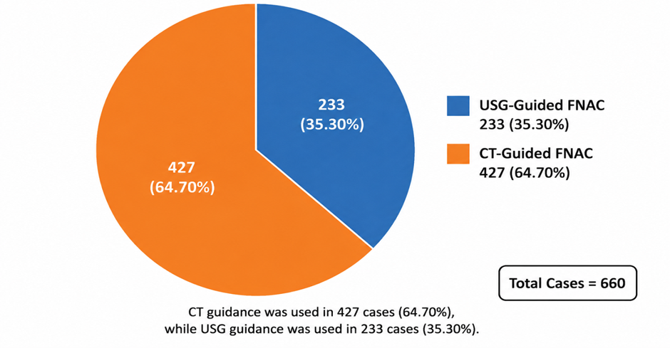
Figure 1. Distribution of image-guided fine-needle aspiration procedures according to the radiological guidance modality
Figure 1 illustrates the distribution of image-guided fine-needle aspiration cytology procedures according to the radiological guidance modality used. Of the total 660 intra-abdominal lesions evaluated, computed tomography-guided FNAC was performed in 427 cases (64.70%), whereas ultrasonography-guided FNAC was performed in 233 cases (35.30%). Thus, CT guidance was used in nearly two-thirds of the procedures and was the predominant imaging modality in this study. Its greater use may be related to its ability to provide accurate localisation of small, deeply situated or anatomically difficult intra-abdominal and retroperitoneal lesions. USG guidance accounted for slightly more than one-third of the procedures and remained useful for lesions that were clearly visualised and accessible through real-time ultrasonographic imaging. These findings demonstrate the complementary roles of CT and USG in selecting an appropriate and safe approach for aspiration of intra-abdominal lesions.
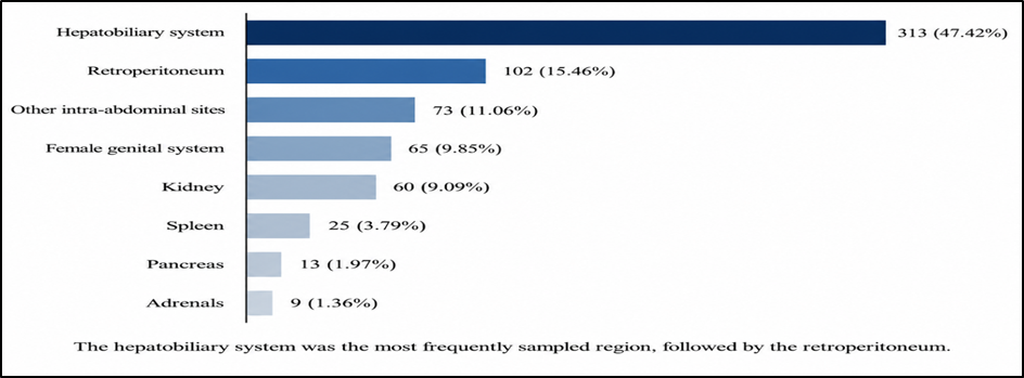
Figure 2. Organ- and anatomical site-wise distribution of intra-abdominal lesions subjected to image-guided fine-needle aspiration cytology
Figure 2 presents the organ- and anatomical site-wise distribution of 660 intra-abdominal lesions subjected to image-guided fine-needle aspiration cytology. The hepatobiliary system was the most frequently sampled region, comprising 313 cases (47.42%), of which 307 involved the liver and three cases each involved the gallbladder and common bile duct. The retroperitoneum was the second most common site, accounting for 102 cases (15.46%), including 70 retroperitoneal lymph-node lesions and 32 other retroperitoneal masses. Other intra-abdominal sites collectively contributed 73 cases (11.06%), followed by the female genital system with 65 cases (9.85%) and the kidneys with 60 cases (9.09%). Less frequently sampled organs included the spleen in 25 cases (3.79%), pancreas in 13 cases (1.97%) and adrenal glands in nine cases (1.36%). These findings demonstrate that the hepatobiliary system, particularly the liver, constituted the principal target for image-guided FNAC, while retroperitoneal lesions represented another important group requiring radiological guidance for safe and accurate sampling.
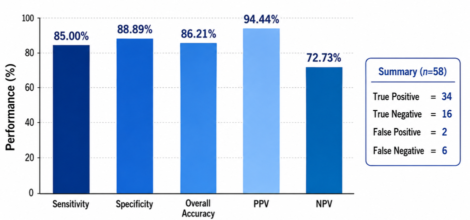
Figure 3. Diagnostic performance of image-guided fine-needle aspiration cytology using histopathology as the reference standard
Figure 3 presents the diagnostic performance of image-guided fine-needle aspiration cytology using histopathological examination as the reference standard. Cytohistopathological correlation was available for 58 cases, including 34 true-positive, 16 true-negative, two false-positive and six false-negative results. FNAC demonstrated a sensitivity of 85.00%, indicating its ability to correctly identify most malignant lesions. The specificity was 88.89%, showing a high capacity to correctly classify non-malignant lesions. The overall diagnostic accuracy was 86.21%. The positive predictive value was 94.44%, suggesting that a malignant cytological diagnosis had a strong likelihood of histopathological confirmation. The negative predictive value was comparatively lower at 72.73%, indicating that a negative or benign cytological result could not completely exclude malignancy.
DISCUSSION:
Image-guided fine-needle aspiration cytology has become an important method for investigating deep-seated intra-abdominal lesions because it combines precise radiological localisation with rapid cytological assessment. The present study included 660 USG- and CT-guided aspirations performed over an eight-year period, representing a large single-centre experience. The main findings were the predominance of patients aged 41–70 years, a higher frequency among males, greater use of CT guidance, predominance of hepatobiliary lesions and a high proportion of malignant cytological diagnoses. Histopathological correlation in the available cases showed good sensitivity, specificity and overall diagnostic accuracy.
The highest frequency of lesions was observed in patients aged 51–60 years, followed by those aged 41–50 and 61–70 years. Overall, 61.36% of the patients were between 41 and 70 years of age. This pattern may be related to the increased occurrence of primary and metastatic malignancies in middle-aged and older adults. However, the presence of paediatric lesions, including hepatoblastoma, Wilms tumour, neuroblastoma and malignant germ-cell tumour, demonstrates that image-guided FNAC also has an important diagnostic role in younger patients. Males constituted 59.39% of the study population, producing a male-to-female ratio of approximately 1.46:1. A similar male predominance was reported by Islam et al., whose study included 75.6% males among patients undergoing USG-guided FNAC of intra-abdominal lesions [11]. Variations in age and sex distribution between different studies may reflect differences in referral patterns, organ distribution and the prevalence of specific malignancies.
CT guidance was used in 427 cases (64.70%), while USG guidance was used in 233 cases (35.30%). The greater use of CT in the present series may be explained by the inclusion of a large number of deep, small and retroperitoneal lesions. Ultrasonography offers real-time needle visualisation, lower cost, repeatability and freedom from ionising radiation. However, bowel gas, obesity, overlying bone and the deep location of some lesions may reduce their visibility on USG. CT provides better anatomical localisation and permits safer planning of the needle pathway when lesions are situated near vessels, bowel loops or other vital structures.
The distribution of guidance modalities differs among published studies. Ghosh et al. reported 500 guided aspirations, of which 266 were performed under USG guidance and 234 under CT guidance. Adequate material was obtained in 91.7% and 87.6% of the USG- and CT-guided procedures, respectively [12]. Sheikh et al. reported that guided FNAC was diagnostic in 93.4% of deep-seated thoracic and abdominal masses and found that the combination of clinical, radiological and cytological information improved diagnostic interpretation [13]. De Filippo et al. obtained a diagnosis in 96% of small abdominal and retroperitoneal lesions using CT-guided aspiration with multiplanar reconstruction and a coaxial technique [14]. These findings support selecting the guidance modality according to lesion visibility and accessibility rather than considering USG and CT as competing techniques.
The hepatobiliary system was the most frequently aspirated anatomical region in the present study, accounting for 313 cases (47.42%). Liver lesions alone represented 307 cases, or 46.52% of the complete study population. This predominance is expected because the liver is readily assessed by radiological imaging and is a common site of primary tumours, infections and metastatic disease. Jan and Mahajan similarly found the liver to be the most frequently sampled organ in their study of intra-abdominal and retroperitoneal masses [10]. The high number of hepatic lesions also reflects the clinical importance of distinguishing primary hepatic malignancy from metastatic carcinoma and non-neoplastic conditions.
Malignant lesions constituted the largest broad cytological category in the present study, accounting for 342 cases (51.82%). Benign lesions constituted 135 cases (20.45%), while 183 aspirates (27.73%) were inconclusive. Islam et al. also reported a predominance of malignancy, with malignant lesions accounting for 52.6% of their intra-abdominal aspirates [11]. In the 500-case series reported by Ghosh et al., malignant lesions constituted a major proportion of adequate CT- and USG-guided aspirates [12]. The high malignant proportion in these studies is likely influenced by the tertiary-care setting, where patients are frequently referred following the radiological detection of lesions suspicious for malignancy.
Among the 307 hepatic lesions, 160 were malignant, 56 were benign and 91 were inconclusive. Metastatic malignancy was the most common malignant hepatic diagnosis, accounting for 83 of the 160 malignant lesions. Hepatocellular carcinoma was the second most frequent diagnosis, with 54 cases. This finding confirms the importance of the liver as a common site for haematogenous metastasis. Reddy et al., in a large series of 755 hepatic aspirates, similarly found metastatic malignancy to be more frequent than primary hepatocellular carcinoma. Their study included 524 hepatic metastases and 148 hepatocellular carcinomas [17].
Adenocarcinoma was the most frequent morphological type among hepatic metastases in the present study and constituted 61.45% of metastatic hepatic lesions. This is clinically reasonable because adenocarcinomas arising from the gastrointestinal tract, pancreas, breast, lung and other organs frequently metastasise to the liver. In 56.63% of hepatic metastases, the primary site was not identified at the time of cytological diagnosis. In such cases, cytomorphology can establish malignancy, but accurate identification of the primary site may require detailed radiological assessment, cell-block preparation and immunocytochemical markers.
The diagnostic performance reported in liver FNAC studies has generally been high. Edoute et al. reported sensitivity of 85.6%, specificity of 98.4%, positive predictive value of 99.1% and negative predictive value of 76.1% in a series of 406 patients with liver lesions [15]. A multicentre study by Hertz et al. involving 602 radiologically guided liver aspirates also supported the diagnostic value of FNAC in focal hepatic lesions [16]. Reddy et al. reported sensitivity of 93%, specificity of 90.9%, positive predictive value of 98.9% and overall accuracy of 92.8% [17]. These studies are broadly consistent with the high positive predictive value observed in the present investigation.
Pyogenic abscess was the most common benign hepatic lesion, accounting for 31 of the 56 benign liver lesions. Other non-neoplastic conditions included tuberculous abscess, amoebic abscess, cysts, granulomatous inflammation and cirrhosis. This finding demonstrates that radiologically detected focal hepatic lesions are not always neoplastic. Inflammatory and infectious lesions may resemble malignancy clinically and radiologically, particularly when they occur as solid or partially necrotic masses. FNAC can provide an early diagnosis and permit appropriate antimicrobial or antitubercular treatment, thereby avoiding unnecessary surgery.
The 25 splenic lesions included 16 benign, six malignant and three inconclusive cases. The predominance of benign and inflammatory lesions indicates the value of FNAC in diagnosing splenic abscesses, granulomatous inflammation, tuberculosis and reactive changes. Splenic aspiration requires particular care because of the vascularity of the organ. In the present methodology, multidirectional needle movement was avoided in splenic lesions, representing a suitable procedural precaution.
Nine of the 13 pancreatic lesions were malignant, and adenocarcinoma was the most frequent diagnosis. Three pancreatic aspirates were inconclusive. Pancreatic lesions may produce non-diagnostic material because of their deep retroperitoneal location, desmoplastic nature, necrosis or the difficulty of safely targeting small viable areas. Gupta reported that image-guided FNAC is useful in the diagnosis of pancreatic malignancy but emphasised that false-negative findings may be reduced by repeating the aspiration when the obtained material is insufficient and clinical suspicion remains high [22].
Among the 60 renal lesions, 29 were malignant, 12 were benign and 19 were inconclusive. Renal-cell carcinoma was the most common specifically classified renal malignancy, followed by Wilms tumour. The benign renal lesions included renal cysts, pyogenic abscesses, tuberculous abscesses, inflammatory lesions, oncocytoma and xanthogranulomatous pyelonephritis. The range of renal diagnoses demonstrates the ability of image-guided FNAC to distinguish malignant tumours from inflammatory and cystic conditions. However, the relatively high inconclusive rate indicates that renal aspirates must be interpreted carefully because oncocytic and clear-cell lesions may show overlapping cytological characteristics.
Seven of the nine adrenal lesions were malignant, including five cases of neuroblastoma. Adrenal aspiration requires careful clinical and biochemical evaluation because an unsuspected pheochromocytoma may cause serious complications during needle sampling. One pheochromocytoma was reported among the benign adrenal lesions in this study. This finding emphasises the need to exclude a functional adrenal tumour before performing percutaneous FNAC whenever clinical or radiological findings suggest pheochromocytoma.
Retroperitoneal lesions constituted the second largest anatomical group, comprising 102 cases. Of the 70 retroperitoneal lymph-node lesions, 50 were malignant. Non-Hodgkin lymphoma was the most frequent primary lymphoid malignancy, followed by metastatic poorly differentiated carcinoma and adenocarcinoma. The retroperitoneum is a diagnostically challenging region because masses may arise from lymph nodes, soft tissues, kidneys, adrenal glands or adjacent organs. Image guidance makes these otherwise inaccessible lesions available for minimally invasive sampling.
Gupta et al. evaluated image-guided aspiration of 112 pelvic and retroperitoneal masses and reported that the technique was valuable for distinguishing malignant from non-malignant lesions, although cell blocks and immunocytochemistry were often required for accurate tumour typing [19]. Mehdi et al. reported excellent diagnostic performance in the histologically correlated retroperitoneal cases in their series and considered guided FNAC an appropriate first-line investigation for selected retroperitoneal masses [20]. Guo et al. obtained satisfactory material in 97.1% of 68 radiologically guided pelvic and retroperitoneal aspirations and reported sensitivity of 90.2% and specificity of 100% for malignancy [21]. These results support the usefulness of guided FNAC in retroperitoneal lesions while recognising that lymphoma and soft-tissue sarcoma may require additional material for complete classification.
Among the 32 non-lymph-node retroperitoneal lesions, 18 were malignant, eight were benign and six were inconclusive. Spindle-cell sarcoma was the most frequent malignant diagnosis. Cytological classification of retroperitoneal spindle-cell lesions can be difficult because several tumours share overlapping cellular patterns. Although FNAC can establish the presence of a malignant mesenchymal neoplasm, definitive subtyping may require a core biopsy, histological architecture and an immunohistochemical panel.
The female genital system accounted for 65 cases, including 52 ovarian and 13 uterine lesions. Of the ovarian aspirates, 19 were malignant, 12 were benign and 21 were inconclusive. Papillary adenocarcinoma was the most frequent malignant ovarian diagnosis. The high inconclusive proportion among ovarian lesions may be related to the cystic nature of many ovarian masses, limited tumour cells in aspirated fluid and sampling from non-representative areas. Percutaneous ovarian FNAC should therefore be reserved for carefully selected patients, particularly those with advanced, recurrent or unresectable disease in whom a cytological diagnosis will influence treatment. In contrast, 12 of the 13 uterine lesions were diagnosed as leiomyoma, demonstrating that image-guided cytology can provide useful information in selected uterine masses.
Histopathological correlation was available in 58 cases. Based on the reported numbers of 34 true-positive, 16 true-negative, two false-positive and six false-negative cases, image-guided FNAC achieved a sensitivity of 85.00%, specificity of 88.89%, positive predictive value of 94.44% and overall accuracy of 86.21%. The high positive predictive value indicates that a malignant cytological diagnosis was highly likely to be confirmed histologically. However, the negative predictive value was lower at 72.73%, showing that a benign or negative aspirate cannot completely exclude malignancy, especially when the clinical and radiological findings remain suspicious.
The overall accuracy in the present study was greater than the 70% reported by Jan and Mahajan [10] but lower than the diagnostic rates reported in some more recent guided-FNAC series [13,14,20,21]. Differences in accuracy may be related to lesion selection, organ distribution, operator experience, needle placement, the number of passes, availability of rapid on-site evaluation, preparation of cell blocks and the proportion of cases with histopathological verification. Direct comparison should therefore be undertaken cautiously because the definition of diagnostic adequacy and the reference standards varied across studies.
The inconclusive rate of 27.73% was a notable finding. It was higher than the 10.3% non-representative rate reported by Islam et al. and the inadequate rates reported by Ghosh et al. for USG- and CT-guided aspirations [11,12]. Inadequate or inconclusive aspirates may result from cystic change, haemorrhage, extensive necrosis, fibrosis, small lesion size, increased depth or failure to target viable tissue. The large number of necrotic and metastatic lesions in the present series may have contributed to this finding. Repeat aspiration from the enhancing or solid component of the lesion may improve the diagnostic yield.
Rapid on-site evaluation by a cytopathologist can identify inadequate samples while the patient is still in the radiology department and permit an immediate additional pass. Ceyhan et al. found that on-site cytopathological evaluation and the combined use of conventional smears and cell blocks improved diagnostic and tumour-subtyping accuracy in hepatic masses. Their combined approach achieved a diagnostic accuracy of 94.5%, while specific classification of malignant tumours improved substantially when cell-block material was added [18]. Incorporating rapid adequacy assessment, cell-block preparation, immunocytochemistry and, where indicated, flow cytometry or molecular testing could reduce the inconclusive rate and improve the classification of lymphoma, poorly differentiated carcinoma and mesenchymal tumours.
An important strength of the present study was its large sample size and inclusion of a broad spectrum of lesions from multiple intra-abdominal and retroperitoneal organs. The use of both USG and CT guidance reflects routine clinical practice and demonstrates the complementary roles of the two imaging modalities. The study also included benign, inflammatory, primary malignant and metastatic lesions, providing a comprehensive picture of intra-abdominal FNAC in a tertiary-care institution.
Several limitations should be considered. The partly retrospective design may have resulted in incomplete clinical or radiological information. Histopathological correlation was available for only 58 of the 660 cases, which may introduce verification bias because surgically treated patients are unlikely to represent the entire study population. Diagnostic performance was not reported separately for USG- and CT-guided FNAC; therefore, a direct comparison of the two modalities was not possible. The effects of lesion size, depth, number of passes and operator experience were also not analysed. Furthermore, rapid on-site evaluation, cell-block preparation and ancillary testing were not routinely documented. The relatively high proportion of inconclusive aspirates and some numerical inconsistencies among the detailed organ-specific tables indicate that the original patient-level data should be rechecked before final manuscript submission.
Despite these limitations, the findings demonstrate that USG- and CT-guided FNAC is a useful and accurate diagnostic technique for intra-abdominal lesions. It was particularly valuable for detecting malignancy, confirming hepatic metastasis, diagnosing primary hepatic and renal tumours, assessing retroperitoneal lymphadenopathy and identifying inflammatory lesions that could be managed without surgery. The high positive predictive value supports its use for confirming malignancy. However, a negative or inconclusive aspirate should be interpreted together with clinical and radiological findings, and repeat aspiration or core biopsy should be considered when suspicion of malignancy persists.
CONCLUSION:
Image-guided fine-needle aspiration cytology is a useful, minimally invasive and reliable diagnostic technique for evaluating intra-abdominal lesions. In the present study of 660 cases, computed tomography guidance was used more frequently than ultrasonography, particularly for deeply located and anatomically difficult lesions. The hepatobiliary system, especially the liver, was the most frequently sampled region, followed by the retroperitoneum.
REFERENCES:
1. Stewart CJR, Coldewey J, Stewart IS. Comparison of fine needle aspiration cytology and needle core biopsy in the diagnosis of radiologically detected abdominal lesions. J Clin Pathol. 2002;55(2):93-97. doi:10.1136/jcp.55.2.93.
2. Vasilj A, Kojić Katović S. Fine needle aspiration cytology of abdominal organs: ten-year single center experience. Acta Clin Croat. 2016;55(1):35-40. doi:10.20471/acc.2016.55.01.6.
3. Sumana BS, Muniyappa B. Ultrasonography guided fine needle aspiration cytology with preparation of cell blocks in the diagnosis of intra-abdominal masses. J Clin Diagn Res. 2015;9(12). doi:10.7860/JCDR/2015/16490.6869.
4. Dosi S, Gupta G, Kawatra M, Chakrabarti PR, Agrawal P, Jain MR. Role of radiological-assisted cytology in intra-abdominal lesions: a 3 years’ experience in a tertiary care center. Int J Appl Basic Med Res. 2016;6(2):101-105. doi:10.4103/2229-516X.179022.
5. Pujani M, Jetley S, Jairajpuri ZS, Khan S, Hassan MJ, Rana S, et al. A critical appraisal of the spectrum of image guided fine needle aspiration cytology: a three year experience from a tertiary care centre in Delhi. Turk Patoloji Derg. 2016;32(1):27-34. doi:10.5146/tjpath.2015.01339.
6. Vinayakamurthy S, Muttath G, Nandeesh BN, Kavita GU. Role of cell block in guided FNAC of abdominal masses. J Clin Diagn Res. 2016;10(3). doi:10.7860/JCDR/2016/17359.7422.
7. Mangal N, Sharma VK, Verma N, Agarwal AK, Sharma SP, Aneja S. Ultrasound guided fine needle aspiration cytology in the diagnosis of retroperitoneal masses: a study of 85 cases. J Cytol. 2009;26(3):97-101. doi:10.4103/0970-9371.59394.
8. Gangopadhyay M, Bhattacharyya NK, Ray S, Chakrabarty S, Pandit N. Guided fine needle aspiration cytology of retroperitoneal masses: our experience. J Cytol. 2011;28(1):20-24. doi:10.4103/0970-9371.76943.
9. Baisakh P, Baisakh MR, Kumari BS, Mohanty BB, Panda DK, Pradhan S. Diagnostic accuracy of ultrasonography-guided fine needle aspiration cytology in abdominopelvic masses: a prospective study. Cureus. 2023;15(6). doi:10.7759/cureus.41228.
10. Jan GM, Mahajan R. Ultrasound guided percutaneous fine needle aspiration biopsy of intraabdominal and retroperitoneal masses. Indian J Gastroenterol. 1989;8(2):99-100.
11. Islam T, Hossain F, Rumpa AP, Sikder NH, Bhuiyan MA, Karim E, et al. Ultrasound guided fine needle aspiration cytology: a sensitive diagnostic tool for diagnosis of intra-abdominal lesions. Bangladesh Med Res Counc Bull. 2013;39:14-17.
12. Ghosh A, Ghartimagar D, Shrestha MK, Tiwari PK, Narasimhan R, Talwar OP. Value of image-guided fine-needle aspiration cytology: a study of 500 cases. Diagn Cytopathol. 2013;41(12):1052-1062. doi:10.1002/dc.22922.
13. Sheikh M, Sawhney S, Dey P, Al-Saeed O, Behbehani A. Deep-seated thoracic and abdominal masses: usefulness of ultrasound and computed tomography guidance in fine needle aspiration cytology diagnosis. Australas Radiol. 2000;44(2):155-160. doi:10.1046/j.1440-1673.2000.00799.x.
14. De Filippo M, Saba L, Azzali E, Milanese G, Mostardi M, Borgia D, et al. CT-guided fine-needle aspiration of abdominal and retroperitoneal small lesions with the coaxial technique using MPR images. Acta Biomed. 2016;87 Suppl 3:57-62.
15. Edoute Y, Tibon-Fisher O, Ben-Haim SA, Malberger E. Imaging-guided and nonimaging-guided fine needle aspiration of liver lesions: experience with 406 patients. J Surg Oncol. 1991;48(4):246-251.
16. Hertz G, Reddy VB, Green L, Spitz D, Massarani-Wafai R, Selvaggi SM, et al. Fine-needle aspiration biopsy of the liver: a multicenter study of 602 radiologically guided FNA. Diagn Cytopathol. 2000;23(5):326-328. doi:10.1002/1097-0339(200011)23:5<326::AID-DC8>3.0.CO;2-1.
17. Reddy CV, Goud YGB, Poornima R, Deshmane V, Madhusudhana BA, Gayathridevi M. Role of FNAC in hepatic lesions: risk of track metastases. South Asian J Cancer. 2015;4(1):35-37. doi:10.4103/2278-330X.149949.
18. Ceyhan K, Kupana SA, Bektas M, Coban S, Tuzun A, Cinar K, et al. The diagnostic value of on-site cytopathological evaluation and cell block preparation in fine-needle aspiration cytology of liver masses. Cytopathology. 2006;17(5):267-274. doi:10.1111/j.1365-2303.2006.00381.x.
19. Gupta RK, Cheung K, AlAnsari AG, Naran S, Lallu S, Fauck R. Value of image-guided needle aspiration cytology in the assessment of pelvic and retroperitoneal masses: a study of 112 cases. Acta Cytol. 2003;47(3):393-398. doi:10.1159/000326539.
20. Mehdi G, Maheshwari V, Afzal S, Ansari HA, Ahmad I. Image-guided fine-needle aspiration of retroperitoneal masses: the role of the cytopathologist. J Cytol. 2013;30(1):36-41. doi:10.4103/0970-9371.107511.
21. Guo Z, Kurtycz DFI, De Las Casas LE, Hoerl HD. Radiologically guided percutaneous fine-needle aspiration biopsy of pelvic and retroperitoneal masses: a retrospective study of 68 cases. Diagn Cytopathol. 2001;25(1):43-49.
22. Gupta RK. Value of image-guided fine-needle aspiration cytology in the diagnosis of pancreatic malignancies. Diagn Cytopathol. 1995;13(2):120-123. doi:10.1002/dc.2840130207.