Cytomorphological Spectrum and Diagnostic Accuracy of Fine-Needle Aspiration Cytology in Intra-Abdominal Lesions.
- Manjula R , Assistant Professor, Department of Pathology, Belgaum Institute of Medical Sciences, Belgaum
- Aditya.S , Consultant Pathologist, Department of Pathology
- Aparna K , Consultant Pathologist, Department of Pathology
Article Information:
Abstract:
Background: Fine-needle aspiration cytology is a rapid, minimally invasive and cost-effective method for evaluating deep-seated intra-abdominal lesions. When performed under radiological guidance, it enables accurate sampling of lesions involving different abdominal organs and may reduce the need for exploratory surgery. Aim: To evaluate the cytomorphological spectrum of intra-abdominal lesions diagnosed by image-guided fine-needle aspiration cytology and to determine its diagnostic accuracy using histopathology as the reference standard wherever available. Materials and Methods: This partly retrospective and partly prospective study was conducted in the Department of Pathology, Goa Medical College, Bambolim, over eight years from 2010 to 2017. A total of 660 intra-abdominal lesions were sampled under ultrasonography or computed tomography guidance using a 22-gauge needle. Smears were stained with haematoxylin and eosin and May–Grünwald–Giemsa stains. Lesions were classified as benign, malignant or inconclusive. Histopathological correlation was available in 58 cases. Sensitivity, specificity, predictive values and overall diagnostic accuracy were calculated. Results: Of the 660 patients, 392 (59.39%) were male and 268 (40.61%) were female. The highest number of cases occurred in the 51–60-year age group. Computed tomography guidance was used in 427 cases (64.70%), while ultrasonography was used in 233 cases (35.30%). The hepatobiliary system was the most frequently sampled region, accounting for 313 cases (47.42%), followed by the retroperitoneum with 102 cases (15.46%). Conclusion: Image-guided FNAC is a useful and reliable first-line technique for diagnosing intra-abdominal lesions. It provides rapid identification of malignancy and supports early clinical management. Inadequate or discordant aspirates should be further evaluated by repeat aspiration, cell-block preparation or histopathological biopsy.
Keywords:
Article :
INTRODUCTION:
Intra-abdominal lesions comprise a diverse group of pathological conditions involving the liver, gallbladder, pancreas, spleen, kidneys, gastrointestinal tract, abdominal lymph nodes, omentum, peritoneum, retroperitoneum and other abdominal structures. These lesions may be inflammatory, infective, cystic, benign neoplastic or malignant. Their clinical presentation is often non-specific and may include abdominal pain, distension, loss of appetite, weight loss, jaundice, fever, altered bowel habits or a palpable abdominal mass. Some lesions remain clinically silent and are detected incidentally during radiological examination. An early and accurate diagnosis is important because the management of inflammatory and benign lesions differs considerably from that of primary or metastatic malignancies.
Imaging techniques such as ultrasonography, computed tomography and magnetic resonance imaging play an important role in identifying the location, size, extent and internal characteristics of intra-abdominal lesions. However, radiological findings may not always provide a definite distinction between benign and malignant conditions. Several inflammatory lesions can resemble neoplasms radiologically, while necrotic or poorly differentiated malignant lesions may show non-specific imaging features. Therefore, obtaining representative cellular or tissue material is usually required to establish a definitive pathological diagnosis. Fine-needle aspiration cytology has become a valuable diagnostic method for evaluating radiologically detected abdominal lesions because it provides a rapid morphological diagnosis with minimal trauma to the patient [1].
Fine-needle aspiration cytology is a simple and minimally invasive procedure in which cellular material is collected from a lesion using a thin needle. The aspirated material is spread on glass slides, stained and examined microscopically. Compared with open surgical biopsy, FNAC is less expensive, requires less preparation, causes limited discomfort and can usually be performed without general anaesthesia. The procedure also provides an early diagnosis, allowing clinicians to plan surgery, chemotherapy, radiotherapy, antimicrobial treatment or further investigations without unnecessary delay. Stewart et al. reported that FNAC was more sensitive than needle-core biopsy for detecting malignancy in radiologically identified abdominal lesions and provided a more rapid diagnosis. However, the combination of cytology and core biopsy may improve diagnostic sensitivity and tumour classification in selected cases [1].
The introduction of radiological guidance has considerably increased the usefulness of FNAC for deep-seated and non-palpable abdominal lesions. Ultrasonography provides real-time visualisation of both the lesion and needle tip and does not expose the patient to ionising radiation. It is commonly used for lesions of the liver, gallbladder, pancreas, kidneys, spleen, omentum and abdominal lymph nodes. Computed tomography guidance is particularly useful for small or deeply situated lesions, poorly visualised masses and lesions located near major blood vessels, bowel loops or other vital structures. Image guidance enables the operator to select a safe needle pathway and target viable areas while avoiding haemorrhagic, cystic or necrotic regions. Dosi et al. demonstrated that ultrasonography- and CT-guided FNAC had an important role in diagnosing both palpable and non-palpable intra-abdominal lesions, with a reported diagnostic yield of 84.5% [2].
The cytomorphological spectrum of intra-abdominal lesions is wide and varies according to the organ involved, age of the patient, clinical setting and referral pattern. Non-neoplastic findings may include acute and chronic inflammation, abscesses, granulomatous inflammation, parasitic lesions, reactive changes and benign cystic conditions. Benign tumours and tumour-like lesions may also be encountered. Malignant lesions include primary carcinomas, lymphomas, germ-cell tumours, soft-tissue sarcomas and metastatic malignancies. Common malignant diagnoses include hepatocellular carcinoma, metastatic adenocarcinoma in the liver, cholangiocarcinoma, gallbladder adenocarcinoma, pancreatic adenocarcinoma, renal cell carcinoma and metastatic carcinoma involving abdominal lymph nodes or the retroperitoneum. Image-guided FNAC enables cytomorphological categorisation of these lesions into inflammatory, benign, suspicious and malignant groups and can often provide a specific diagnosis when the aspirate is sufficiently cellular [3].
The hepatobiliary system is frequently sampled because the liver is a common site of primary malignancies, metastatic deposits, infections and other focal lesions. In a study of radiologically assisted cytology, more than half of the intra-abdominal lesions were located in the hepatobiliary region, followed by adnexal, pancreatic, lymph-node, renal and retroperitoneal lesions [2]. The distribution of lesions may differ between institutions depending on disease prevalence, patient characteristics and availability of imaging and oncology services. Careful assessment of the cytological pattern, cellular arrangement, nuclear features, background material and clinical-radiological findings is essential for identifying the origin and nature of a lesion.
The diagnostic performance of FNAC depends on the quality and representativeness of the collected material. Sumana and Muniyappa reported a diagnostic yield of 96.77% and an overall diagnostic accuracy of 96.43% for ultrasonography-guided FNAC of intra-abdominal masses. The reported sensitivity was 95.35%, while specificity was 100% for differentiating benign and non-neoplastic lesions from malignant lesions [4]. Another prospective study reported diagnostically adequate material in 75.7% of intra-abdominal aspirations and identified the liver as the most frequently sampled organ [5]. A more recent study of abdominopelvic masses reported an overall diagnostic accuracy of 90.97%, supporting the reliability of guided FNAC when cytological findings are compared with histopathological diagnoses [6].
Despite its advantages, FNAC has certain limitations. Inadequate cellularity may occur when the lesion is small, deeply located, extensively necrotic, cystic, haemorrhagic or fibrotic. Sampling errors may also arise when the needle fails to enter the representative area of a heterogeneous lesion. Some well-differentiated malignancies may closely resemble benign lesions, whereas severe reactive atypia may produce a false-positive interpretation. Cytology may identify a lesion as malignant but may not always permit precise tumour typing, grading or assessment of tissue invasion. These limitations make close clinicoradiological correlation and histopathological confirmation necessary in inconclusive, suspicious or discordant cases.
Cell-block preparation from residual aspirated material can improve the morphological assessment of intra-abdominal lesions. It preserves cellular fragments in a paraffin-embedded form and provides sections that resemble routine histopathology. Cell blocks may reveal architectural features that are not clearly visible on conventional smears and can be used for special stains and immunohistochemistry. Vinayakamurthy et al. found that cell-block examination contributed to more precise cytopathological diagnosis and helped diagnose cases in which routine smears were unsatisfactory [7]. The combined use of conventional smears and cell blocks has also been reported to improve sensitivity and specificity in the diagnosis of intra-abdominal masses [8].
Recent evidence continues to support the combined use of image-guided FNAC, rapid assessment and cell-block preparation. Bohara et al. reported that guided FNAC provided a conclusive diagnosis in 88.7% of liver and gallbladder masses, while cell blocks assisted in diagnosing and typing malignancy in selected cases. Inadequate cellularity remained the main cause of inconclusive diagnosis [9]. These findings highlight the importance of appropriate radiological targeting, careful smear preparation, adequate sampling and the use of ancillary techniques when required.
Considering the wide range of inflammatory, benign and malignant conditions affecting the abdominal organs, an evaluation of their cytomorphological patterns is essential for understanding the diagnostic utility of FNAC. Correlation with histopathology provides an objective method for assessing false-positive and false-negative results and permits the calculation of sensitivity, specificity, predictive values and overall diagnostic accuracy. The present study was therefore undertaken to determine the cytomorphological spectrum of intra-abdominal lesions diagnosed by image-guided FNAC and to evaluate its diagnostic accuracy using histopathological examination as the reference standard wherever available.
MATERIALS AND METHODS:
Study design and setting
This partly retrospective and partly prospective cytopathological study was conducted in the Department of Pathology, Goa Medical College, Bambolim, Goa, India. The study covered an eight-year period from January 2010 to December 2017. It evaluated the cytomorphological spectrum of radiologically detected intra-abdominal lesions and the diagnostic performance of fine-needle aspiration cytology using histopathology as the reference standard wherever available.
A total of 660 image-guided fine-needle aspiration procedures performed for intra-abdominal lesions were included. The study population comprised patients of all age groups and both sexes who underwent ultrasonography-guided or computed tomography-guided FNAC of an intra-abdominal mass during the study period.
Eligibility criteria
Patients with a clinically or radiologically identified intra-abdominal lesion that was considered suitable for percutaneous FNAC were included. Lesions involving the hepatobiliary system, spleen, pancreas, kidneys, adrenal glands, female genital tract, retroperitoneum, gastrointestinal tract, mesentery, omentum, psoas region, undescended intra-abdominal testis and paravesical region were evaluated.
Before aspiration, coagulation parameters, including bleeding time, clotting time, platelet count and prothrombin time, were reviewed. The procedure was undertaken only when these parameters were within acceptable limits. Aspiration was withheld in patients with an uncorrected coagulation abnormality or when a safe needle pathway could not be identified radiologically. Cases for which the aspirated material was scanty, haemorrhagic, necrotic or otherwise insufficient for a definite interpretation were retained in the study and classified as inconclusive rather than excluded.
Pre-procedural assessment
The relevant clinical history, examination findings and radiological details were recorded. Written informed consent was obtained before the procedure. The patient was transferred to the imaging room, and baseline pulse rate and blood pressure were documented. The most appropriate patient position and needle route were selected according to the location, size and accessibility of the lesion.
The lesion was localised under either ultrasonography or computed tomography guidance. Ultrasonography was used for 233 procedures, whereas computed tomography was used for 427 procedures. A skin marker was applied when required to identify the entry site. Local anaesthesia was not routinely administered.
Fine-needle aspiration procedure
FNAC was performed using a 22-gauge, 90-mm lumbar puncture needle attached to a 10-mL disposable syringe mounted on a modified Cameco syringe holder. The skin over the selected entry site was cleaned with povidone-iodine and spirit and allowed to dry.
The needle was advanced into the lesion under continuous or intermittent radiological guidance. When required, the patient was instructed to suspend respiration during needle insertion. The position of the needle tip within the target lesion was confirmed using ultrasonography or computed tomography. Negative pressure was created by withdrawing the syringe plunger, and the needle was moved gently in different directions within the lesion to obtain representative cellular material. Multidirectional movement was avoided during aspiration of splenic lesions because of the increased risk of haemorrhage.
Suction was released before withdrawing the needle to minimise contamination of the needle track. Firm pressure was applied over the puncture site to control bleeding, after which the site was sealed with tincture benzoin. Patients were observed for 6–24 hours following the procedure for pain, bleeding, haemodynamic instability or other procedure-related complications.
Smear preparation and staining
The aspirated material was expelled onto three to five clean glass slides, depending on the amount obtained. Thin smears were prepared by gently placing a second slide over the aspirated material and spreading it evenly without excessive pressure.
Most smears were immediately wet-fixed in a solution containing equal volumes of ether and absolute ethyl alcohol for a minimum of two hours and subsequently stained with haematoxylin and eosin. Additional smears were air-dried and stained using May–Grünwald–Giemsa stain. When fluid material was aspirated, it was centrifuged, and smears were prepared from the resulting sediment.
Cytological evaluation and diagnostic categorisation
All smears were examined by light microscopy. Cytological interpretation was made by assessing cellularity, cell arrangement, nuclear and cytoplasmic characteristics, background material, inflammation, necrosis and other organ-specific morphological features. Cytological findings were interpreted in conjunction with the available clinical and radiological information.
The cases were categorised as benign, malignant or inconclusive. The benign category included non-neoplastic and benign neoplastic lesions. Malignant lesions were further classified as primary, metastatic or positive for malignancy when precise tumour typing was not possible. Aspirates containing inadequate material, atypical cells, only blood, extensive necrosis, normal epithelial elements or features insufficient to establish benignity or malignancy were classified as inconclusive.
Histopathological correlation
Histopathological correlation was performed in patients who subsequently underwent biopsy, surgical resection or excision of the lesion. Histopathological diagnoses were available for 58 cases. For diagnostic performance analysis, histopathology was regarded as the reference standard. Cytological and histopathological findings were classified as true positive, true negative, false positive or false negative for the detection of malignancy.
Statistical analysis
Data were summarised using frequencies, percentages, age distributions and sex ratios. Organ-wise and cytomorphological distributions were calculated using descriptive statistics. Diagnostic sensitivity, specificity, positive predictive value, negative predictive value and overall accuracy were calculated from the 2 × 2 contingency table. Sensitivity was calculated as TP/(TP + FN), specificity as TN/(TN + FP), positive predictive value as TP/(TP + FP), negative predictive value as TN/(TN + FN), and diagnostic accuracy as (TP + TN)/(TP + TN + FP + FN). All values were expressed as percentages.
RESULTS:
A total of 660 patients with radiologically detected intra-abdominal lesions underwent image-guided fine-needle aspiration cytology during the eight-year study period. The patients ranged in age from 19 days to 89 years. The largest proportion belonged to the 51–60-year age group, accounting for 153 cases (23.18%), followed by the 41–50-year age group with 134 cases (20.30%) and the 61–70-year age group with 118 cases (17.88%).
There were 392 males (59.39%) and 268 females (40.61%), giving a male-to-female ratio of approximately 1.5:1. Male predominance was particularly evident in patients aged 51–70 years.
Table 1. Age- and sex-wise distribution of patients undergoing image-guided FNAC
|
Age group, years |
Male, n |
Female, n |
Total, n |
Percentage, % |
|
0–10 |
21 |
6 |
27 |
4.09 |
|
11–20 |
10 |
14 |
24 |
3.64 |
|
21–30 |
25 |
32 |
57 |
8.64 |
|
31–40 |
53 |
40 |
93 |
14.09 |
|
41–50 |
67 |
67 |
134 |
20.30 |
|
51–60 |
104 |
49 |
153 |
23.18 |
|
61–70 |
79 |
39 |
118 |
17.88 |
|
>70 |
33 |
21 |
54 |
8.18 |
|
Total |
392 |
268 |
660 |
100.00 |
Computed tomography was the predominant radiological guidance modality and was used in 427 procedures (64.70%). Ultrasonography-guided FNAC was performed in 233 cases (35.30%). Thus, approximately two-thirds of the intra-abdominal lesions were sampled under CT guidance.
Table 2. Distribution of FNAC procedures according to radiological guidance
|
Guidance modality |
Number of cases |
Percentage, % |
|
Ultrasonography |
233 |
35.30 |
|
Computed tomography |
427 |
64.70 |
|
Total |
660 |
100.00 |
The hepatobiliary system was the most frequently sampled anatomical region, contributing 313 cases (47.42%). Most hepatobiliary aspirations were obtained from the liver. Retroperitoneal lesions represented the second-largest group, accounting for 102 cases (15.46%), including 70 retroperitoneal lymph-node lesions and 32 other retroperitoneal masses.
Other intra-abdominal lesions accounted for 73 cases (11.06%), whereas the female genital system and kidneys contributed 65 cases (9.85%) and 60 cases (9.09%), respectively. Splenic, pancreatic and adrenal lesions were less frequent.
Table 3. Organ- and anatomical site-wise distribution of intra-abdominal lesions
|
Organ or anatomical region |
Number of cases |
Percentage, % |
|
Hepatobiliary system |
313 |
47.42 |
|
Retroperitoneum |
102 |
15.46 |
|
Other intra-abdominal sites |
73 |
11.06 |
|
Female genital system |
65 |
9.85 |
|
Kidney |
60 |
9.09 |
|
Spleen |
25 |
3.79 |
|
Pancreas |
13 |
1.97 |
|
Adrenal gland |
9 |
1.36 |
|
Total |
660 |
100.00 |
The hepatobiliary group included 307 liver lesions, three gallbladder lesions and three common bile duct lesions. Retroperitoneal lesions included 70 lymph-node lesions and 32 non-nodal masses. The group categorised as other intra-abdominal sites included lesions of the gastrointestinal tract, mesentery, omentum, psoas region, undescended intra-abdominal testis and paravesical region.
Adequate cellular material permitting a definite cytological interpretation was obtained in 477 of 660 procedures, producing an overall diagnostic yield of 72.27%. The remaining 183 aspirates (27.73%) were classified as inconclusive.
Malignant lesions formed the largest diagnostic group, with 342 cases (51.82%). These comprised 200 primary malignancies, 114 metastatic malignancies and 28 cases reported as positive for malignancy without precise tumour subtyping. Benign lesions accounted for 135 cases (20.45%), including 102 non-neoplastic lesions and 33 benign neoplasms.
Among the inconclusive aspirates, 157 cases contained inadequate or non-representative material, while 26 showed atypical cells or cytological features in which malignancy could not be confidently excluded.
Table 4. Overall cytological spectrum of intra-abdominal lesions
|
Broad category |
Cytological subcategory |
Number of cases |
Percentage of total, % |
|
Benign |
Non-neoplastic lesion |
102 |
15.45 |
|
Benign |
Benign neoplasm |
33 |
5.00 |
|
Malignant |
Primary malignancy |
200 |
30.30 |
|
Malignant |
Metastatic malignancy |
114 |
17.27 |
|
Malignant |
Positive for malignancy |
28 |
4.24 |
|
Inconclusive |
Inadequate or non-representative material |
157 |
23.79 |
|
Inconclusive |
Atypical or indeterminate cytology |
26 |
3.94 |
|
Total |
660 |
100.00 |
Overall, malignant lesions were 2.5 times more frequent than benign lesions. Definitive benign or malignant categorisation was possible in nearly three-quarters of the aspirations.
The distribution of benign, malignant and inconclusive diagnoses differed considerably according to the organ or anatomical region. Malignancy was particularly frequent in adrenal, pancreatic, retroperitoneal and hepatobiliary lesions. By contrast, benign lesions predominated in the spleen.
Within the hepatobiliary system, 165 of 313 lesions were malignant. Among the 160 malignant hepatic lesions, metastatic malignancies were the most frequent, accounting for 83 cases (51.88%), followed by hepatocellular carcinoma in 54 cases (33.75%). Seventeen hepatic aspirates were reported as positive for malignancy, while hepatoblastoma, lymphoma and solid-pseudopapillary epithelial neoplasm were less frequent.
Among the 56 benign hepatic lesions, pyogenic abscess was the leading diagnosis, accounting for 31 cases (55.36%), followed by tuberculous abscess in eight cases (14.29%). Other benign hepatic diagnoses included amoebic abscess, cystic lesions, granulomatous inflammation, vascular lesions, cirrhosis and focal nodular hyperplasia.
Of the 83 hepatic metastases, adenocarcinoma was the most frequent cytomorphological type, accounting for 51 cases (61.45%). This was followed by poorly differentiated carcinoma in 16 cases (19.28%), small-cell carcinoma in eight cases (9.64%), infiltrating duct carcinoma in five cases (6.02%), squamous-cell carcinoma in two cases (2.41%) and multiple myeloma in one case (1.20%). The primary tumour site was unknown at the time of aspiration in 47 of the 83 metastatic hepatic lesions (56.63%).
Pancreatic adenocarcinoma was identified in eight of the 13 pancreatic lesions. Renal cell carcinoma was the most frequent specific malignant renal diagnosis. Neuroblastoma predominated among adrenal malignancies, while papillary adenocarcinoma was the most frequent ovarian malignancy. Non-Hodgkin lymphoma was the leading primary malignancy of retroperitoneal lymph nodes, and adenocarcinoma predominated among gastrointestinal lesions.
Table 5. Organ-system-wise cytological classification
|
Organ or anatomical region |
Total, n |
Benign, n (%) |
Malignant, n (%) |
Inconclusive, n (%) |
|
Hepatobiliary system |
313 |
56 (17.89) |
165 (52.72) |
92 (29.39) |
|
Spleen |
25 |
16 (64.00) |
3 (12.00) |
6 (24.00) |
|
Pancreas |
13 |
1 (7.69) |
9 (69.23) |
3 (23.08) |
|
Kidney |
60 |
12 (20.00) |
29 (48.33) |
19 (31.67) |
|
Adrenal gland |
9 |
1 (11.11) |
7 (77.78) |
1 (11.11) |
|
Female genital system |
65 |
24 (36.92) |
20 (30.77) |
21 (32.31) |
|
Retroperitoneum |
102 |
14 (13.73) |
68 (66.67) |
20 (19.61) |
|
Other intra-abdominal sites |
73 |
11 (15.07) |
41 (56.16) |
21 (28.77) |
|
Total |
660 |
135 (20.45) |
342 (51.82) |
183 (27.73) |
Values are expressed as number and percentage within each anatomical region. The hepatobiliary category includes the liver, gallbladder and common bile duct. The retroperitoneal category includes lymph-node and non-nodal retroperitoneal lesions.
Histopathological follow-up was available in 58 cases, representing 8.79% of the entire study population. Of these, 34 were true-positive, 16 were true-negative, two were false-positive and six were false-negative cytological diagnoses.
FNAC demonstrated a sensitivity of 85.00% and specificity of 88.89% for detecting malignancy. The positive predictive value was 94.44%, indicating that most lesions diagnosed cytologically as malignant were confirmed on histopathological examination. The negative predictive value was 72.73%, while the overall diagnostic accuracy was 86.21%.
Table 6. Diagnostic performance of FNAC using histopathology as the reference standard
|
Diagnostic measure |
Calculation |
Value, % |
|
Sensitivity |
34/(34 + 6) |
85.00 |
|
Specificity |
16/(16 + 2) |
88.89 |
|
Positive predictive value |
34/(34 + 2) |
94.44 |
|
Negative predictive value |
16/(16 + 6) |
72.73 |
|
Overall diagnostic accuracy |
(34 + 16)/58 |
86.21 |
TP = 34; TN = 16; FP = 2; FN = 6.
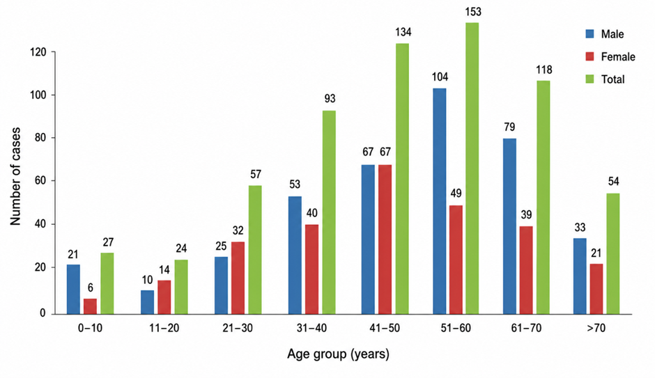
Figure 1. Age- and sex-wise distribution of patients undergoing image-guided fine-needle aspiration cytology for intra-abdominal lesions
Figure 1 presents the age- and sex-wise distribution of 660 patients who underwent image-guided fine-needle aspiration cytology for intra-abdominal lesions. The highest number of cases was recorded in the 51–60-year age group, comprising 153 patients (23.18%), followed by the 41–50-year age group with 134 patients (20.30%) and the 61–70-year age group with 118 patients (17.88%). The lowest number of cases was observed in the 11–20-year age group, with 24 patients (3.64%). Overall, males constituted 392 cases (59.39%), while females accounted for 268 cases (40.61%), demonstrating a clear male predominance. The predominance of cases in the middle-aged and older age groups suggests that intra-abdominal lesions were more commonly identified during the fifth to seventh decades of life.
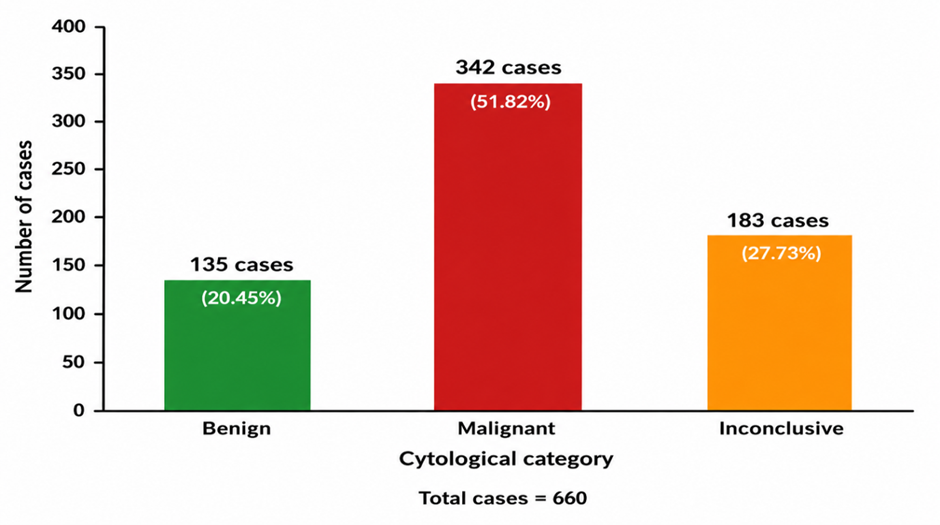
Figure 2. Overall cytological classification of intra-abdominal lesions
Figure 2 illustrates the overall cytological classification of 660 intra-abdominal lesions evaluated by image-guided fine-needle aspiration cytology. Malignant lesions formed the largest category, comprising 342 cases (51.82%), followed by inconclusive aspirates with 183 cases (27.73%). Benign lesions accounted for 135 cases (20.45%). The findings show that more than half of the aspirated intra-abdominal lesions were malignant, while approximately one-quarter remained inconclusive because of inadequate or indeterminate cytological material.
DISCUSSION:
Image-guided fine-needle aspiration cytology is an established minimally invasive technique for evaluating deep-seated abdominal lesions. It provides rapid cellular diagnosis and can differentiate inflammatory, benign and malignant lesions without requiring exploratory surgery in many patients. The present study evaluated 660 intra-abdominal lesions over eight years and demonstrated the broad cytomorphological spectrum encountered in a tertiary-care setting. A definite benign or malignant diagnosis was possible in 477 cases, while histopathological follow-up showed that FNAC had good sensitivity, specificity and overall diagnostic accuracy.
The patients ranged from 19 days to 89 years, confirming that intra-abdominal lesions requiring cytological evaluation may occur across all age groups. The largest number of cases was observed in the 51–60-year age group, followed by the 41–50-year and 61–70-year groups. Thus, more than 60% of the lesions occurred between the fourth and seventh decades of life. This concentration among middle-aged and older patients may be explained by the increasing prevalence of primary and metastatic malignancies with advancing age. Vasilj and Kojić Katović also evaluated patients across a wide age range and reported that image-guided FNAC could be safely performed in both paediatric and adult patients [10]. Their study included patients aged 10–83 years, indicating that age alone is not a limitation when an appropriate image-guided approach is available.
A male predominance was observed in the present study, with 392 males (59.39%) and 268 females (40.61%), producing a male-to-female ratio of approximately 1.5:1. Vasilj and Kojić Katović similarly reported 54% males and 46% females among 1,084 abdominal FNAC procedures [10]. Sattar et al. also found a marked male predominance among patients undergoing FNAC of focal hepatic lesions [14]. The higher proportion of males in the present series may reflect the predominance of hepatobiliary and retroperitoneal malignancies, differences in exposure to relevant risk factors and possible sex-related differences in healthcare utilisation. However, the study was hospital based, and this distribution should not be interpreted as the population incidence of intra-abdominal lesions.
Computed tomography was used to guide 427 aspirations (64.70%), whereas ultrasonography was used in 233 cases (35.30%). The greater use of CT guidance in this series was probably related to the inclusion of deeply located retroperitoneal, gastrointestinal, adrenal and small abdominal lesions. CT provides detailed anatomical localisation and allows the operator to assess the relationship of a lesion to bowel loops, vascular structures and adjacent organs. Sheikh et al. demonstrated the usefulness of both ultrasonography and CT guidance for sampling deep-seated thoracic and abdominal masses, including lesions of the liver, abdominal lymph nodes, pancreas, gastrointestinal tract and kidneys [11]. In contrast, several published series relied mainly or entirely on ultrasonographic guidance because it is rapid, inexpensive, radiation free and provides real-time needle visualisation [10,13]. Therefore, the choice of guidance modality should depend on lesion visibility, depth, anatomical accessibility and the safest available needle pathway rather than on a fixed preference for one method.
The hepatobiliary system was the most frequently aspirated region, accounting for 313 cases (47.42%), of which 307 arose in the liver. This finding is consistent with previous studies in which the liver was the most commonly sampled intra-abdominal organ. In the 10-year study by Vasilj and Kojić Katović, the liver accounted for 53% of all abdominal aspirations [10]. Sheikh et al. also reported that the liver was the predominant abdominal site among image-guided aspirations [11]. The high frequency of liver FNAC can be attributed to the large size and relative accessibility of the organ, as well as its involvement by primary tumours, metastatic deposits, abscesses and other focal lesions.
Malignant lesions represented the largest cytological category, comprising 342 cases (51.82%). Benign lesions accounted for 135 cases (20.45%), while 183 aspirates (27.73%) were inconclusive. The proportion of malignancy closely resembles the 52% malignant rate reported by Vasilj and Kojić Katović [10]. The high malignant yield in the present series is likely to reflect the referral pattern of a tertiary hospital, where patients with radiologically suspicious, advanced or inaccessible lesions are more likely to undergo image-guided sampling. Consequently, the observed distribution represents the cytology workload of the institution and not the true prevalence of malignant intra-abdominal disease in the general population.
Primary malignancies accounted for 200 cases, metastatic malignancies for 114 cases and 28 cases were reported as positive for malignancy without precise tumour classification. The inability to subtype some malignant lesions is an important limitation of conventional FNAC. Poorly differentiated tumours, extensively necrotic lesions and malignancies with overlapping cytomorphological features may permit recognition of malignancy but not accurate determination of histogenesis. Clinical history, radiological findings, cell-block preparation and immunocytochemistry may therefore be needed for definitive classification. Image-guided FNAC of deep lymph nodes has also been shown to provide useful diagnostic material, although ancillary studies may be required for accurate lymphoma classification and for distinguishing lymphoma from metastatic malignancy [12].
Among hepatic malignancies, metastatic tumours were the most frequent, accounting for 83 of 160 malignant hepatic lesions, followed by hepatocellular carcinoma in 54 cases. Metastatic adenocarcinoma was the most common morphological type of hepatic metastasis. This pattern agrees with the recognised role of the liver as a major site of haematogenous metastasis from gastrointestinal, pancreatic, breast, pulmonary and other primary malignancies. Vasilj and Kojić Katović found metastatic carcinoma in 50% of liver aspirates classified by tumour type, while primary hepatic tumours accounted for 27% [10]. Similarly, Sattar et al. reported that metastatic lesions were more frequent than hepatocellular carcinoma among 450 liver aspirations [14]. Their study included 183 metastatic deposits and 137 hepatocellular carcinomas, with metastatic adenocarcinoma forming the largest metastatic group. These findings support the diagnostic value of FNAC in distinguishing primary hepatic carcinoma from secondary deposits, particularly when cytomorphology is interpreted with imaging and clinical information.
Pyogenic abscess was the most frequent benign hepatic diagnosis, followed by tuberculous and amoebic abscesses. This finding demonstrates that image-guided FNAC is useful not only for identifying neoplastic lesions but also for diagnosing infective and inflammatory processes that can radiologically mimic malignancy. Sattar et al. similarly found abscesses to be the leading benign hepatic diagnosis in their series [14]. In such cases, aspiration may have both diagnostic and therapeutic importance and may provide material for microbiological investigations. Nevertheless, inflammatory lesions with extensive necrosis should be carefully evaluated because necrotic malignancies can yield predominantly inflammatory or acellular material.
Retroperitoneal lesions formed the second most frequently sampled anatomical category and accounted for 102 cases. Malignant lesions predominated in this region, including lymphomas, metastatic carcinomas, germ-cell tumours and sarcomas. Retroperitoneal lymph nodes alone contributed 70 cases. Chakrabarti et al. reported that lymph-node lesions formed the largest group of malignant retroperitoneal masses in their study and observed a 95.83% agreement between cytological and histopathological diagnoses among correlated neoplastic cases [15]. CT-guided FNAC has also demonstrated high diagnostic performance in retroperitoneal tumours, with reported sensitivity and specificity of 86% and 96%, respectively [16]. These findings support the use of image-guided cytology as an initial diagnostic procedure for retroperitoneal lesions, particularly when surgical biopsy would be technically difficult or associated with greater morbidity.
The diagnostic yield in the present study was 72.27%, while 27.73% of aspirates were categorised as inconclusive. This inadequacy or indeterminate rate was higher than the 17.7% rate reported by Vasilj and Kojić Katović [10]. The difference may be related to the wider range of organs sampled in the present study, greater use of CT for difficult deep-seated lesions, cystic and necrotic masses, the use of conventional smears without routine cell-block preparation and the absence of documented rapid on-site adequacy assessment. Inadequate aspirates are commonly associated with small lesions, deep location, extensive necrosis, fibrosis, haemorrhage, cystic change and inaccurate targeting of viable tumour. The particularly high inconclusive rates in ovarian, renal and hepatic lesions may also reflect cystic contents, haemorrhagic material and tumour heterogeneity.
The diagnostic limitations of FNAC should be considered when an aspirate is negative or inconclusive despite strong clinical or radiological suspicion of malignancy. O’Connell et al. found a higher insufficient sampling rate for FNAC than for fine-needle trucut biopsy in solid abdominal tumours and reported that core biopsy produced a higher diagnostic yield [17]. Nyman et al. similarly found that automated core biopsy improved diagnostic accuracy compared with fine-needle aspiration without increasing the complication rate [18]. These observations do not reduce the clinical value of FNAC; rather, they suggest that FNAC and core biopsy should be viewed as complementary techniques. Repeat image-guided aspiration, cell-block preparation or core biopsy should be considered when the initial FNAC is inadequate or when tissue architecture is required for tumour grading, lymphoma classification, immunohistochemistry or molecular analysis.
Histopathological correlation was available in 58 cases. FNAC achieved a sensitivity of 85.00%, specificity of 88.89%, positive predictive value of 94.44% and overall diagnostic accuracy of 86.21% for the detection of malignancy. The high positive predictive value indicates that a cytological diagnosis of malignancy was highly reliable. Ahsan et al. also reported a significant association between ultrasonography-guided FNAC and histopathological diagnosis in abdominal masses [13]. The diagnostic performance in the present study was also comparable with CT-guided retroperitoneal FNAC reported by Das et al. [16].
The negative predictive value was lower than the positive predictive value, indicating that a benign or negative FNAC result should be interpreted cautiously when imaging remains suspicious. False-negative results may occur because of sampling of necrotic, cystic or non-representative regions, low cellularity or failure to recognise well-differentiated malignant cells. False-positive results may result from severe reactive atypia, granulomatous inflammation or degenerative changes. Close correlation among the cytopathologist, radiologist and treating clinician is therefore essential, and discordant cases should undergo repeat aspiration or histological biopsy.
The study has several limitations. Histopathological confirmation was available for only 58 of the 660 cases, which may have introduced verification bias because lesions selected for surgery or biopsy may not represent the entire study population. The retrospective component limited the availability of uniform clinical, radiological and follow-up information. Cell-block preparation, immunocytochemistry and rapid on-site evaluation were not routinely documented. Furthermore, the data were collected from a single tertiary-care institution over an extended period, during which imaging equipment, sampling practices and cytological reporting may have changed.
Despite these limitations, the large sample size and inclusion of a broad range of abdominal organs provide a comprehensive overview of the cytomorphological spectrum encountered in image-guided FNAC. The findings demonstrate that the technique is particularly effective for detecting malignancy and distinguishing primary from metastatic lesions in many cases. Improvements in pre-procedural radiological planning, targeting of viable tissue, immediate assessment of specimen adequacy, preparation of cell blocks and selective use of immunocytochemistry or core biopsy could further reduce the inconclusive rate and improve specific tumour classification.
Overall, image-guided FNAC was a useful, rapid and relatively accurate diagnostic method for intra-abdominal lesions. Its high positive predictive value supports its role in confirming malignancy and guiding early clinical management. However, inadequate, negative and indeterminate aspirates should not be considered definitive when clinical or radiological suspicion persists. A multidisciplinary and multimodal diagnostic approach remains necessary to achieve the highest diagnostic accuracy.
CONCLUSION:
Image-guided fine-needle aspiration cytology proved to be a useful, rapid and minimally invasive diagnostic technique for evaluating intra-abdominal lesions. In the present study, malignant lesions formed the largest cytological category, accounting for 342 of 660 cases, while benign lesions and inconclusive aspirates constituted 135 and 183 cases, respectively. The hepatobiliary system, particularly the liver, was the most frequently sampled anatomical region, followed by the retroperitoneum. Metastatic malignancy was the most common malignant hepatic lesion, whereas pyogenic abscess was the leading benign hepatic diagnosis.
REFERENCES:
1. Stewart CJR, Coldewey J, Stewart IS. Comparison of fine needle aspiration cytology and needle core biopsy in the diagnosis of radiologically detected abdominal lesions. J Clin Pathol. 2002;55(2):93-97. doi:10.1136/jcp.55.2.93.
2. Dosi S, Gupta G, Kawatra M, Chakrabarti PR, Agrawal P, Jain MR. Role of radiological-assisted cytology in intra-abdominal lesions: a 3 years’ experience in a tertiary care center. Int J Appl Basic Med Res. 2016;6(2):101-105. doi:10.4103/2229-516X.179022.
3. Pujani M, Jetley S, Jairajpuri ZS, Khan S, Hassan MJ, Rana S, et al. A critical appraisal of the spectrum of image guided fine needle aspiration cytology: a three year experience from a tertiary care centre in Delhi. Turk Patoloji Derg. 2016;32(1):27-34. doi:10.5146/tjpath.2015.01339.
4. Sumana BS, Muniyappa B. Ultrasonography guided fine needle aspiration cytology with preparation of cell blocks in the diagnosis of intra-abdominal masses. J Clin Diagn Res. 2015;9(12):EC08-EC12. doi:10.7860/JCDR/2015/16490.6869.
5. Melese SS, Getaneh FB. Yield of ultrasound guided fine-needle aspiration of intraabdominal masses and lymph nodes: a prospective cross-sectional study. Ethiop J Health Sci. 2021;31(6):1241-1246. doi:10.4314/ejhs.v31i6.20.
6. Baisakh P, Baisakh MR, Kumari BS, Mohanty BB, Panda DK, Pradhan S. Diagnostic accuracy of ultrasonography-guided fine needle aspiration cytology in abdominopelvic masses: a prospective study. Cureus. 2023;15(6):e41228. doi:10.7759/cureus.41228.
7. Vinayakamurthy S, Manoli N, Shivajirao P, Manjunath, Jothady S. Role of cell block in guided FNAC of abdominal masses. J Clin Diagn Res. 2016;10(3):EC01-EC05. doi:10.7860/JCDR/2016/17359.7422.
8. Bhowmik S, Chakrabarti I, Ghosh P, Bera P, Banik T. Comparative evaluation of cell block method and smear cytology in fine needle aspiration cytology of intra-abdominal mass lesions. Iran J Pathol. 2018;13(2):179-187.
9. Bohara S, Shukla P, Shah S, Chaturvedi R, Singh K. Utility of guided FNAC and cell block preparation from liver and gall bladder masses: learning experience from a tertiary care center. J Cancer Res Ther. 2024;20(5):1459-1463. doi:10.4103/jcrt.jcrt_172_23.
10. Vasilj A, Kojić Katović S. Fine needle aspiration cytology of abdominal organs: ten-year single center experience. Acta Clin Croat. 2016;55(1):35-40. doi:10.20471/acc.2016.55.01.6.
11. Sheikh M, Sawhney S, Dey P, al-Saeed O, Behbehani A. Deep-seated thoracic and abdominal masses: usefulness of ultrasound and computed tomography guidance in fine needle aspiration cytology diagnosis. Australas Radiol. 2000;44(2):155-160. doi:10.1046/j.1440-1673.2000.00799.x.
12. Saikia UN, Khirdwadkar N, Saikia B, Sood B, Goldsmith R, Dey P, et al. Image-guided fine-needle aspiration cytology of deep-seated enlarged lymph nodes. Acta Radiol. 2002;43(2):230-234. doi:10.1080/028418502127347844.
13. Ahsan MK, Khan AH, Rahman Z, Hye MA, Alam SM, Bardhan H, et al. Role of USG-guided fine needle aspiration cytology in the diagnosis of abdominal mass. Mymensingh Med J. 2015;24(1):89-93.
14. Sattar A, Khan AM, Anjum S, Naqvi A. Role of ultrasound guided fine needle aspiration cytology in diagnosis of space occupying lesions of liver. J Ayub Med Coll Abbottabad. 2014;26(3):334-336.
15. Chakrabarti I, Bhowmik S, Sinha MGM, Bera P. Ultrasound-guided aspiration cytology of retroperitoneal masses with histopathological corroboration: a study of 71 cases. J Cytol. 2014;31(1):15-19. doi:10.4103/0970-9371.130629.
16. Das C, Sengupta M, Mukhopadhyay M, Saha AK. Critical clinical appraisal of the role of computed tomography-guided minimally invasive aspiration cytology in evaluation of retroperitoneal masses. Indian J Med Paediatr Oncol. 2014;35(1):60-65. doi:10.4103/0971-5851.133723.
17. O’Connell AM, Keeling F, Given M, Logan M, Lee MJ. Fine-needle trucut biopsy versus fine-needle aspiration cytology with ultrasound guidance in the abdomen. J Med Imaging Radiat Oncol. 2008;52(3):231-236. doi:10.1111/j.1440-1673.2008.01952.x.
Nyman RS, Cappelen-Smith J, Brismar J, von Sinner W, Kagevi I. Yield and complications in ultrasound-guided biopsy of abdominal lesions: comparison of fine-needle aspiration biopsy and 1.2-mm needle core biopsy using an automated biopsy gun. Acta Radiol. 1995;36(5):485-490. doi:10.1080/02841859509