Patient Outcomes with Intracranial Hemorrhage and Hematoma – A Retrospective Analysis.
- Geetha J. , Professor, Department of Anaesthesia, Dhanalakshmi Srinivasan Medical College and Hospital, Siruvachur, Perambalur, Tamil Nadu, India.
- Logesh Ram J. , Postgraduate, Department of Anaesthesia, Dhanalakshmi Srinivasan Medical College and Hospital, Siruvachur, Perambalur, Tamil Nadu, India.
- Harini D. , CRMI, Department of Anaesthesia, Dhanalakshmi Srinivasan Medical College and Hospital, Siruvachur, Perambalur, Tamil Nadu, India.
- Indhra Pradeepa A , CRMI, Department of Anaesthesia, Dhanalakshmi Srinivasan Medical College and Hospital, Siruvachur, Perambalur, Tamil Nadu, India.
- Varsha. , CRMI, Department of Anaesthesia, Dhanalakshmi Srinivasan Medical College and Hospital, Siruvachur, Perambalur, Tamil Nadu, India.
Article Information:
Abstract:
Background: India estimates 1.5 to 2 million cases of traumatic brain injury (TBI) with nearly one million fatalities per year. Large studies conducted in 2025 in India showed a prevalence of head injury of 25.7 per thousand people. Rapid urbanisation and motorisation have led to a rise in road traffic accidents (RTA). This short observational retrospective study analyses the outcome of 50 patients admitted into our critical care unit during the study period. Materials And Methods: The Retrospective analysis study was conducted in the Critical Care Unit, DSMCH from March 2026 – May 2026 with Sample size 50. Patients admitted with head injury into the critical care unit. 50 complete case records as available from the medical records section of patients admitted with head injury into the intensive care unit were scrutinised. Glasgow Coma Scale on admission was mentioned as E for eye movements, V for verbal response, T for intubated patients, and M for motor response. The radiological imaging diagnosis for the clinical presentation was noted as hematoma (C, for clot), hemorrhage (H), or both hemorrhage and hematoma (C & H), for all 50 patients. The outcome of the patients from the intensive care unit was classified as F for Improved, E for Expired, and patients who could not continue treatment were classified as A for AMA (against medical advice) or R for those patients discharged at request for continuation of improvement towards further domiciliary care. The patients on endotracheal intubation, mentioned as T, were four in number and received surgical intervention. The observations were tabulated and analysed. Observations And Results: The age group of patients included in this study ranged from 20 to 80 years, with a mean of 43.8 years. Hematoma was more common in the younger age group and hemorrhage in the elderly in our study sample. 64% of patients improved, 6% expired, and those discharged at request or against medical advice comprised 30%. Hematoma was present in 46% of cases, hemorrhage in 38%, and both were present together in 16% of cases. 80% of the head injury patients were males. GCS across outcome groups by the Kruskal-Wallis test showed a p value of 0.1. Hemorrhage versus hematoma outcome, on applying the Chi square test, showed a p value of 0.409. Conclusion: The goal of this study was to stress a comprehensive approach to TBI among medical undergraduates. The management of TBI revolves around maintenance of cerebral perfusion pressure by optimising MAP and ICP, and to recognise, remember, and execute all components of the FAST HUGS BID mnemonic in intensive care. Public awareness and enforcement of road safety measures and traffic rules, and innovations in safety devices in vehicles to prevent fatalities, are emphasised with the completion of this study. Abbreviations: RTA – Road Traffic Accident, TBI – Traumatic Brain Injury, GCS – Glasgow Coma Scale, ICP – Intracranial Pressure, MAP – Mean Arterial Pressure, CPP – Cerebral Perfusion Pressure.
Keywords:
Article :
INTRODUCTION:
Head injuries constitute a common cause of mortality in the ever-increasing road traffic accidents. The prevalence of head injury has been estimated as 259 per one lakh population in the U.S. The estimated prevalence of head injury in India was found to be 25.7 per 1000 population in a meta-analysis which surveyed 85,720 patients.[1]
The patients who survive in a vegetative or partially vegetative state following an intracranial event comprise a social and domestic burden. Mortality and morbidity due to head injury are mainly due to an acute rise in intracranial pressure subsequent to cerebral edema. A hemorrhage or hematoma, according to the site or activity, perilesional reaction may compress the brain stem and affect the vital centers, which is gradual and can lead from morbidity to mortality.
This short retrospective study was planned in the critical care section to analyse and compare the patient outcomes with intracranial hemorrhage and hematoma, their GCS scores, and to examine the gravity of either of the two pathologies that in turn influenced the patient's improvement or mortality.
Aim
To evaluate patient outcomes in intracranial hemorrhage and hematoma.
Objectives
● To enumerate patient demographics and Glasgow Coma Scale (GCS) on admission.
● To confirm hemorrhage or hematoma from CT/MRI reports.
● To analyse outcome as improved and discharged, or otherwise.
MATERIALS AND METHODS:
Study
Retrospective analysis.
Study Place
Critical Care Unit, DSMCH.
Study Period
March 2026 – May 2026.
Study Sample
50.
Study Population
Patients admitted with head injury into the critical care unit. 50 complete case records as available from the medical records section of patients admitted with head injury into the intensive care unit were scrutinised. Glasgow Coma Scale on admission was mentioned as E for eye movements, V for verbal response, T for intubated patients, and M for motor response. The radiological imaging diagnosis for the clinical presentation was noted as hematoma (C, for clot), hemorrhage (H), or both hemorrhage and hematoma (C & H), for all 50 patients.
The outcome of the patients from the intensive care unit was classified as F for Improved, E for Expired, and patients who could not continue treatment were classified as A for AMA (against medical advice) or R for those patients discharged at request for continuation of improvement towards further domiciliary care. The patients on endotracheal intubation, mentioned as T, were four in number and received surgical intervention. The observations were tabulated and analysed.
RESULTS:
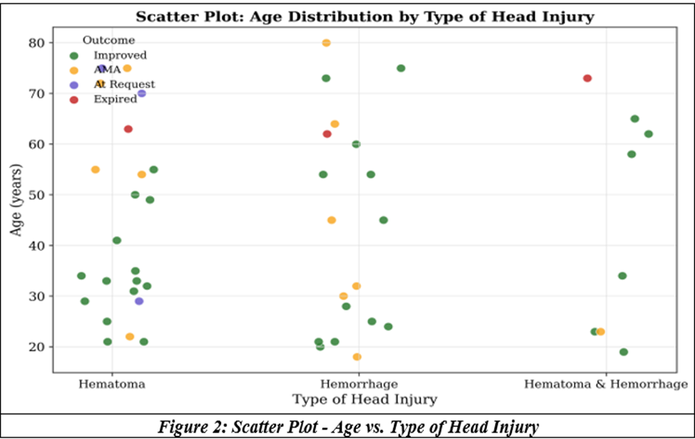
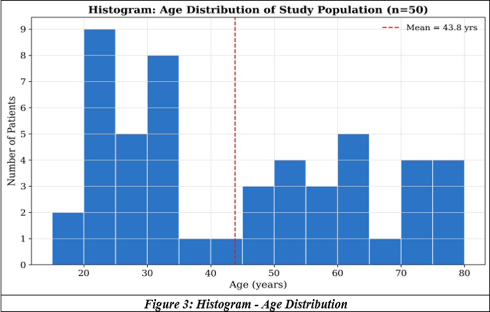
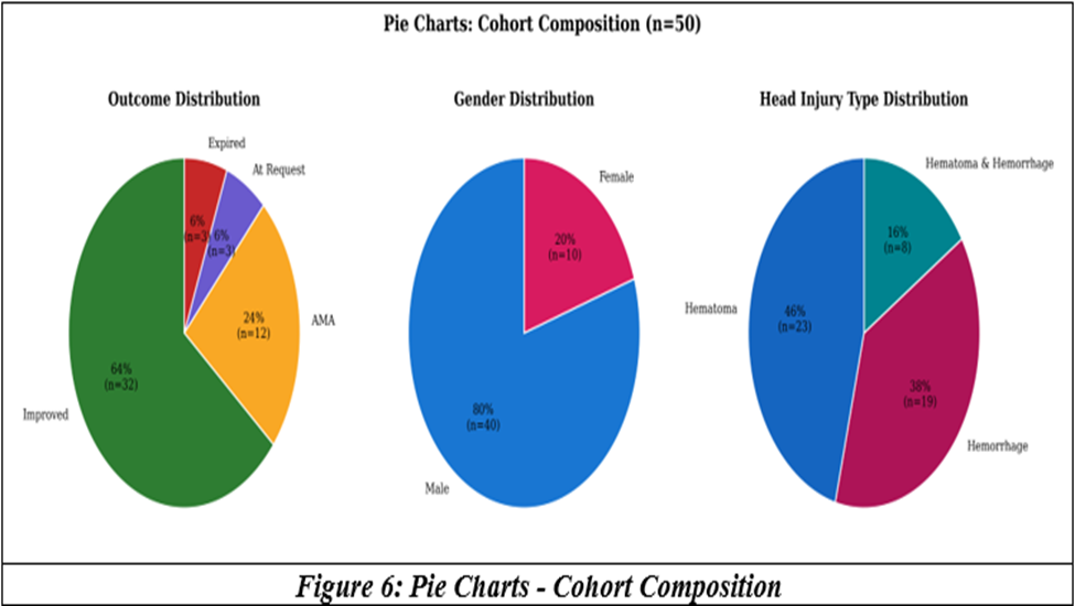
The age group of patients included in this study ranged from 20 to 80 years, with a mean of 43.8 years. Hematoma was more common in the younger age group and hemorrhage in the elderly in our study sample. 64% of patients improved, 6% expired, and those discharged at request or against medical advice comprised 30%.
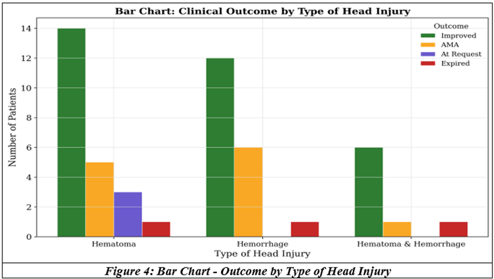
Hematoma was present in 46% of cases, hemorrhage in 38%, and both were present together in 16% of cases. 80% of the head injury patients were males. GCS across outcome groups by the Kruskal-Wallis test showed a p value of 0.1. Hemorrhage versus hematoma outcome, on applying the Chi square test, showed a p value of 0.409.
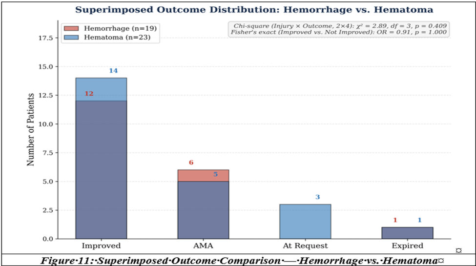
Chi square test on injury and outcome (2 x 4) shows statistical significance, p = 0.409.
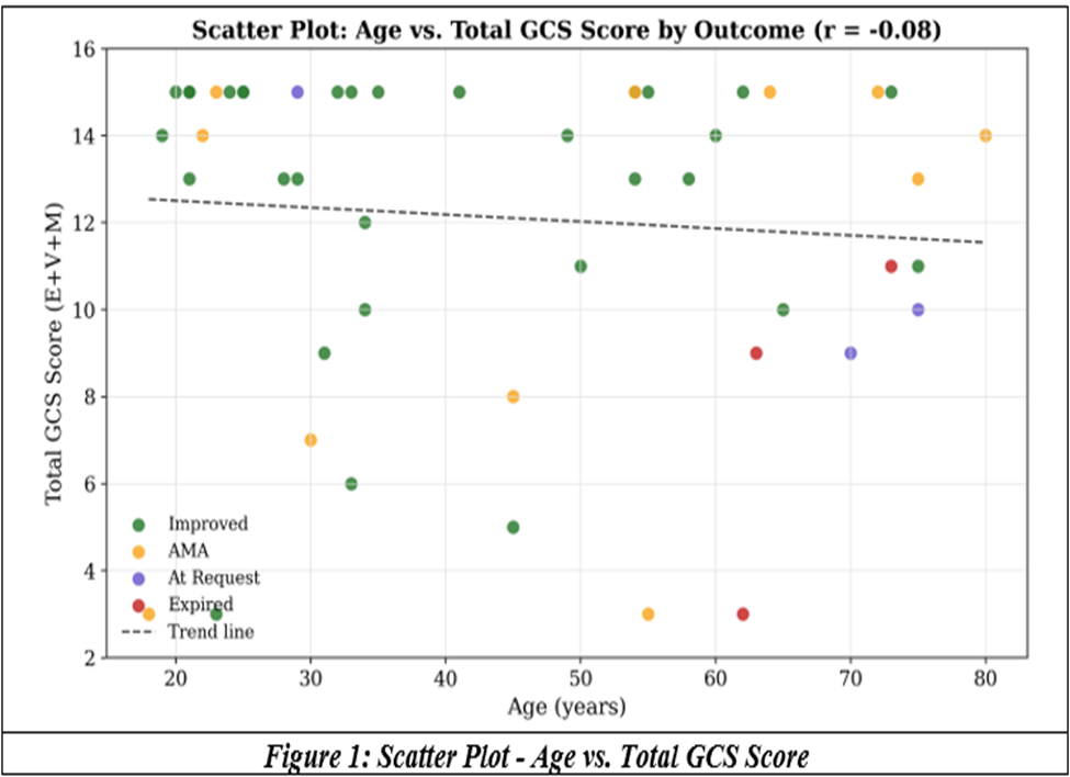
1. Scatter Plot - Age vs. Total GCS Score:
Comparing patient age against Total Glasgow Coma Scale, stratified by outcome
2. Scatter Plot - Age vs. Type of Head Injury
Age distribution within each injury category, colored by outcome
3. Histogram - Age Distribution
Frequency distribution of patient age across the full cohort (n = 50)
4. Bar Chart - Outcome by Type of Head Injury
Comparing clinical outcome distribution across the three injury categories
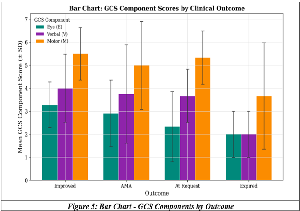
5. Bar Chart - GCS Components by Outcome
Mean Eye, Verbal, and Motor GCS scores compared across outcome groups
6. Pie Charts - Cohort Composition
Proportional breakdown of outcome, gender, and head injury type
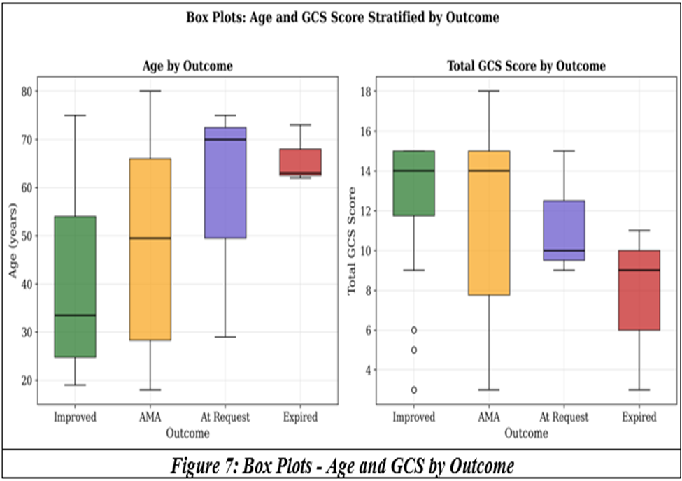
7. Box Plots — Age and GCS by Outcome
Median, interquartile range, and spread of Age and Total GCS across outcome groups
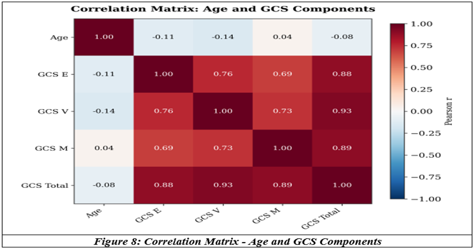
8. Correlation Matrix — Age and GCS Components
Pairwise Pearson correlation coefficients (matrix / heatmap graph)
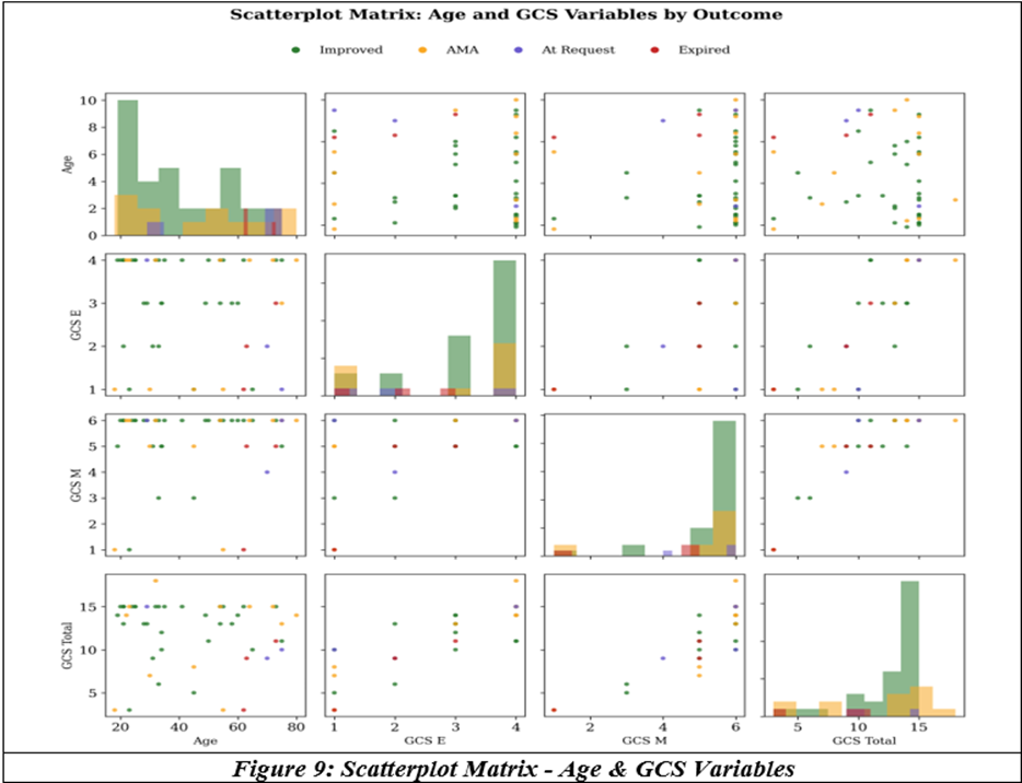
9. Scatterplot Matrix — Age & GCS Variables
Pairwise relationships and distributions of Age, GCS-E, GCS-M, and Total GCS, by outcome
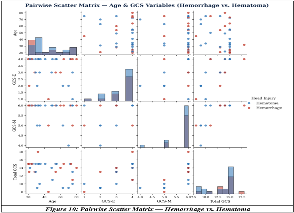
10. Pairwise Scatter Matrix — Hemorrhage vs. Hematoma
Age, GCS-E, GCS-M, and Total GCS restricted to the two dominant injury types, colored by Head Injury
11. Superimposed Outcome Comparison — Hemorrhage vs. Hematoma
Overlaid outcome counts, n = 19 Hemorrhage vs. n = 23 Hematoma ("Hematoma & Hemorrhage" cases excluded).
DISCUSSION:
Fatalities due to road traffic accidents have decreased in high income countries, of which head injury fatality declined by 54.3% according to data from the German In-Depth Accident Study (GIDAS).[2] A total of 24,405 accidents reported from 1991 to 2011 were examined, and a relative decrease in mortality of 68.8% was noted. The World Health Organisation has initiated the Decade of Action for Road Safety because it is predicted that fatality due to RTA could become the fifth leading cause of death by 2030. Another retrospective study over a 4-year period, 2009–2012, of 694 patients with head injuries[3] was analysed. Extracranial pathology was present in 65% of cases, and 97.6% of patients required intubation. The study concluded that the cause of mortality (28.5%) was due to GCS < 8 on admission and subdural hematoma with secondary systemic insults such as respiratory, circulatory and metabolic derangements. Head injury includes craniocerebral trauma that might present as focal or diffuse, acute or chronic, sudden or gradual,[4] and the patients could be classified as low risk group, moderate risk group and high risk group. Traumatic brain injury is a heterogeneous disorder, and the severity is classified based on the Glasgow Coma Scale on admission,[5] with GCS ranging from 13–15 classified as mild, 9–12 as moderate, and GCS < 8 as the severe category.
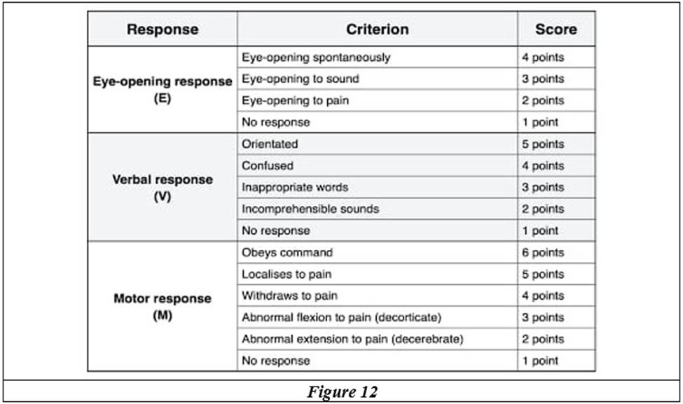
The mild form indicates mortality of less than 1%, the moderate category up to 15%, and a poor GCS indicates a mortality of up to 40%.[6] The focal intracranial damage may be epidural and subdural hematomas along with parenchymal contusions, or diffuse, such as axonal injury or cerebral edema.
Traumatic brain injury is a dynamic process resulting in alterations of structure and function of all elements of the brain. The grading of severity of TBI depends mainly on the GCS motor score, which remains one of the strongest predictors.[7] The pathology and pathophysiology of craniocerebral trauma can be discussed under the following headings:
a) Skull fractures – linear, depressed, comminuted.
b) Cerebral concussion and axonal shearing.
c) Cerebral edema
d) Parenchymal contusion and hemorrhage: Parenchymal hematomas are in the deep white matter, whereas contusions are cortical. In the absence of secondary hemorrhage, small contusions have a good recovery as outcome.
e) Subdural hematoma: These may be acute or chronic and occur due to tearing and stretching of the veins that drain the surface of the brain into the venous sinuses. CT reveals a high-density crescentic collection. The size, situation, extent, and compression on adjacent structures are the factors that decide medical or surgical intervention as well as outcome.
f) Epidural hematoma: This occurs in less than 1% of cases, with tears of the middle meningeal artery or dural sinuses. A lucid interval with relapse into coma or hemiplegia as the hematoma expands carries a high risk of mortality.
g) Subarachnoid hemorrhage: This is of little clinical importance if small, but if large, is a poor prognostic sign.
The diagnosis could be based on history, clinical features and examination, and finally radiography and imaging.
History
The circumstances of the event, force and location of head impact, vomiting, confusion or progressive seizure activity, alcohol intoxication, medicolegal registration, and entry into the accident register with proper legal intimation as per institutional protocol are all essential.
Examination
General examination and trauma survey should be followed by detailed physical and neurological examinations. Bloody discharge or clear fluid (testing positive for sugar) from the nose may indicate CSF rhinorrhea with fracture of the base of the skull. The biochemistry of CSF, with a glucose concentration of 30 mg/dl compared to that of lacrimal or nasal secretions with a value of less than 5 mg/dl of glucose, must be remembered. Eye movements, pupils, oculocephalic and oculovestibular reflexes, and motor examination are all performed in a methodical manner.
Most often, focal and diffuse traumatic brain injuries can lead to similar clinical pictures:
a) Decreased consciousness and coma could be due to lesions with localised mass effect on the diencephalon or brainstem, or primary brainstem lesions. The same clinical picture may be due to diffuse axonal injury to the diencephalon or brainstem, as well as diffuse edema with compression of mesencephalic or diencephalic structures. Hypometabolic inactivation or excitotoxic effects may cause temporary functional effects presenting in a similar manner.
b) Dysexecutive syndrome or memory dysfunction could be due to concussion of the more vulnerable frontal and temporal lobes, or axonal injury to fibre bundles such as the uncinate fasciculus and corona radiata, or widespread Wallerian degeneration.
c) Post-traumatic amnesia (inability to count numbers in reverse, inability to recall, and poor attention) could occur due to focal medial temporal lobe lesions, or with focal compression of the medial temporal lobe, or diffuse axonal injury affecting the memory network.
d) Motor weakness could occur with focal lesions with mass effect comprising the motor pathways, or hemorrhage of the basal ganglia, while diffuse axonal injury or hypoxic ischemic injury of the corticospinal tract may also present a similar picture.
e) The differentiation is by radiography and imaging. Computerised tomography of the brain provides unsurpassed sensitivity for detecting intracranial blood, in addition to skull fractures. An MRI detects subtle injury to the brain but may not be accessible due to time and expense in emergency situations.
CT brain is thus diagnostic and prognostic, indicates the need for emergency surgery, and assesses the progress of response to treatment; serial CTs may detect a rebleed or an expanding hematoma. This information also helps guide decisions for emergency surgical intervention.
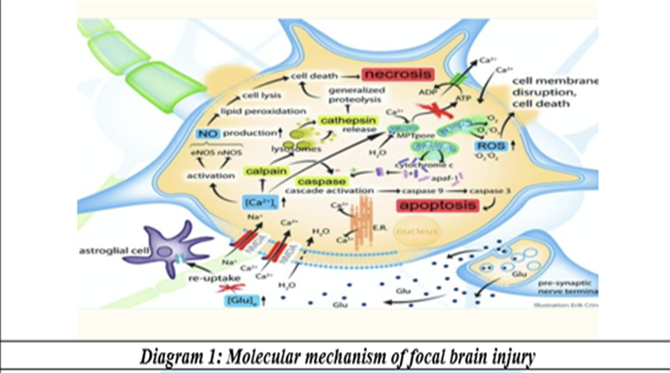
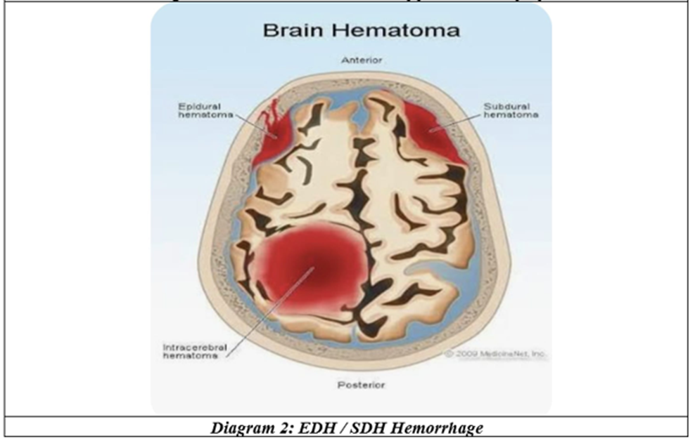
Molecular and cellular mechanisms of focal brain injury have been extensively studied. The impact on the head and sequential energy transfer to cerebral tissues cause depolarisation of nerve cells, resulting in uncontrolled excessive release of excitatory neurotransmitters leading to excitotoxicity. Glutamate levels increase up to 50-fold in focal parenchymal contusions.[8] Human microdialysis studies demonstrate that raised extracellular glutamate levels are associated with poor prognosis.[9] Glutamate is released by presynaptic vesicles after depolarisation, but astrocytes perform the reuptake through an ATP-dependent sodium co-transport system. Excessive extracellular glutamate initiates a massive influx of calcium and sodium ions into neurons and glial cells.[10] These activate voltage-dependent calcium channels, followed by passive water movement leading to neuronal swelling. Protein phosphorylation is disrupted, and activation of calcium-dependent enzymes calpain 1 and 2 result in protein and enzyme destruction. Peroxynitrite, which forms from the nitric oxide pathway, results in lipid peroxidation, cell lysis, and DNA fragmentation.[11] The eventual cascade to cell death is played out through mitochondrial dysfunction.[12]
Imaging TBI
Visualisation of the full extent of damage after TBI is complex. CT is the choice in acute-phase work-up. In chronic-phase work-up, MRI techniques such as T2-weighted imaging or attenuated inversion recovery are preferred to detect non-hemorrhagic contusions and edema. Diffuse axonal injury is accompanied by small punctate hemorrhages; hence T2 gradient echo MRI is used. Diffusion tensor imaging produces in vivo information of brain tissue integrity by yielding an image on the basis of diffusion of water molecules.[13]
Outcome
Mild category of TBI with GCS 13–15 with less than 8% abnormalities on CT almost never needed neurosurgical intervention.[14] Moderate category of TBI with GCS 9–12 had an average of 64.7% CT abnormalities and needed neurosurgical intervention at the rate of 16.5%.[15] Severe TBI presenting with GCS less than or equal to 8 showed more than 90% abnormalities on CT, with 36.3% needing neurosurgical intervention.[16]
Summary of Results
This short study was initiated focussing on spreading awareness and post-speciality knowledge for undergraduate medical interns, to enable them to initiate management in the casualty and intensive care unit. The study group involved a convenient sample available during the study period. A higher GCS on admission was related to an improved outcome (p = 0.1). There existed a common assumption that intracranial hemorrhage had acute, grave consequences while intracranial hematoma followed a more gradual course of events. This was disproved in this study, which had patients with smaller hematoma who had acute grave consequences; four patients in the series had large hematoma with hemorrhage that needed decompression. The contained hemorrhage and small hematomata had similar outcomes. In patients with intracranial hemorrhage versus hematoma, the association with outcome was moderately significant (p = 0.409). Age and total GCS did not show a significant correlation with intracranial pathology.
Management
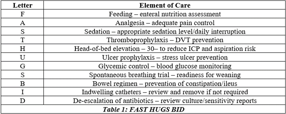
The holistic care during the course of the patient's admission in the intensive care unit greatly affects the outcome. This is discussed under two headings: management of elevated ICP, and the mnemonic FAST HUGS BID.
Management of ICP (Mayer SA, Dennis L, 1998)
The goal of maintaining an adequate cerebral perfusion pressure is attained by managing ICP. This includes:
1. Evacuation of mass effect, either by CSF drainage or surgical removal of hematoma.
2. Achieving immobility to reduce energy requirement and sympathetic response.
3. Pressor infusion if CPP < 70 mmHg, and antihypertensives or vasodilators if CPP > 120 mmHg (CPP = MAP − ICP; normal 70–90 mmHg).
4. Hyperventilation to maintain pCO₂ at 28–32 mmHg.
5. Consider pentobarbital and hypothermia (33°C) to reduce cerebral oxygen consumption and for neuroprotection.
6. Head-end elevation by 30°.
The essential aspects of care in the ICU that are usually overlooked, but greatly contribute to patient outcome, have been developed as a mnemonic tool for easy remembrance and practice by medical and paramedical personnel.[17]
CONCLUSION:
Fatalities in RTA are largely due to head injuries. Early hospital admission, quick evaluation, aggressive intervention, and a holistic approach across all systems all have a significant role to play in achieving a successful outcome. The severity of injury, multisystem involvement, poor GCS on admission, an expanding intracranial hematoma, a progressive or rebleeding hemorrhage, involvement of the brain stem, and a poor motor score on admission all indicate a high risk of mortality. The nature of the intracranial blood, whether hematoma or hemorrhage, appears less important than its size, situation, and mass effect. Focal pathologies may have a time-limited recovery, while diffuse brain injuries have an unpredictable outcome.
A comprehensive approach, including enforcement of road safety policies, innovations in car engineering, implementation of protocols in emergency medicine, alcohol level control, and safety measures such as helmets and seat belts alongside other technical advances, is the road to preventing these injuries.
Limitations
Small sample, complete radiological details inaccessible.
REFERENCES:
1. Saishree, Ranjan JK, Asthana HS, et al. Prevalence of Head Injury in India: A Systematic Review and Meta-analysis of Cross-sectional Community-based Surveys. Ann Neurosci 2025;33(2):237-46.
2. rnstberger A, Joeris A, Daigl M, et al. Decrease of morbidity in road traffic accidents in a high income country - an analysis of 24,405 accidents in a 21 year period. Injury 2015;46 Suppl 4:S135-43.
3. Chelly H, Bahloul M, Ammar R, et al. Clinical characteristics and prognosis of traumatic head injury following road traffic accidents admitted in ICU "analysis of 694 cases". Eur J Trauma Emerg Surg 2019;45(2):245-53.
4. Mayer SA, Rowland LP. Head injury. Sec. IV, Trauma, Chap- 63. In: Merritt's neurology. Wolters Kluwer Health 2021: p. 311.
5. Andriessen TMJC, Jacobs B, Vos PE. Clinical characteristics and pathophysiological mechanisms of focal and diffuse traumatic brain injury. J Cell Mol Med 2010;14(10):2381-92.
6. Cremer OL, Moons KG, van Dijk GW, et al. Prognosis following severe head injury: Development and validation of a model for prediction of death, disability, and functional recovery. J Trauma 2006;61(6):1484-91.
7. Steyerberg EW, Mushkudiani N, Perel P, et al. Predicting outcome after traumatic brain injury: development and international validation of prognostic scores based on admission characteristics. PLoS Med 2008;5(8):e165.
8. Bullock R, Zauner A, Myseros JS, et al. Evidence for prolonged release of excitatory amino acids in severe human head trauma: relationship to clinical events. Ann N Y Acad Sci 1995;765:86.
9. Chamoun R, Suki D, Gopinath SP, et al. Role of extracellular glutamate measured by cerebral microdialysis in severe traumatic brain injury. J Neurosurg 2010;113:564-70.
10. Choi DW. Ionic dependence of glutamate neurotoxicity. J Neurosci 1987;7:369-79.
11. Hall ED, Detloff MR, Johnson K, et al. Peroxynitrite-mediated protein nitration and lipid peroxidation in a mouse model of traumatic brain injury. J Neurotrauma 2004;21:9-20.
12. Xiong Y, Gu Q, Peterson PL et al. Mitochondrial dysfunction and calcium perturbation induced by traumatic brain injury. J Neurotrauma 1997;14:23-34.
13. Le Bihan D. Looking into the functional architecture of the brain with diffusion MRI. Nat Rev Neurosci 2003;4:469-80.
14. Geijerstam JL, Britton M. Mild head injury – mortality and complication rate: meta-analysis of findings in a systematic literature review. Acta Neurochir 2003;145:843-50.
15. Fabbri A, Servadei F, Marchesini G, et al. Early predictors of unfavourable outcome in subjects with moderate head injury in the emergency room. J Neurol Neurosurg Psychiatry 2008;79:567-73.
16. Marshall LF, Gautille T, Klauber MR, et al. The outcome of severe closed head injury. J Neurosurg 1991;75:S28-36.
17. Nair AS, Naik VM, Rayani BK. FAST HUGS BID for the surgical patient. Indian J Crit Care Med 2017;21(10):713-4.