Analysis Of Functional Outcome For Unstable Distal Radius Fracture Treated With Closed Reduction And Percutaneous K Wire Fixation With Casting And Closed Reduction With Casting : A Comparative Study.
- Sridhara Venkappa Badiger , Assistant Professor, Department Of Orthopaedics K H Patil Institute Of Medical Sciences Gadag, Karnataka, India.
- Santosh Sangam , Senior Resident Department Of Orthopaedics KH Patil Institute of medical sciences Gadag Karnataka India.
- Prajwal J , Assistant Professor Department of Orthopaedic Nandi Medical College and Research Institute.
Article Information:
Abstract:
Background: Distal radius fractures are among the most common fractures of the upper limb and can result in pain, deformity, loss of wrist function, and prolonged disability. Unstable distal radius fractures are particularly challenging because reduction maintained by cast immobilization alone may be lost due to deforming forces. Closed reduction with percutaneous Kirschner wire (K-wire) fixation and casting may provide greater radiological stability and facilitate earlier functional recovery. This study was undertaken to compare the functional and radiological outcomes of closed reduction and cast immobilization (CRC) with closed reduction and percutaneous K-wire fixation followed by casting (CRPP+C) in patients with unstable distal radius fractures. Materials and Methods: A prospective comparative study was conducted in the Department of Orthopaedics, Al-Ameen Medical College and Hospital, Vijayapura, over a period of two years. Fifty adult patients with unstable distal radius fractures were enrolled and divided into two treatment groups: patients treated with closed reduction and cast immobilization (CRC) and patients treated with closed reduction, percutaneous K-wire fixation, and casting (CRPP+C). Radiological parameters, pain, functional recovery, grip strength, and return to occupational activity were assessed at regular follow-up intervals up to 12 weeks. The incidence of secondary displacement and procedure-related complications was also evaluated. Results: The study population had a mean age of approximately 51 years, with females comprising 54% of the cohort. AO type A3 fractures were the predominant fracture pattern. Patients treated with CRPP+C demonstrated better maintenance of radiological alignment and greater resistance to secondary displacement compared with those treated with cast immobilization alone. The K-wire group also showed earlier pain relief, improved grip strength, and faster functional recovery during the early follow-up period. At six weeks, 72% of patients in the CRC group continued to report moderate pain, whereas the CRPP+C group demonstrated a more favorable pain distribution. By 12 weeks, pain outcomes between the groups showed convergence, with 44% of the CRC group and 68% of the CRPP+C group reporting no pain. Although both groups progressed toward fracture union and functional improvement, the advantages of K-wire fixation were most evident during the early recovery period. Conclusion: Closed reduction with percutaneous K-wire fixation followed by casting is an effective and reliable treatment for unstable distal radius fractures. Compared with closed reduction and cast immobilization alone, CRPP+C provides better radiological stability, reduces the risk of secondary displacement, and promotes earlier functional recovery and pain relief. While closed reduction and casting remain appropriate for selected patients, percutaneous K-wire fixation should be considered particularly in unstable fracture patterns with a high risk of loss of reduction.
Keywords:
Article :
INTRODUCTION:
Together with the distal ulna and the triangular fibrocartilage complex (TFCC), the distal radius, which is the final 3 cm of the radial bone, forms the primary surface of the radiocarpal joint. In terms of biomechanics, this area is essential for shifting loads from the hand to the forearm, allowing the wrist to flex, extend, move radially and ulnarly, and support forearm rotation.
This intricate structure is disrupted by a distal radius fracture (DRF), which results in discomfort, deformity, and loss of function. The traditional mechanism, called a fall onto an outstretched hand (FOOSH), drives the carpal bones, especially the lunate, into the distal radial articular surface by applying axial compression and hyperextension forces (Corsino et al., 2023). Reduction is the first stage of treating fractures and is based on the concepts of Reduction, Retention, and Rehabilitation. The non-operative realignment of fracture fragments using manual traction and manipulation, known as "closed reduction" (CR), makes use of ligamentotaxis, which uses intact soft-tissue hinges to help guide fragments back into position.
This is typically accomplished with adequate pain treatment, such as sedation, a hematoma block, or a Bier's block. In contrast, Open Reduction (OR) entails making a surgical incision to directly view the fracture site. This enables precise anatomical restoration, which is frequently required for complex intra-articular fractures or when soft tissue interposition prevents closed techniques from working. Either the outside or the interior can be used to continue the decrease. The conventional method of immobilizing a cast entails creating a sturdy sleeve that securely fastens the cast using fiberglass or molded plaster. One type of minimal internal fixation is percutaneous Kirschner Wire (K-wire) fixation. K-wires are threaded or smooth metal pins that have diameters between 0.8 and 2.0 mm. While the image is being enhanced, they are subjected to fracture fragments. They are frequently used in conjunction with a cast and function similarly to internal splints to increase the stability of the reduction (Chattopadhyay et al., 2017).
Depending on how the fracture appears, several configurations are chosen, such as radial styloid pins, Kapandji's intrafocal pins, cross-pinning, or multi-planar structures (Sanil & Sajan, 2024; Sable & Purane, 2024). In comparison to patients treated with Closed Reduction and Casting alone, it is predicted that patients with unstable distal radius fractures undergoing Closed Reduction and Percutaneous K-wire Fixation with Casting will show statistically significant better radiographic outcomes and a lower incidence of secondary displacement. Both groups are expected to improve in terms of functional outcomes, but the CRPP+C group is expected to exhibit more early functional recovery (at 6–12 weeks). However, during a longer follow-up period (6–12 months), this difference might lessen. The study anticipates that the two groups will have unique profiles of procedure-specific problems.
This comprehensive study, carried out at the Department of Orthopedics at Al-Ameen Medical College Hospital, aims to contribute definitive local evidence to the international orthopaedic discourse, ultimately guiding surgeons toward more customized and effective treatment approaches for patients with unstable distal radius fractures.
MATERIALS AND METHODS:
Study Design
• Type: Comparative longitudinal study
• Design: Prospective interventional study comparing two treatment modalities
Study Setting
• Al-Ameen Medical College and Hospital, Vijayapura, Karnataka
• Department of Orthopaedics
Study Duration
January 2024 to January 2026 (including patient recruitment, intervention, follow-up, and data analysis)
Sample Size
• Total: 50 patients
• Group 1 (Closed Reduction + Casting): 25 patients
• Group 2 (Closed Reduction + K-wire Fixation + Casting): 25 patients
Inclusion Criteria:
• Age ≥ 18 years
• Extra-articular distal radius fractures amenable to both closed reduction with casting and K-wire fixation
• Patients unwilling for surgical intervention included in casting group
Exclusion Criteria:
• Patients dependent on others for basic activities
• Complex intra-articular fractures with more than one sagittal fragment
• Open fractures
• Fractures with neurovascular injuries
• Associated musculoskeletal injuries to the ipsilateral upper limb
Methodology
Standard closed manipulation procedures were used to reduce fractures in patients assigned to the closed reduction and cast immobilization group. Hematoma block anesthetic was used throughout the surgery. Longitudinal traction was used to disimpact the fracture fragments while the patient was comfortably positioned with the injured limb supported. With the help of an attendant, counter-traction was administered at the arm with the elbow flexed while traction was maintained by holding the injured hand's thumb, index finger, and middle finger.
Translation reduction techniques were used to correct the misplaced distal piece when sufficient traction was obtained. Flexion and ulnar deviation movements were used to decrease the distal fragment while maintaining traction at the fracture site. By using mild pronation, flexion, and ulnar deviation forces, the fracture was finally locked in the decreased position. Throughout the procedure, caution was used to avoid using too much power and to stop more soft tissue damage.
RESULTS:
Table 1. Patient Distribution By Sex
|
Sex |
K-Wire Fixation and Casting |
Cast Immobilization |
|
Male |
12 |
11 |
|
Female |
13 |
14 |
|
Total |
25 |
25 |
|
Grand Total |
- |
50 |
Table 1 depicts the sex-wise distribution of patients included in the present comparative study evaluating functional and anatomical outcomes of unstable distal radius fractures managed by two different treatment modalities, namely closed reduction with percutaneous K-wire fixation and casting, and closed reduction with cast immobilization alone. A total of 50 patients were enrolled in the study, with equal allocation of 25 patients to each treatment group, thereby ensuring uniform group sizes and minimizing allocation bias.In the K-wire fixation and casting group, 12 patients (48%) were male and 13 patients (52%) were female.
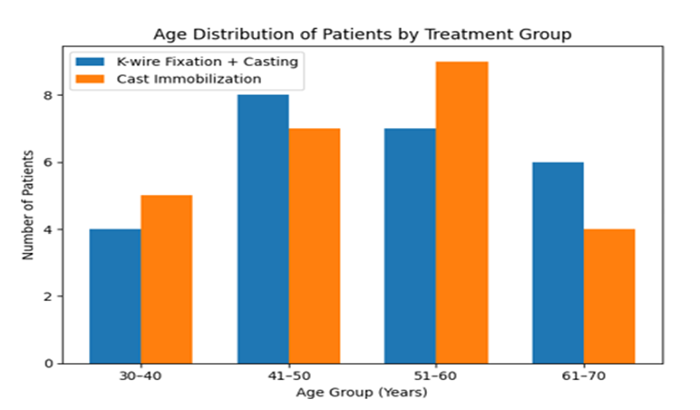
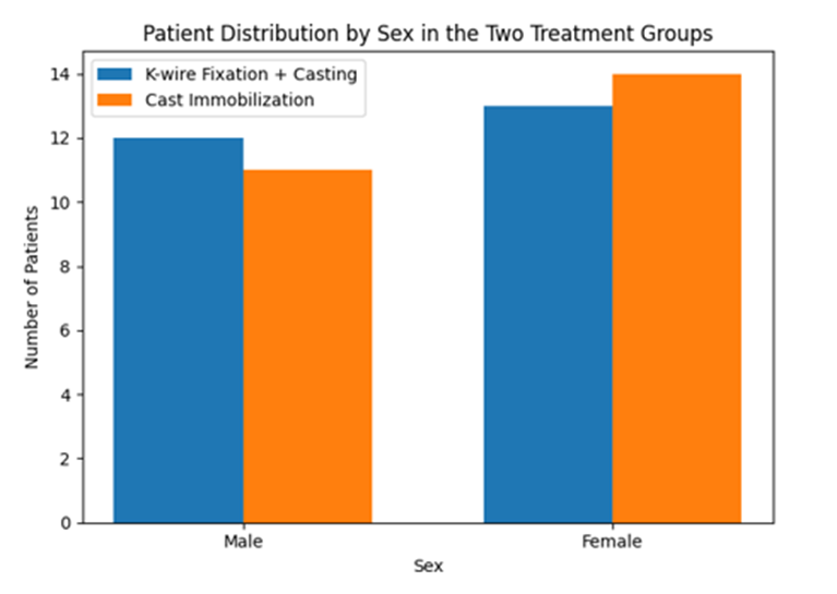
Table 2. AGE OF PATIENTS
|
Age in Years |
K-Wire Fixation and Casting – Number of Patients |
|
Cast Immobilization – Number of Patients |
|
|
|
Male |
Female |
Male |
Female |
|
30 – 40 |
2 |
2 |
2 |
3 |
|
41 – 50 |
4 |
4 |
3 |
4 |
|
51 – 60 |
2 |
5 |
4 |
5 |
|
61 – 70 |
4 |
2 |
2 |
2 |
|
Mean Age (Years) |
51.4 |
52.30 |
51.09 |
51.14 |
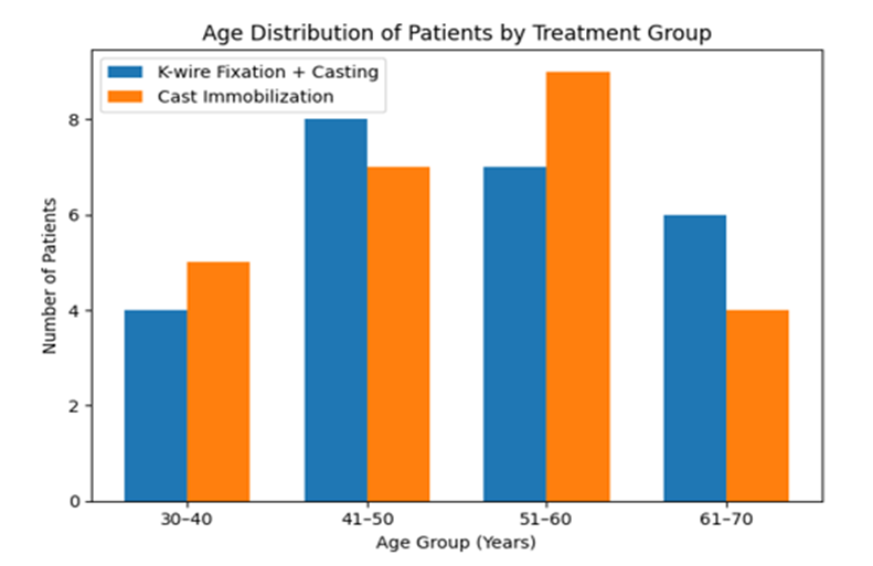
Table 2 illustrates the age distribution of patients included in the present study comparing outcomes of unstable distal radius fractures treated by closed reduction with percutaneous K-wire fixation and casting versus closed reduction with cast immobilization alone. The patients were categorized into four age groups ranging from 30 to 70 years, and the distribution was analyzed separately for males and females in both treatment groups. The mean age values were also calculated to understand the overall age profile of the study population.
Table 3. SIDE OF INJURY
|
Side of Injury |
K-Wire Fixation and Casting(No. of Patients) |
Cast Immobilization (No.of Patients) |
|
Right |
11 |
12 |
|
Left |
14 |
13 |
|
% Dominant Side Injury |
44% |
48% |
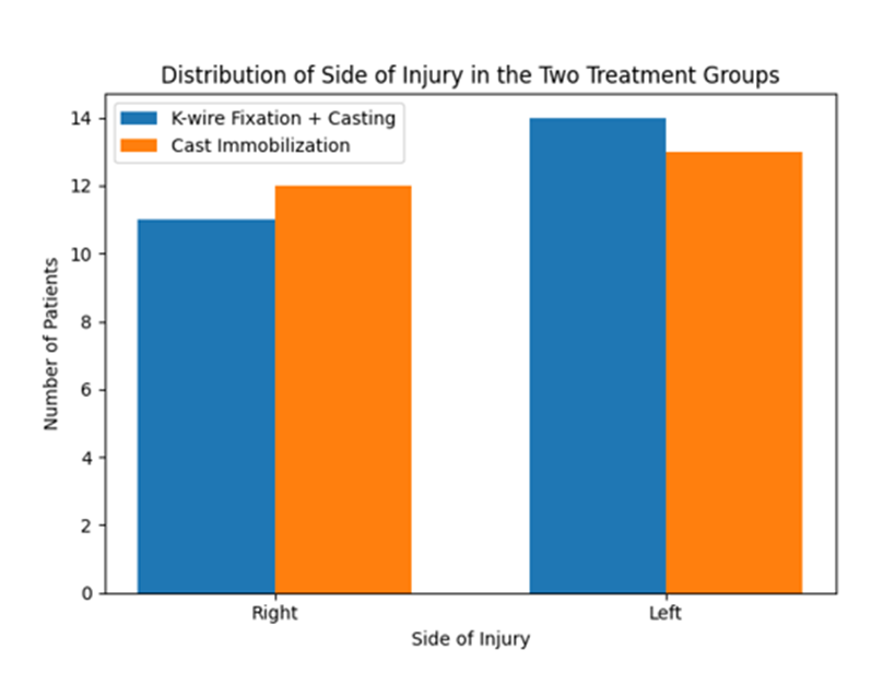
Table 3 presents the distribution of patients based on the side of wrist involvement in both treatment groups of the present study. Assessment of the side of injury is an important demographic and functional parameter, as involvement of the dominant extremity may significantly influence functional impairment, rehabilitation demands, patient-reported outcomes, and the urgency of return to occupational and daily activities.
Table 4. FRACTURE TYPE BY AO CLASSIFICATION
|
AO Type |
K-Wire Fixation and Casting(No. of Patients) |
Cast Immobilization(No. of Patients) |
|
A2 |
8 |
9 |
|
A3 |
14 |
13 |
|
C1 |
3 |
3 |
|
Total |
25 |
25 |
Table 3 presents the distribution of patients based on the side of wrist involvement in both treatment groups of the present study. Assessment of the side of injury is an important demographic and functional parameter, as involvement of the dominant extremity may significantly influence functional impairment, rehabilitation demands, patient-reported outcomes, and the urgency of return to occupational and daily activities.
Table 4. FRACTURE TYPE BY AO CLASSIFICATION
|
AO Type |
K-Wire Fixation and Casting(No. of Patients) |
Cast Immobilization(No. of Patients) |
|
A2 |
8 |
9 |
|
A3 |
14 |
13 |
|
C1 |
3 |
3 |
|
Total |
25 |
25 |
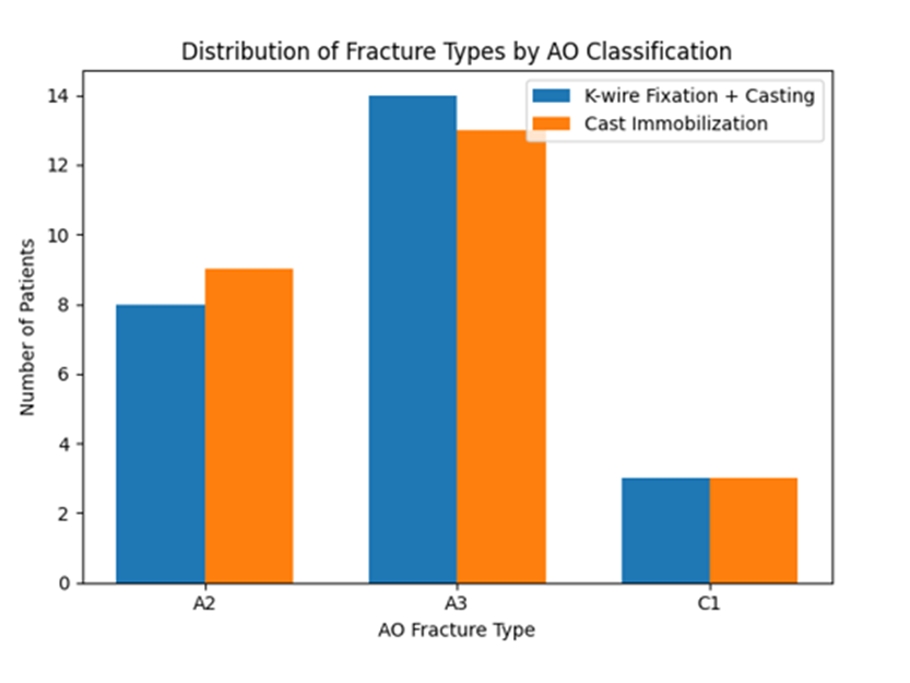
On the basis of the AO classification system, Table 4 shows the distribution of unstable distal radius fractures in the study population. The AO classification is used to classify distal radius fractures by complexity, number of fragments, and joint involvement to assess severity and create a treatment plan. Only displaced extra-articular and simple intra-articular unstable fractures from AO types A2, A3, and C1 met the inclusion criteria, so this study focused on them.In the cohort that received K-wire fixation and casting, 14 patients (56%) had AO type A3 fractures. Three patients (12%) had AO type C1 fractures, while eight (32%) had A2 fractures.
Table 5. PAIN
|
Pain Severity |
K-Wire Fixation and Casting(No. of Patients at) |
|
|
Cast Immobilization(No. of Patients at) |
|
|
|
|
6 Weeks |
9 Weeks |
12 Weeks |
6 Weeks |
9 Weeks |
12 Weeks |
|
Nil |
6 |
12 |
16 |
4 |
6 |
15 |
|
Mild |
11 |
9 |
7 |
3 |
11 |
9 |
|
Moderate |
8 |
4 |
2 |
18 |
8 |
1 |
|
Severe |
– |
– |
– |
– |
– |
– |
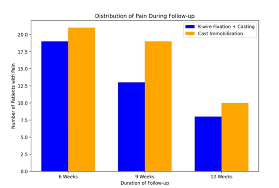
Table 5 illustrates the distribution of pain severity among patients in both treatment groups—K-wire fixation with casting and cast immobilization alone—at follow-up intervals of 6, 9, and 12 weeks. Pain was categorized into four grades: nil, mild, moderate, and severe. At the 6-week follow-up, the K-wire fixation group demonstrated comparatively lower pain levels. In this group, 6 patients reported no pain, 11 patients had mild pain, and 8 patients experienced moderate pain. In contrast, the cast immobilization group showed a higher proportion of moderate pain, with 18 patients reporting moderate pain, 3 patients mild pain, and 4 patients no pain. This suggests that patients treated with percutaneous K-wire fixation experienced earlier reduction in pain compared with those managed with casting alone.
Table 6. GRIP STRENGTH
|
% of Opposite Side |
K-Wire Fixation and Casting(No. of Patients at) |
|
|
Cast Immobilization (No. of Patients at) |
|
|
|
|
6 Weeks |
9 Weeks |
12 Weeks |
6 Weeks |
9 Weeks |
12 Weeks |
|
76– 100% |
6 (24%) |
11(44%) |
15(60%) |
3 (12%) |
8(32%) |
12(48%) |
|
51–75% |
9 (36%) |
8(32%) |
5 (20%) |
11 (44%) |
9(36%) |
8(32%) |
|
26–50% |
10 (40%) |
6(24%) |
5 (20%) |
11 (44%) |
8(32%) |
5(20%) |
|
0–25% |
– |
– |
– |
– |
– |
– |
Table 6 presents the distribution of grip strength recovery expressed as a percentage of the opposite (uninjured) side in patients treated with K-wire fixation with casting and cast immobilization alone at 6, 9, and 12 weeks of follow-up. At 6 weeks, patients treated with K-wire fixation and casting demonstrated better grip strength recovery compared with those treated with cast immobilization. In the K-wire group, 24% of patients achieved 76–100% grip strength, while 36% were in the 51–75% range and 40% were in the 26–50% range.
DISCUSSION:
In order to assess and compare the functional and radiological results of two commonly used treatments for unstable distal radius fractures—closed reduction with cast immobilization (CRC) and closed reduction with percutaneous Kirschner wire fixation followed by casting (CRPP+C)—the current prospective comparative study was created. Al-Ameen Medical College and Hospital enrolled fifty adult patients over the course of two years.
Vijayapura, and monitored for up to twelve weeks after the intervention at regular intervals. While conceding that cast immobilization is still a suitable and acceptable treatment option when carefully chosen, the results generally support the idea that percutaneous K-wire fixation gives quantifiable advantages in early functional recovery and radiological stability. These findings contribute to an increasing amount of research on the best ways to treat this extremely prevalent upper limb injury, which makes up almost three-quarters of all forearm fractures and over 18% of all adult fractures (Corsino et al., 2023). Because prolonged functional impairment directly translates into loss of productive capability and poor quality of life, distal radius fractures incur a disproportionately severe socioeconomic impact, especially in a working-age population. This cohort's fracture pattern was primarily AO type A3, which is recognized as intrinsically unstable because it lacks cortical buttressing and is characterized by extra-articular metaphyseal comminution (Vos et al., 2015).
With a mean age of roughly 51 years and a slight female predominance of 54%, the study sample's demographic profile and the high prevalence of this fracture subtype are both consistent with patterns found in epidemiological studies that identify postmenopausal women as the group most susceptible to low-energy fragility fractures at the distal radius (Søsborg-Würtz et al., 2018). Potential confounders resulting from dominant-hand engagement were distributed equally throughout the cohorts since the side distribution was generally equivalent across the two groups.
A quicker trajectory of pain remission in the early post-treatment phase was one of the most immediately noticeable benefits seen in the percutaneous K-wire fixation group. In contrast to the CRPP+C group, which had a more favorable distribution with a higher percentage reporting either no pain or only mild discomfort, the majority of patients in the CRC group (72%) continued to feel moderate pain at the six-week assessment. By the twelve-week period, both groups had converged toward similar levels of pain, with 44% of the cast group and 68% of the K-wire group reporting no pain. This temporal pattern is biologically realistic, with surgical stabilization providing an earlier pain benefit that progressively diminishes. Fracture micromotion is a known cause of inflammatory discomfort in the periosteal and endosteal compartments, and internal fixation with K-wires removes the micromotion at the fracture site that happens when an unstable fracture is treated with cast immobilization alone.
These findings are consistent with those of Rodríguezw et al. (2019), who examined pain perception in patients undergoing preoperative closed reduction for unstable dorsally displaced distal radius fractures and found that sufficient initial reduction significantly reduces pain burden in the early post-injury phase. The additional fixation in Group II prevented the recurrence of deforming forces that may re-stress the fracture and cause continuous nociceptive stimulation, suggesting that the same mechanism was at work in the immediate post-procedural period in the current investigation.
In a comparison of CRPP and traditional casting for extra-articular distal radius fractures, Singh et al. (2018) found that the K-wire fixation cohort had better early pain control; this pattern is consistent with the trajectory seen during the six- and nine-week evaluations in the current study. The notable convergence of pain scores at twelve weeks in both treatment groups indicates that the biological basis for varying pain levels decreases after fracture consolidation is sufficiently advanced to withstand deforming forces, even in the cast-treated group. Although K-wire fixation speeds up early comfort, it does not necessarily change the final pain resolution endpoint at three months, which makes this observation clinically significant.
When considered collectively, the results of this study show that closed reduction with percutaneous K-wire fixation and casting offers clinically significant benefits over cast immobilization alone in the treatment of unstable distal radius fractures, especially with regard to radiological stability, early pain relief, recovery of grip strength, and return to occupational activity. The K-wire group's superiority in preserving anatomical parameters over the course of the twelve-week observation period is mechanistically consistent with the known pathophysiology of unstable metaphyseal fractures, where cast-dependent reduction in comminuted patterns is invariably compromised by deforming muscular and gravitational forces.
The well-known phenomenon that the wrist has a remarkable capacity for functional adaptation even in the context of mild-to-moderate residual malalignment, at least within the short-term follow-up window, is reflected in the functional outcome score differences, which were modest in absolute magnitude but consistently directionally favorable to the K-wire group. These findings provide useful advice for the practicing orthopaedic surgeon at a district or tertiary care hospital in the Indian subcontinent, where operational resources are accessible yet patient willingness and expense may occasionally favor conservative management. The data supports the addition of percutaneous K-wire fixation to closed reduction as a method that lowers the risk of secondary displacement and speeds up functional recovery for fractures that meet instability criteria, especially AO type A3 patterns with dorsal comminution, radial shortening exceeding 5 mm, and dorsal angulation exceeding 20°. For patients in whom surgical fixation is declined or contraindicated, closed reduction and casting continue to play a significant role. The current data confirms that, with careful cast application and close monitoring to identify and address early loss of reduction, satisfactory, if somewhat inferior, outcomes can be achieved. The information produced here highlights the significance of individualized patient assessment in choosing the best treatment strategy for this frequent and functionally significant injury and adds to the expanding regional evidence base for orthopaedic decision-making.
CONCLUSION:
An efficient and dependable technique for treating unstable distal radius fractures is closed reduction with percutaneous K-wire fixation in conjunction with casting. When compared to cast immobilization alone, this method offers superior fracture stability, better anatomical alignment maintenance, and quicker functional recovery.K-wire fixation has major advantages in terms of early functional outcomes and lower likelihood of secondary displacement, even if both treatment approaches eventually lead to fracture union.
REFERENCES:
1. Vos, L., Strackee, S., Goslings, J., Schep, N., & Walenkamp, M. (2015). The Unstable distal radius Fracture—How do we define it? A systematic review. Journal of Wrist Surgery, 04(04), 307–316. https://doi.org/10.1055/s-0035-1556860
2. Sekar, P. (2020). Unstable intra-articular fracture distal radius managed by combined external fixation and internal fixation by volar plating. Journal of Orthopedics and Joint Surgery, 2(1), 31–37. https://doi.org/10.5005/jp-journals-10079-1025
3. Chawla, S., Gupta, A., Pasupuleti, A., Karnati, A. K. R., & Gupta, M. (2023).Unstable distal radius fractures fixation with a 2.4 mm volar variable-angle locking plate: radiological and functional assessment. International Journal of Research in Orthopaedics, 9(4), 669–674. https://doi.org/10.18203/issn.2455-4510.intjresorthop20231436
4. Putnam, M. D., & Fischer, M. D. (1997). Treatment of unstable distal radius fractures: Methods and comparison of external distraction and ORIF versus external distraction-ORIF neutralization. The Journal of Hand Surgery, 22(2), 238–251. https://doi.org/10.1016/s0363-5023(97)80158-x
5. Corsino, C. B., Reeves, R. A., & Sieg, R. N. (2023, August 8). Distal radius fractures. StatPearls - NCBI Bookshelf. https://www.ncbi.nlm.nih.gov/books/NBK536916/
6. Søsborg-Würtz, H., Gellert, S. C., Erichsen, J. L., & Viberg, B. (2018). Closed reduction of distal radius fractures: a systematic review and meta-analysis. EFORT Open Reviews, 3(4), 114–120. https://doi.org/10.1302/2058-5241.3.170063
7. Sanil, N., & Sajan, A. (2024). The Kapandji pinning: Technique of closed reduction for distal end radius fracture in adults. Indian Journal of Orthopaedics Surgery, 10(2), 149–153. https://doi.org/10.18231/j.ijos.2024.025
8. Anderson, A. B., & Tintle, S. M. (2021). Closed reduction techniques for distal radius fractures and appropriate casting methods. Hand Clinics, 37(2), 239–245. https://doi.org/10.1016/j.hcl.2021.02.006
9. Sb, N. K., Pereira, N. J., & Rm, N. S. (2024). A study on the functional outcome of closed reduction and K-wire fixation in the treatment of extra-articular distal radius fractures. Asian Journal of Medical Sciences, 15(11), 155–160. https://doi.org/10.71152/ajms.v15i11.4179
10. Singh, G., Rastogi, P., Kodi, H., Vishwakarma, R., & Singh, A. (2018). Comparative analysis of closed reduction and percutaneous pinning versus conventional closed reduction with casting in extra-articular fractures of distal end radius. International Surgery Journal, 5(11), 3651. https://doi.org/10.18203/2349-2902.isj20184639
11. Özkan, S., Westenberg, R., Helliwell, L., & Mudgal, C. S. (2018). Distal radius fractures: evaluation of closed reduction and percutaneous Kirschner wire pinning. Journal of Hand and Microsurgery, 10(3), 134–138. https://doi.org/10.1055/s-0038-1648334
12. Beyer, F., Oppermann, J., Prasse, T., Müller, L. P., Eysel, P., & Bredow, J. (2024). How preoperative closed reduction and time to surgery impact postoperative palmar inclination in distal radius fractures. Journal of Clinical Medicine, 13(8), 2316. https://doi.org/10.3390/jcm13082316
13. Nazari, R., Mousavi, S., Fakoor, M., Dargah, A. H., Mayan, B. H., Taheri, H., Mousavinezhad, S. M., Senobari, N., Gooshvar, M., & Ebrahimi, P. (2025). Comparative analysis of closed reduction with external fixation versus closed reduction with percutaneous pinning for distal radius fractures. Scientific Reports, 15(1), 19147. https://doi.org/10.1038/s41598-025-04001-8
14. Hassellund, S. S., Oftebro, I., Williksen, J. H., Søreide, E., Madsen, J. E., & Frihagen, F. (2023). Closed reduction of dorsally displaced distal radius fractures in the elderly provided improved final radiographic results. Journal of Orthopaedic Surgery and Research, 18(1), 247. https://doi.org/10.1186/s13018-023-03733-5
15. Handoll, H. H., & Madhok, R. (2003). Closed reduction methods for treating distal radial fractures in adults. Cochrane Database of Systematic Reviews, 2010(1), CD003763. https://doi.org/10.1002/14651858.cd003763
16. Löw, S., Papay, M., & Eingartner, C. (2019). Pain Perception following Initial Closed Reduction in the Preoperative Care of Unstable, Dorsally Displaced Distal Radius Fractures. Journal of Hand and Microsurgery, 11(2), 111–116. https://doi.org/10.1055/s-0039-1688681
17. Neidenbach, P., Audigé, L., Wilhelmi-Mock, M., Hanson, B., & De Boer, P. (2009). The efficacy of closed reduction in displaced distal radius fractures. Injury, 41(6), 592–598. https://doi.org/10.1016/j.injury.2009.10.055
18. Chattopadhyay, A., Banerjee, U., Sinha, P. K., Misra, S., Chattopadhyay, A., & Veeragandham, P. (2017). A prospective study of distal radius fracture management by close reduction, percutaneous Kirschner wire fixation and plaster immobilization. International Journal of Research in Orthopaedics, 3(3), 537. https://doi.org/10.18203/issn.2455-4510.intjresorthop20171898
19. Sanders, L., Johnson, N., & Dias, J. J. (2021). Kirschner Wire fixation in dorsally displaced distal radius fractures: a biomechanical evaluation. Journal of Wrist Surgery, 11(01), 021–027. https://doi.org/10.1055/s-0041-1729761
20. Vipin, R., Rengarajan, N., Manoharan, M., & Kesavan, K. (2021). A novel 5-pin fixation for distal radius fractures and its functional assessment. Journal of Orthopedics and Joint Surgery, 3(1), 13–17. https://doi.org/10.5005/jp-journals-10079-1045
21. Thaware, A. D., Garg, R. N., Patil, S., & Kuris, J. (2024). Assessment of clinical, functional and radiological outcomes in patients with fracture of distal end radius managed with close reduction and internal fixation with multiple percutaneous K wires: a prospective, single centre study. International Journal of Research in Orthopaedics,10(2), 274–280. https://doi.org/10.18203/issn.2455-4510.intjresorthop20240407
22. Ali, T., Baketh, M. S., & Mir, M. A. (2020). Closed reduction and percutaneous k-wire fixation for distal end radius fractures. International Journal of Research in Medical Sciences, 8(7), 2400. https://doi.org/10.18203/2320-6012.ijrms20202564
23. Brennan, S. A., Kiernan, C., Beecher, S., O‘Reilly, R. T., Devitt, B. M., Kearns, S. R., & O‘Sullivan, M. E. (2015). Volar plate versus k-wire fixation of distal radius fractures. Injury, 47(2), 372–376. https://doi.org/10.1016/j.injury.2015.08.040
24. Gandhi, S., Safiullah, M., Reddy, V. N., Khan, A., & M, M. R. P. (2025). A Case Series of Functional and Radiological Outcome of Comminuted Distal Radius Fractures Treated with Bridging External Fixator with Optional Percutaneous K-wires. Journal of Orthopaedic Case Reports, 15(8), 270–274. https://doi.org/10.13107/jocr.2025.v15.i08.5966
25. Van Aaken, J., Beaulieu, J., Della Santa, D., Kibbel, O., & Fusetti, C. (2008). High rate of complications associated with extrafocal kirschner wire pinning for distal radius fractures. Chirurgie De La Main, 27(4), 160–166. https://doi.org/10.1016/j.main.2008.05.005.