Role of Neutrophil to Lymphocyte Ratio as an Inflammatory Biomarker in Severity of Coronary Artery Disease in Acute Myocardial Infraction.
- Madhavi Kulkarni , Professor, Department of Pathology,Navodaya Medical College and Research Centre, Raichur, Karnataka, India.
- Kruthikashree K M , Resident, Department of Pathology,Navodaya Medical College and Research Centre, Raichur, Karnataka, India.
- Ajith V. Kulkarni , Associate Professor, Department of Medicine,Navodaya Medical College and Research Centre, Raichur, Karnataka, India.
- A. S. Aanand , Professor & Head, Department of Pathology,Navodaya Medical College and Research Centre, Raichur, Karnataka, India.
Article Information:
Abstract:
Background: Atherosclerosis and acute myocardial infarction are now well established as inflammation-driven processes. The neutrophil-to-lymphocyte ratio (NLR) is a simple, inexpensive inflammatory marker derived from routine blood counts and has been proposed as a surrogate marker of disease severity and prognosis in acute coronary syndromes. However, existing literature shows variable results regarding its correlation with angiographic severity of coronary artery disease. Objective: To evaluate the association between the neutrophil-to-lymphocyte ratio measured at the time of admission and the angiographic severity of coronary artery disease, as assessed by the SYNTAX score, in patients with acute myocardial infarction. Methods: This prospective observational study included 30 adult patients with acute coronary syndrome. Peripheral venous blood samples were obtained at admission prior to any invasive intervention. Complete blood counts were analysed using an automated haematology analyser, and NLR was calculated from absolute neutrophil and lymphocyte counts. All patients underwent coronary angiography, and CAD severity was quantified using the SYNTAX scoring system. Statistical analysis was performed to assess the correlation between NLR and angiographic severity. Results: A total of 30 patients with acute coronary syndrome who underwent PTCA were included in the study. The mean age of the subjects was 51.40 ± 14.33 years, with a marked male predominance (83.3%). The mean SYNTAX score was 18.45 ± 10.79. The mean neutrophil percentage was 79.77 ± 8.69, mean lymphocyte percentage was 14.63 ± 7.99, and mean neutrophil–lymphocyte ratio (NLR) was 8.41 ± 8.35. Low SYNTAX score was observed in 63.3% of patients, while 36.7% belonged to the intermediate and high SYNTAX score group. ROC curve analysis showed poor diagnostic performance of NLR in predicting angiographic severity (AUC = 0.411, p = 0.426). Pearson’s correlation analysis demonstrated no significant correlation between SYNTAX score and neutrophil percentage (r = 0.00, p = 0.999), lymphocyte percentage (r = −0.02, p = 0.918), or NLR (r = −0.09, p = 0.635). Conclusion: Acute coronary syndrome reflects an underlying inflammatory atherothrombotic process. Although elevated neutrophil counts and NLR in the present study indicate a systemic inflammatory response in acute myocardial infarction, no significant correlation was observed between NLR and SYNTAX score. This suggests that peripheral blood inflammatory indices may not reliably represent the structural complexity and burden of coronary atherosclerosis. The findings highlight that angiographic severity is a manifestation of chronic vascular pathology, whereas NLR reflects a dynamic systemic inflammatory state. Larger studies integrating hematological and tissue-level inflammatory markers are required to clarify the pathological relevance of NLR in assessing coronary artery disease severity.
Keywords:
Article :
INTRODUCTION:
Cardiovascular diseases (CVDs) are the leading cause of death worldwide and a significant contributor to disability. In 2019, approximately 17.9 million people died from CVDs, accounting for 32% of all global deaths. Of these, 85% were caused by myocardial infarction and stroke. Notably, around three-quarters of CVD-related deaths occur in low- and middle-income countries.[1]
Although coronary artery disease (CAD) has long been understood to begin with cell proliferation linked to endothelial injury, recent research on atherosclerotic plaques reveals a more complex pathophysiology, with inflammation playing a key role in both the initiation and progression of the disease. It is well established that elevated levels of inflammatory markers are associated with a higher incidence of cardiac events in patients with CAD.[2]
The Neutrophil to Lymphocyte Ratio (NLR) has emerged as a potential prognostic marker in patients with acute myocardial infarction (MI). Various international studies have investigated the association between NLR and the severity of coronary artery disease (CAD), consistently showing that NLR levels are elevated in cases of more severe CAD.[3]
However, study results remain conflicting, as neutrophil counts can be affected by other coexisting risk factors. Therefore, it is still unclear whether neutrophils serve as an independent prognostic marker in acute cardiovascular disease or offer additional value beyond total WBC count and CRP levels. Hence, the present study aims to assess the relationship between the neutrophil-to-lymphocyte ratio and severity of coronary artery disease.[4].
MATERIALS AND METHODS:
This was a hospital-based, observational, cross-sectional study that was conducted in the Department of Pathology, in collaboration with the Department of Cardiology, Navodaya Medical College Hospital and Research Centre, Raichur, on patients diagnosed with acute coronary syndrome (ACS) who underwent percutaneous transluminal coronary angioplasty (PTCA). The study was carried out over a defined study period after approval had been obtained from the Institutional Ethics Committee. A total of 30 consecutive patients with a clinical and electrocardiographic diagnosis of ACS who had undergone coronary angiography followed by PTCA were included in the study
Inclusion Criteria:
Patients aged ≥18 years diagnosed with ACS [ST-elevation myocardial infarction (STEMI), non-ST-elevation myocardial infarction (NSTEMI), or unstable angina] based on clinical, ECG, and biochemical markers.
Exclusion Criteria:
Patients aged < 18 years
Patients with active infections, chronic inflammatory or autoimmune diseases, malignancy, hematologic disorders, recent trauma or surgery (<30 days), or on immunosuppressive therapy.
Sample Size:
Sample size is calculated using formula
n= (Z1-α+Z 1-β) 2 /0.5loge((1+r)/(1-r)) 2 +3
Correlation coefficient, r=0.5(from parent article correlation b/w NLR and vessel score)
• Zα/2 = value corresponding to the desired confidence level (e.g.1.96 for a 95% confidence level)
• Zβ = value corresponding to the desired power (e.g.0.84 for a 80% power of test)
Mean and SD are taken from parent article1
Z1-α Confidence level for 95% 1.64
Z 1-β 80%power of test 0.84
r Correlation coefficient 0.5
0.5loge((1+r)/(1-r)) Fisher’s transformation 0.55
n Sample size 30
By substituting the above values in the formula, the sample size is 30
Sample Collection and Preparation:
Data Collection ¬
Demographic and clinical data including age, sex, risk factors such as diabetes, hypertension, smoking and lipid profile of the patients, were recorded.
Laboratory Analysis
Venous blood samples were collected at the time of hospital admissionin a tube containingEDTA, before any invasive procedure or therapy. Hematological parameters wereassessed using 5 part automated hematology analyzer (Horiba Yumizen H500). Complete blood count (CBC) was performed, including hemoglobin (Hb), Red blood cell (RBC) count, white blood cell (WBC) count, platelets count, differential leukocyte count (Neutrophils, Eosinophils, Basophils, Monocytes and Lymphocytes), Mean corpuscular volume (MCV), Mean corpuscular Hemoglobin(MCH) andMean corpuscular hemoglobin concentration (MCHC), Neutrophil-to-Lymphocyte ratio (NLR) will be calculated as:
NLR = Absolute Neutrophil Count / Absolute LymphocyteCount
Absolute neutrophil and lymphocyte counts were derived from the total leukocyte count and differential leukocyte count obtained from the automated hematology analyzer, and NLR was calculated using these absolute counts.
Coronary Angiogram
A coronary angiogram was performed to evaluate the extent and severity of coronary artery disease. The angiographic findings were analyzed using the SYNTAX score, which quantitatively assessed the complexity of coronary lesions based on parameters such as lesion location, morphology, and the number of affected vessels. This scoring system aided in determining the appropriate therapeutic strategy and predicting clinical outcomes.”
STATISTICAL ANALYSIS
SPSS (Statistical Package for Social Sciences) Version 23 (IBM Corp. IBM SPSS Statistics for Windows, Version 23.0. Armonk, NY: IBM Corp; 2015.]) was used to perform the statistical analysis
· Data were entered in the excel spread sheet.
· Descriptive statistics of the explanatory and outcome variables were calculated by mean, standard deviation for quantitative variables, frequency and proportions for qualitative variables.
· Inferential statistics like
· Receiver Operating Characteristic (ROC) curve analysis was performed to determine the diagnostic performance of neutrophil-to-lymphocyte ratio (NLR) in predicting the severity of coronary artery disease. The optimal cut-off value, sensitivity, specificity, and area under the curve (AUC) were calculated.
· Pearson’s correlation analysis was performed to assess the correlation between SYNTAX score and neutrophil percentage, lymphocyte percentage, and neutrophil-to-lymphocyte ratio (NLR)
· The level of significance is set at 5%
Ethics:
The study was conducted after obtaining approval from the Institutional Ethics Committee of Navodaya Medical College Hospital and Research Centre, Raichur, Karnataka. All procedures performed in this study involving human participants were in accordance with the ethical standards of the institutional committee and with the principles of the Helsinki Declaration (1975), as revised in 1983. Written informed consent was obtained from all participants prior to inclusion in the study. Patient confidentiality was maintained throughout, and no names, initials, or hospital numbers were used in the study records or illustrative material.
RESULTS:
Table 1: Mean Age Distribution of the Subjects
|
|
N |
Minimum |
Maximum |
Mean |
S. D |
|
AGE |
30 |
20.0 |
76.0 |
51.40 |
14.33 |
Table 1 shows the mean age distribution of the study subjects. A total of 30 participants were included in the study. The age of the subjects ranged from 20 to 76 years, with a mean age of 51.40 years and a standard deviation of 14.33 years.
Table 2: Distribution of the Subjects Based On Age Groups
|
Age Groups |
Frequency |
Percent |
|
20 to 30 yrs |
3 |
10.0 |
|
31 to 40 yrs |
3 |
10.0 |
|
41 to 50 yrs |
9 |
30.0 |
|
51 to 60 yrs |
8 |
26.7 |
|
61 to 70 yrs |
4 |
13.3 |
|
71 to 76 yrs |
3 |
10.0 |
|
Total |
30 |
100.0 |
Table 2 depicts the distribution of the study subjects according to age groups. Among the 30 participants, the highest proportion belonged to the 41–50 years age group (30.0%), followed by those aged 51–60 years (26.7%). Smaller proportions of subjects were observed in the 61–70 years age group (13.3%), while 20–30 years, 31–40 years, and 71–76 years age groups each constituted 10.0% of the study population. Overall, the majority of participants were in the middle-aged group, indicating a predominance of subjects between 41 and 60 years of age.
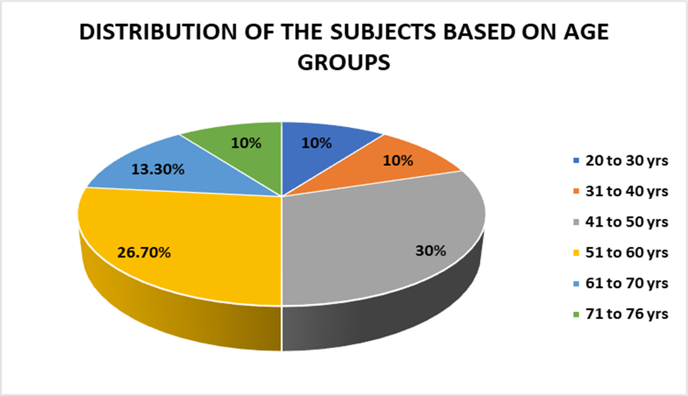
Table 3: Distribution of The Subjects Based On Gender
|
Gender |
Frequency |
Percent |
|
Females |
5 |
16.7 |
|
Males |
25 |
83.3 |
|
Total |
30 |
100.0 |
Table 3 shows the gender-wise distribution of the study subjects. Out of the total 30 participants, the majority were males (83.3%), while females constituted 16.7% of the study population, indicating a marked male predominance in the study sample.
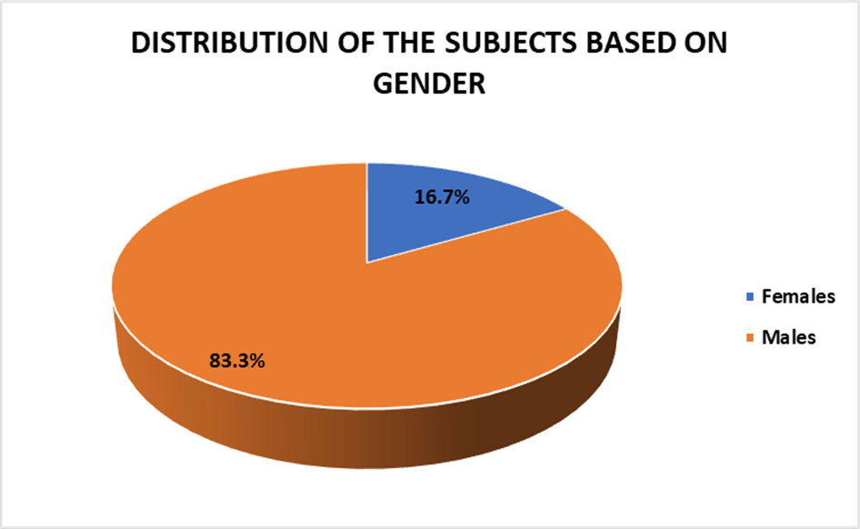
Table 4: Distribution of the Subjects Based on Diagnosis
|
Diagnosis |
Frequency |
Percent |
|
ACS S/P PTCA TO LAD |
15 |
50.0 |
|
ACS S/P PTCA TO LAD AND LCX |
4 |
13.3 |
|
ACS S/P PTCA TO LCX |
4 |
13.3 |
|
ACS S/P PTCA TO LM |
1 |
3.3 |
|
ACS S/P PTCA TO RCA AND LCX |
1 |
3.3 |
|
ACS S/P PTCA TO RCA AND OM2 |
1 |
3.3 |
|
ACS S/P PTCA TO RCA |
4 |
13.3 |
|
Total |
30 |
100.0 |
Table 4 presents the distribution of the study subjects based on diagnosis. Among the 30 participants, ACS status post PTCA to the LAD was the most common diagnosis, accounting for 50.0% of cases. This was followed by ACS status post PTCA to the LAD and LCX, LCX alone, and RCA alone, each constituting 13.3% of the study population. Less frequent diagnoses included ACS status post PTCA to the left main (LM), RCA and LCX, and RCA and OM2, each representing 3.3% of the subjects. Overall, PTCA involving the LAD was the most prevalent among the study participants.
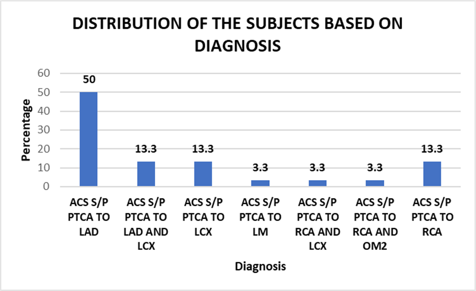
Table 5: Mean Syntax Score, Neutrophils In%, Lymphocytes In %, NLR
|
|
N |
Minimum |
Maximum |
Mean |
S. D |
|
Syntax score 1 |
30 |
3.0 |
50.0 |
18.45 |
10.79 |
|
Neutrophils in % |
30 |
59.0 |
93.0 |
79.77 |
8.69 |
|
Lymphocytes in % |
30 |
2.0 |
36.0 |
14.63 |
7.99 |
|
NLR |
30 |
1.63 |
46.00 |
8.41 |
8.35 |
Table 5 illustrates the descriptive statistics of the SYNTAX score, neutrophil percentage, lymphocyte percentage, and neutrophil–lymphocyte ratio (NLR) among the study subjects. The mean SYNTAX score was 18.45 ± 10.79, with values ranging from 3.0 to 50.0. The mean neutrophil percentage was 79.77 ± 8.69, ranging from 59.0% to 93.0%, while the mean lymphocyte percentage was 14.63 ± 7.99, with a range of 2.0% to 36.0%. The mean NLR was 8.41 ± 8.35, with values ranging from 1.63 to 46.00, reflecting a wide variation in inflammatory status among the subjects.
TABLE 6: Distribution of the Subjects Based On Syntax Score
|
Syntax Score |
Frequency |
Percent |
|
Low |
19 |
63.3 |
|
High and Intermediate |
11 |
36.7 |
|
Total |
30 |
100.0 |
Table 6 depicts the distribution of the study subjects based on SYNTAX score categories. Among the 30 participants, the majority (63.3%) had a low SYNTAX score, while 36.7% of the subjects belonged to the intermediate and high SYNTAX score group.
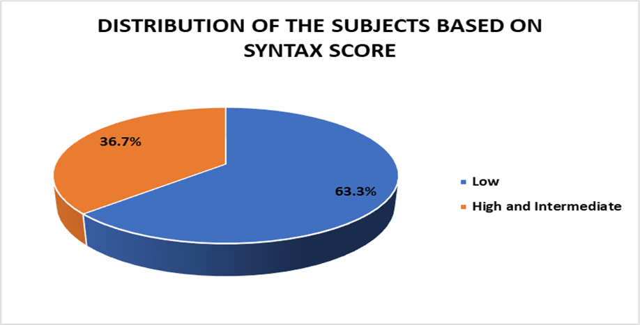
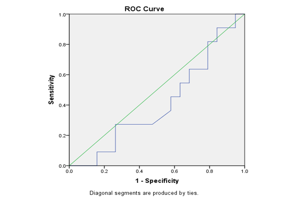
Table 7: Roc Curve Analysis for Predicting Severity of Coronary Artery Disease Using NLR as A Predictor
|
Area Under the Curve |
|||||
|
Test Result Variable(s) |
Area |
Std. Error |
p value |
Asymptotic 95% Confidence Interval |
|
|
Lower Bound |
Upper Bound |
||||
|
NLR |
.411 |
.107 |
.426 |
.202 |
.621 |
The ROC curve analysis for NLR as a predictor of CAD revealed an area under the curve (AUC) of 0.411, indicating weak diagnostic accuracy (p = 0.426). The optimal cutoff value for predicting CAD was determined to be 5.62, with a sensitivity of 45.5% and a specificity of 42.1%.
Table 8: Pearson’s Correlation between Syntax Score and Neutropihils, Lymphocytes and NLR Ratio
|
|
Syntax Score Vs |
||
|
|
NEUTROPHILS IN % |
LYMPHOCYTES IN % |
NLR |
|
r value |
0 |
-0.02 |
-0.09 |
|
p value |
0.999 |
0.918 |
0.635 |
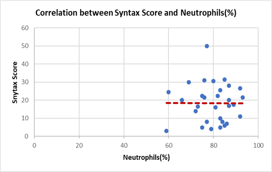
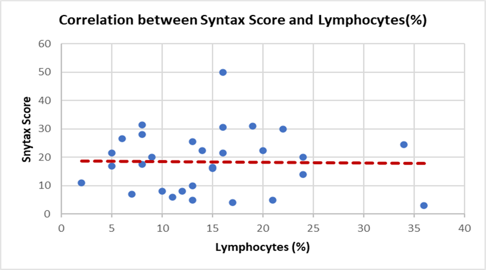
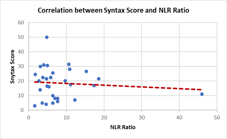
Table 8 shows the Pearson’s correlation between SYNTAX score and inflammatory parameters, including neutrophil percentage, lymphocyte percentage, and neutrophil–lymphocyte ratio (NLR). The correlation between SYNTAX score and neutrophil percentage was negligible (r = 0.00) and statistically not significant (p = 0.999). Similarly, a weak negative correlation was observed between SYNTAX score and lymphocyte percentage (r = −0.02) and between SYNTAX score and NLR (r = −0.09); however, these associations were not statistically significant (p = 0.918 and p = 0.635, respectively). Overall, no significant correlation was found between SYNTAX score and the evaluated inflammatory markers.
DISCUSSION:
In the present study, the mean age of the study population was 51.4 years, with the majority of patients belonging to the 41–60-year age group. This finding indicates that acute myocardial infarction predominantly affected individuals during their economically productive years in our study population. Cardiovascular diseases remain the leading cause of mortality worldwide, accounting for an estimated 17.9 million deaths annually, with heart attacks and strokes contributing to approximately 85% of these deaths.[1] The relatively younger age observed in our study reflects the substantial burden of coronary artery disease in low- and middle-income countries and underscores the importance of early identification and management of cardiovascular risk factors.
The relatively younger age of presentation in the present study is consistent with the pattern observed in developing countries, where coronary artery disease often manifests earlier due to a higher prevalence of metabolic risk factors such as diabetes mellitus, hypertension, smoking, and dyslipidaemia. Chronic endothelial injury and inflammation play a crucial role in the development and progression of atherosclerotic plaques. In this context, Kaya et al. demonstrated that inflammatory markers such as the neutrophil-to-lymphocyte ratio are significantly associated with the presence and severity of coronary artery disease, highlighting the importance of systemic inflammation in the pathogenesis of atherosclerosis.[2]
The present study demonstrated a marked male predominance (83.3%). This finding is consistent with previous epidemiological studies. Betai et al. reported that coronary heart disease occurs more frequently in men than in women, with prevalence rates of approximately 8.3% in males compared with 6.1% in females, and that men tend to develop cardiovascular disease at an earlier age.[5]
In the present study, PTCA involving the left anterior descending artery (LAD) was the most commonly involved culprit vessel, accounting for 50% of cases, whereas left main (LM) artery involvement, along with RCA–LCX and RCA–OM2 lesions, was among the least commonly encountered, each accounting for 3.3% of cases. A similar predominance of LAD as the most commonly involved culprit vessel and relatively infrequent left main coronary artery involvement have also been reported by Haider et al.[6] The predominance of LAD involvement may be attributed to the extensive myocardial territory supplied by this artery, including the anterior wall and interventricular septum, making it the vessel most frequently responsible for clinically significant myocardial infarction following atherosclerotic plaque rupture and thrombotic occlusion.[7]
The mean SYNTAX score in the present study was 18.45 ± 10.79, with 63.3% of patients having a low SYNTAX score and 36.7% having intermediate or high scores. According to Sianos G et al., the SYNTAX score is widely used to assess the anatomical complexity of coronary artery disease and guide therapeutic decision-making, with higher scores indicating more complex coronary lesions and greater atherosclerotic burden.[8]
ROC curve analysis in the present study showed an AUC of 0.411, indicating poor discriminatory ability of NLR in predicting the severity of coronary artery disease. The optimal cut-off value for NLR was 5.62, with low sensitivity and specificity.
These findings suggest that although NLR reflects systemic inflammation, it alone may not reliably predict the anatomical complexity of coronary artery disease.
In the present study, the mean neutrophil percentage was 79.77 ± 8.69, while the mean lymphocyte percentage was 14.63 ± 7.99, resulting in a mean NLR of 8.41 ± 8.35. Elevated NLR reflects the presence of systemic inflammation associated with acute coronary events.
Neutrophils contribute to atherosclerosis progression and plaque rupture through the release of reactive oxygen species, inflammatory cytokines, and proteolytic enzymes, while lymphocyte reduction is associated with physiological stress and neurohormonal activation during acute myocardial infarction.
Several studies have reported a significant association between elevated NLR and coronary artery disease severity. Kaya et al. demonstrated that patients with higher NLR values had significantly more severe coronary artery disease compared with patients with lower NLR levels.[2]
Several investigators, including Easmin et al., Reda et al., Kandibendla et al., Shahsanaei et al., Ha et al., and Chen et al., have consistently demonstrated that an elevated neutrophil-to-lymphocyte ratio (NLR) is associated with increased severity of coronary artery disease and adverse clinical outcomes in patients with acute coronary syndrome.[3,4,9-12] These studies suggest that NLR correlates with greater angiographic disease severity, myocardial injury, cardiac dysfunction, poor prognosis, and unfavorable in-hospital and long-term outcomes, supporting its utility as a simple, inexpensive, and readily available inflammatory biomarker for risk stratification in acute coronary syndrome. [3,4,9-12]
In the present study, Pearson’s correlation analysis showed no statistically significant correlation between SYNTAX score and neutrophil percentage, lymphocyte percentage, or NLR.
Although a weak negative correlation was observed between NLR and SYNTAX score (r = −0.09), the relationship was not statistically significant.
This discrepancy may be explained by the fact that NLR represents an acute inflammatory response, whereas the SYNTAX score reflects long-term structural complexity of coronary artery lesions. Therefore, the inflammatory status at admission may not directly correlate with the anatomical severity of coronary artery disease.
Additionally, the relatively small sample size and inter-individual variability in NLR values may have limited the statistical power to detect significant correlations.
The findings of the present study are consistent with those of Dziedzic et al., who reported that the neutrophil-to-lymphocyte ratio was not significantly associated with the angiographic severity of coronary artery disease in patients with a previous acute coronary syndrome. [13] They suggested that although NLR reflects systemic inflammation, it may not reliably indicate the anatomical severity of coronary artery disease.
Similarly, Maleki et al. observed that although NLR initially demonstrated an association with the SYNTAX score in patients with non-ST-segment elevation acute coronary syndrome, it was not an independent predictor after adjustment for established clinical risk factors, including the TIMI risk score.[7] This finding suggests that the predictive value of NLR may be influenced by other clinical variables and should be interpreted in conjunction with established risk assessment tools rather than as an isolated marker.
The present study was limited by its relatively small sample size and single-centre design, which may have reduced the statistical power and generalizability of the findings. In addition, the predominance of patients with low SYNTAX scores resulted in an unequal distribution of disease severity, which may have influenced the ability to detect a significant association between neutrophil-to-lymphocyte ratio and angiographic complexity. Furthermore, NLR was measured only once at hospital admission and may not have reflected the dynamic inflammatory changes occurring during acute myocardial infarction. As an observational cross-sectional study, the influence of residual confounding factors cannot be completely excluded. Therefore, larger prospective multicentre studies with balanced representation of disease severity and serial assessment of inflammatory markers are warranted to validate these findings.
CONCLUSION:
The present study evaluated the association between the neutrophil-to-lymphocyte ratio (NLR), a hematological marker of systemic inflammation, and the angiographic severity of coronary artery disease assessed by the SYNTAX score in patients presenting with acute coronary syndrome. Elevated neutrophil counts and increased NLR values observed in the present study are consistent with the systemic inflammatory response associated with acute myocardial infarction. However, no statistically significant correlation was identified between NLR and SYNTAX score, and ROC analysis demonstrated poor diagnostic performance of NLR in predicting angiographic disease severity. These findings suggest that while inflammatory markers reflect the inflammatory milieu of acute myocardial infarction, NLR alone was not a reliable predictor of coronary artery disease severity in the present study. Therefore, although NLR is a simple and inexpensive inflammatory biomarker derived from routine hematological investigations, it should not be used as an isolated predictor of angiographic severity of coronary artery disease.
REFERENCES:
1. Cardiovascular diseases (CVDs) [Internet]. Geneva: World Health Organization; [cited 2021 Dec 30]. Available from: https://www.who.int/news-room/fact-sheets/detail/cardiovascular-diseases-(cvds)
2. Kaya A, Kurt M, Tanboga IH, Is T, Sival Y, Kaya Y. Relation of neutrophil to lymphocyte ratio with the presence and severity of stable coronary artery disease. Clin Appl Thromb Hemost. 2014;20(5):473–4.
3. Easmin T, Khalequzzaman M, Ahmed M, Hasan MM. Association between neutrophil to lymphocyte ratio and severity of coronary artery disease in acute myocardial infarction patients. Bangladesh Heart J. 2024 Jan;39(1):1–9.
4. Reda AA, Salama MZ, Ibrahim WA. Predictive prognostic value of neutrophil-lymphocyte ratio in acute coronary syndrome. Menoufia Med J. 2019;32(1):1–7.
5. Betai D, Ahmed AS, Saxena P, Rashid H, Patel H, Shahzadi A, Mowo-Wale AG, Nazir Z. Gender Disparities in Cardiovascular Disease and Their Management: A Review. Cureus. 2024 May 5;16(5):e59663. doi: 10.7759/cureus.59663. PMID: 38836150; PMCID: PMC11148660.
6. Haider KH, Alshoabi SA, Alharbi IA, Gameraddin M, Abdulaal OM, Gareeballah A, et al. Clinical presentation and angiographic findings of acute myocardial infarction in young adults in Jazan region. BMC Cardiovasc Disord. 2023;23(1):302. doi:10.1186/s12872-023-03335-3.
7. Maleki M, Tajlil A, Separham A, Sohrabi B, Pourafkari L, Roshanravan N, et al. Association of neutrophil to lymphocyte ratio (NLR) with angiographic SYNTAX score in patients with non-ST-segment elevation acute coronary syndrome (NSTE-ACS). J Cardiovasc Thorac Res. 2021;13(3):216-221. doi:10.34172/jcvtr.2021.40.
8. Sianos G, Morel MA, Kappetein AP, Morice MC, Colombo A, Dawkins K, et al. The SYNTAX Score: an angiographic tool grading the complexity of coronary artery disease. EuroIntervention. 2005;1:219-227.
9. Ha ET, Yee A, Peterson SJ, Kobayashi Y, Sacchi T, Parikh M, Brener SJ. Neutrophil-to-lymphocyte ratio and prognosis in patients undergoing percutaneous coronary intervention for acute coronary syndrome. Am J Cardiol. 2023;203:1–7.
10. Chen C, Cong BL, Wang M, Abdullah M, Wang XL, Zhang YH, Xu SJ, Cui L. Neutrophil to lymphocyte ratio as a predictor of myocardial damage and cardiac dysfunction in acute coronary syndrome patients. Integr Med Res. 2018;7(2):192–199.
11. Kandibendla V, Prakash GT, Bose SC, Dhewle P. Correlation of neutrophil to lymphocyte ratio to severity of coronary artery disease and in-hospital clinical outcomes in patients with acute coronary syndrome: A prospective observational study. J Acute Dis. 2024;13(1):14–19
12. Shahsanaei F, Abbaszadeh S, Behrooj S, Rahimi Petrudi N, Ramezani B. The value of neutrophil-to-lymphocyte ratio in predicting severity of coronary involvement and long-term outcome of percutaneous coronary intervention in patients with acute coronary syndrome: a systematic review and meta-analysis. BMC Cardiovasc Disord. 2023;23(1):342.
13. Dziedzic EA, Gąsior JS, Tuzimek A, Dąbrowski M, Jankowski P. Neutrophil-to-lymphocyte ratio is not associated with severity of coronary artery disease and is not correlated with vitamin D level in patients with a history of acute coronary syndrome. Biology. 2022;11:1001.