Clinical and Imaging Outcomes of Arthroscopic Rotator Cuff Repair Augmented with Leukocyte-Poor PRP Versus Bone Marrow Aspirate Concentrate: A Comparative Observational Study.
- Bharath M. , Consultant Orthopaedician, Department of Health and Family Welfare, Mysuru, Karnataka, India.
- Muruli Manohar Das , Assistant Professor, Department of Orthopaedics, Sri Chamundeshwari Medical College Hospital & Research Institute, Channapatna, Karnataka, India.
- Avinash G.C. , Assistant Professor, Department of Orthopaedics, Sri Chamundeshwari Medical College Hospital & Research Institute, Channapatna, Karnataka, India.
Article Information:
Abstract:
Background: Biological augmentation is increasingly used during arthroscopic rotator cuff repair, yet the relative clinical and structural performance of different orthobiologic strategies remains uncertain. Objective: To compare 24-month clinical outcomes and 12-month magnetic resonance imaging (MRI) findings after arthroscopic rotator cuff repair augmented with leukocyte-poor platelet-rich plasma (LP-PRP) or bone marrow aspirate concentrate (BMAC). Methods: This comparative observational study included 50 patients, with 25 in the LP-PRP group and 25 in the BMAC group. American Shoulder and Elbow Surgeons (ASES) score, Constant score, visual analogue scale (VAS) for pain, and forward flexion were assessed preoperatively and at 24 months. Repair integrity was evaluated by MRI at 12 months using the Sugaya classification. Results: Baseline characteristics were comparable between groups. ASES score improved from 45.20±6.65 to 83.96±6.91 in LP-PRP and from 44.76±7.18 to 84.72±7.00 in BMAC (both p<0.001). Constant score improved from 50.16±7.08 to 79.48±7.35 and from 49.76±7.66 to 80.28±7.53, respectively (both p<0.001). VAS decreased from 6.97±0.95 to 1.99±0.92 in LP-PRP and from 7.02±0.96 to 1.94±0.93 in BMAC (both p<0.001). Forward flexion increased from 120.84±17.43° to 157.28±13.64° and from 119.72±18.40° to 157.96±13.67°, respectively (both p<0.001). At 24 months, between-group differences in ASES (p=0.701), Constant score (p=0.706), VAS (p=0.831), and forward flexion (p=0.861) were not significant. Median Sugaya grade was 2 (IQR 2-3) in both groups (p=0.480). Conclusion: LP-PRP and BMAC augmentation were associated with substantial clinical improvement after arthroscopic rotator cuff repair. Two-year clinical outcomes and 12-month MRI grades were broadly comparable, with no clear endpoint superiority of either augmentation strategy.
Keywords:
Article :
INTRODUCTION:
Rotator cuff tears are common in middle-aged and older adults and may progressively affect pain, strength, overhead function, and activities of daily living. In a population-based study, Yamamoto et al. identified rotator cuff tears in 20.7% of examined shoulders, with prevalence increasing with age.[1] Arthroscopic repair can restore function in appropriately selected patients, but the biological quality of tendon-to-bone healing remains an important determinant of structural durability.
Clinical recovery after repair is commonly assessed using shoulder-specific functional instruments. The American Shoulder and Elbow Surgeons (ASES) assessment provides a standardized measure of pain and function,[2] while the Constant-Murley score combines pain, activity, motion, and strength into a broader functional evaluation.[3] Structural assessment is equally relevant because satisfactory clinical recovery does not always parallel tendon integrity. Sugaya et al. established an MRI-based classification that grades repaired tendon morphology from preserved thickness to structural discontinuity.[4]
Biological augmentation has therefore attracted considerable attention in rotator cuff surgery. Platelet-rich plasma delivers concentrated platelet-derived mediators to the repair environment, although clinical results have varied between studies. Early randomized work by Randelli et al. suggested short-term pain and functional advantages without a persistent two-year clinical difference.[5] Bone marrow aspirate concentrate represents a different biological strategy by providing marrow-derived cellular and soluble components. Hernigou et al. reported improved tendon healing and longer-term integrity after marrow-derived cell augmentation in a case-controlled study.[6]
Leukocyte-poor platelet-rich plasma (LP-PRP) attempts to deliver platelet-derived factors while reducing the leukocyte component, whereas BMAC provides a cellular concentrate with a broader regenerative profile. Both approaches are used to support tendon-bone healing, yet a clinically useful comparison should consider function, pain, motion, and structural imaging rather than a single endpoint. The present study compared 24-month clinical outcomes and 12-month MRI findings after arthroscopic rotator cuff repair augmented with LP-PRP or BMAC.
MATERIALS AND METHODS:
Study Design and Setting
A hospital-based comparative observational study was conducted in the Department of Orthopaedics in a Tertiary Care Hospital, South India. For a period of one and a half years.
Study Population
The study included 50 patients who underwent arthroscopic rotator cuff repair with biological augmentation and had complete clinical and imaging follow-up data. Twenty-five patients received LP-PRP augmentation and 25 received BMAC augmentation.
Eligibility for the Analysis
Patients were included in the analytical cohort when the augmentation strategy was recorded as LP-PRP or BMAC and complete values were available for demographic characteristics, tear size, preoperative clinical assessment, 24-month clinical outcomes, 12-month MRI Sugaya grade, and postoperative complications.
Study Groups
Participants were classified for comparison according to the augmentation received during arthroscopic repair: LP-PRP or BMAC. The study was observational and the augmentation strategy was analysed as recorded in clinical care.
Outcome Measures
Clinical outcomes comprised the ASES score, Constant score, VAS pain score, and active forward flexion. These parameters were recorded preoperatively and at 24-month follow-up. MRI evaluation of repair integrity was performed at 12 months and recorded using the Sugaya classification. Age, gender, BMI, operated side, tear size, and recorded complications were also evaluated.
Statistical Analysis
Continuous variables were summarized as mean±standard deviation and categorical variables as frequency and percentage. Normality was assessed using the Shapiro-Wilk test. Normally distributed between-group continuous variables were compared using the independent-samples t-test. Paired preoperative-to-postoperative changes were assessed using the Wilcoxon signed-rank test, and change scores between groups were compared using the Mann-Whitney U test where the change-score distribution was non-normal. Categorical variables were compared using the chi-square test or Fisher's exact test as appropriate. Sugaya grade was treated as an ordinal variable and compared between groups using the Mann-Whitney U test. A two-sided p-value <0.05 was considered statistically significant. Statistical analysis was carried out using standard statistical software.
Ethical Considerations
The study was conducted after obtaining approval from the Institutional Ethics Committee. Written informed consent was obtained from all participants. Patient confidentiality and anonymity were maintained throughout the study. The study involved evaluation of clinical and imaging outcomes without any additional research-specific intervention.
RESULTS:
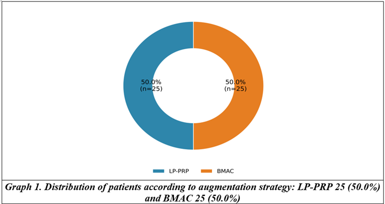
A total of 50 patients were analysed, with 25 (50.0%) in the LP-PRP group and 25 (50.0%) in the BMAC group (Graph 1). Complete 24-month clinical outcome data and 12-month MRI Sugaya grades were available for all included patients.
Baseline demographic, tear-size, and clinical characteristics were closely comparable between groups (Table 1). Mean age was 56.44±6.27 years in the LP-PRP group and 56.16±6.29 years in the BMAC group (p=0.875). Mean BMI was 28.52±1.95 kg/m² and 28.66±2.24 kg/m², respectively (p=0.814). Both groups contained 13 males and 12 females and had the same distribution of tear sizes: five small, nine medium, seven large, and four massive tears. No significant baseline difference was observed in ASES score, Constant score, VAS, or forward flexion.
Table 1. Baseline demographic, tear-size, and clinical characteristics of the study groups
|
Variable |
LP-PRP (n=25) |
BMAC (n=25) |
p-value |
|
Age (years) |
56.44±6.27 |
56.16±6.29 |
0.875 |
|
BMI (kg/m²) |
28.52±1.95 |
28.66±2.24 |
0.814 |
|
Gender, male/female, n |
13 / 12 |
13 / 12 |
1.000 |
|
Side, right/left, n |
13 / 12 |
13 / 12 |
1.000 |
|
Tear size, small/medium/large/massive, n |
5 / 9 / 7 / 4 |
5 / 9 / 7 / 4 |
1.000 |
|
Pre-op ASES |
45.20±6.65 |
44.76±7.18 |
0.823 |
|
Pre-op Constant |
50.16±7.08 |
49.76±7.66 |
0.849 |
|
Pre-op VAS |
6.97±0.95 |
7.02±0.96 |
0.871 |
|
Pre-op forward flexion (°) |
120.84±17.43 |
119.72±18.40 |
0.826 |
|
Values are mean±SD unless otherwise stated. LP-PRP: leukocyte-poor platelet-rich plasma; BMAC: bone marrow aspirate concentrate; BMI: body mass index; ASES: American Shoulder and Elbow Surgeons; VAS: visual analogue scale |
|||
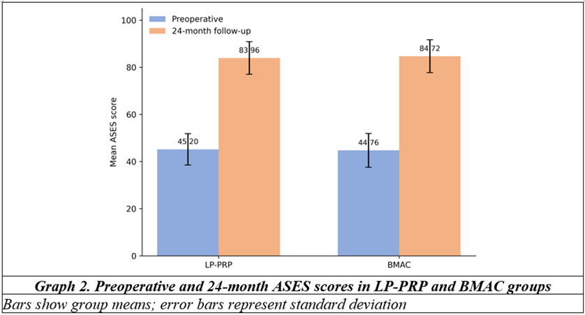
Both augmentation groups demonstrated marked improvement in clinical outcomes at 24 months (Table 2). Mean ASES score increased from 45.20±6.65 to 83.96±6.91 in LP-PRP and from 44.76±7.18 to 84.72±7.00 in BMAC (p<0.001 within each group). The pattern of ASES recovery is shown in Graph 2.
Table 2. Preoperative and 24-month clinical outcomes within each augmentation group
|
Outcome |
LP-PRP pre-op |
LP-PRP 24-month |
p-value |
BMAC pre-op |
BMAC 24-month |
p-value |
|
ASES |
45.20±6.65 |
83.96±6.91 |
<0.001 |
44.76±7.18 |
84.72±7.00 |
<0.001 |
|
Constant |
50.16±7.08 |
79.48±7.35 |
<0.001 |
49.76±7.66 |
80.28±7.53 |
<0.001 |
|
VAS |
6.97±0.95 |
1.99±0.92 |
<0.001 |
7.02±0.96 |
1.94±0.93 |
<0.001 |
|
Forward flexion (°) |
120.84±17.43 |
157.28±13.64 |
<0.001 |
119.72±18.40 |
157.96±13.67 |
<0.001 |
|
Within-group p-values were obtained using the Wilcoxon signed-rank test |
||||||
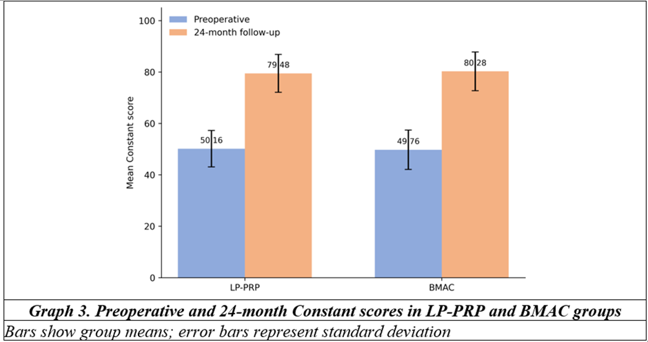
Mean Constant score improved from 50.16±7.08 to 79.48±7.35 in the LP-PRP group and from 49.76±7.66 to 80.28±7.53 in the BMAC group (p<0.001 within each group). Graph 3 demonstrates the corresponding functional improvement. Mean VAS pain score declined from 6.97±0.95 to 1.99±0.92 in LP-PRP and from 7.02±0.96 to 1.94±0.93 in BMAC (p<0.001 for both groups).
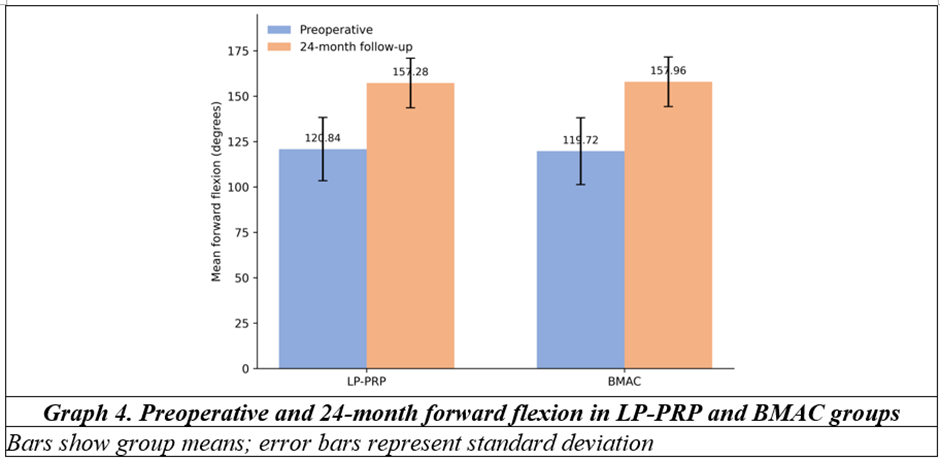
Forward flexion increased from 120.84±17.43° to 157.28±13.64° in the LP-PRP group and from 119.72±18.40° to 157.96±13.67° in the BMAC group (p<0.001 within each group), as illustrated in Graph 4.
At 24 months, the two groups had similar endpoint outcomes (Table 3). Between-group p-values were 0.701 for ASES score, 0.706 for Constant score, 0.831 for VAS, and 0.861 for forward flexion. Analysis of change from baseline showed slightly greater improvement with BMAC for ASES score (39.96±1.93 versus 38.76±1.54; p=0.032), Constant score (30.52±1.73 versus 29.32±1.52; p=0.014), and VAS reduction (5.08±0.16 versus 4.98±0.20; p=0.033). The absolute differences were small. Improvement in forward flexion did not differ significantly between groups (38.24±5.40° versus 36.44±5.12°; p=0.157).
Table 3. Comparison of 24-month outcomes and magnitude of improvement between LP-PRP and BMAC groups
|
Outcome |
LP-PRP (n=25) |
BMAC (n=25) |
p-value |
|
24-month ASES |
83.96±6.91 |
84.72±7.00 |
0.701 |
|
Improvement in ASES |
38.76±1.54 |
39.96±1.93 |
0.032 |
|
24-month Constant |
79.48±7.35 |
80.28±7.53 |
0.706 |
|
Improvement in Constant |
29.32±1.52 |
30.52±1.73 |
0.014 |
|
24-month VAS |
1.99±0.92 |
1.94±0.93 |
0.831 |
|
Reduction in VAS |
4.98±0.20 |
5.08±0.16 |
0.033 |
|
24-month flexion (°) |
157.28±13.64 |
157.96±13.67 |
0.861 |
|
Improvement in flexion (°) |
36.44±5.12 |
38.24±5.40 |
0.157 |
|
Endpoint outcomes were compared using independent-samples t-tests. Change scores were compared using the Mann-Whitney U test |
|||
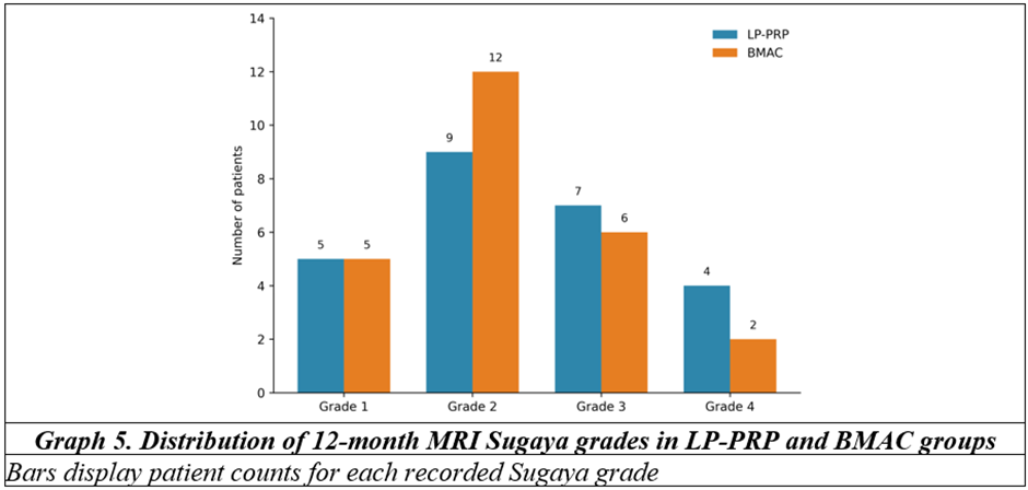
MRI at 12 months showed Sugaya grades I, II, III, and IV in 5, 9, 7, and 4 LP-PRP patients, respectively, compared with 5, 12, 6, and 2 BMAC patients (Table 4 and Graph 5). Median Sugaya grade was 2 (IQR 2-3) in both groups, and the ordinal distributions did not differ significantly (p=0.480). No Sugaya grade V was recorded.
Table 4. Twelve-month MRI Sugaya grades according to augmentation strategy
|
Sugaya Grade |
LP-PRP (n=25) |
BMAC (n=25) |
Total (n=50) |
|
Grade I |
5 (20.0%) |
5 (20.0%) |
10 (20.0%) |
|
Grade II |
9 (36.0%) |
12 (48.0%) |
21 (42.0%) |
|
Grade III |
7 (28.0%) |
6 (24.0%) |
13 (26.0%) |
|
Grade IV |
4 (16.0%) |
2 (8.0%) |
6 (12.0%) |
|
Median (IQR) |
2 (2-3) |
2 (2-3) |
— |
|
Ordinal comparison by Mann-Whitney U test: p=0.480. Overall grade distribution by chi-square test: p=0.760 |
|||
At 24 months, the two groups had similar endpoint outcomes (Table 3). Between-group p-values were 0.701 for ASES score, 0.706 for Constant score, 0.831 for VAS, and 0.861 for forward flexion. Analysis of change from baseline showed slightly greater improvement with BMAC for ASES score (39.96±1.93 versus 38.76±1.54; p=0.032), Constant score (30.52±1.73 versus 29.32±1.52; p=0.014), and VAS reduction (5.08±0.16 versus 4.98±0.20; p=0.033). The absolute differences were small. Improvement in forward flexion did not differ significantly between groups (38.24±5.40° versus 36.44±5.12°; p=0.157).
Table 3. Comparison of 24-month outcomes and magnitude of improvement between LP-PRP and BMAC groups
|
Outcome |
LP-PRP (n=25) |
BMAC (n=25) |
p-value |
|
24-month ASES |
83.96±6.91 |
84.72±7.00 |
0.701 |
|
Improvement in ASES |
38.76±1.54 |
39.96±1.93 |
0.032 |
|
24-month Constant |
79.48±7.35 |
80.28±7.53 |
0.706 |
|
Improvement in Constant |
29.32±1.52 |
30.52±1.73 |
0.014 |
|
24-month VAS |
1.99±0.92 |
1.94±0.93 |
0.831 |
|
Reduction in VAS |
4.98±0.20 |
5.08±0.16 |
0.033 |
|
24-month flexion (°) |
157.28±13.64 |
157.96±13.67 |
0.861 |
|
Improvement in flexion (°) |
36.44±5.12 |
38.24±5.40 |
0.157 |
|
Endpoint outcomes were compared using independent-samples t-tests. Change scores were compared using the Mann-Whitney U test |
|||
MRI at 12 months showed Sugaya grades I, II, III, and IV in 5, 9, 7, and 4 LP-PRP patients, respectively, compared with 5, 12, 6, and 2 BMAC patients (Table 4 and Graph 5). Median Sugaya grade was 2 (IQR 2-3) in both groups, and the ordinal distributions did not differ significantly (p=0.480). No Sugaya grade V was recorded.
Table 4. Twelve-month MRI Sugaya grades according to augmentation strategy
|
Sugaya Grade |
LP-PRP (n=25) |
BMAC (n=25) |
Total (n=50) |
|
Grade I |
5 (20.0%) |
5 (20.0%) |
10 (20.0%) |
|
Grade II |
9 (36.0%) |
12 (48.0%) |
21 (42.0%) |
|
Grade III |
7 (28.0%) |
6 (24.0%) |
13 (26.0%) |
|
Grade IV |
4 (16.0%) |
2 (8.0%) |
6 (12.0%) |
|
Median (IQR) |
2 (2-3) |
2 (2-3) |
— |
|
Ordinal comparison by Mann-Whitney U test: p=0.480. Overall grade distribution by chi-square test: p=0.760 |
|||
Postoperative complications were uncommon (Table 5). In the LP-PRP group, one patient developed transient stiffness and one had a superficial infection that resolved. One patient in the BMAC group developed transient stiffness. Overall complication frequency was 8.0% in LP-PRP and 4.0% in BMAC (p=1.000).
Table 5. Recorded postoperative complications according to augmentation strategy
|
Complication |
LP-PRP (n=25) |
BMAC (n=25) |
Total (n=50) |
|
None |
23 (92.0%) |
24 (96.0%) |
47 (94.0%) |
|
Transient stiffness |
1 (4.0%) |
1 (4.0%) |
2 (4.0%) |
|
Superficial infection, resolved |
1 (4.0%) |
0 |
1 (2.0%) |
|
Any complication |
2 (8.0%) |
1 (4.0%) |
3 (6.0%) |
|
Fisher's exact test for any complication: p=1.000 |
|||

DISCUSSION:
The main finding of this study is that both LP-PRP and BMAC augmentation were followed by substantial clinical recovery after arthroscopic rotator cuff repair. ASES score, Constant score, pain, and forward flexion improved significantly in both groups over 24 months. At the final clinical assessment, however, the groups were remarkably similar. MRI assessment at 12 months also showed no statistically significant difference in Sugaya grade. These findings do not support clear endpoint superiority of either augmentation strategy in this cohort.
The change-score analysis deserves a cautious reading. BMAC showed statistically greater improvement in ASES and Constant scores and a slightly larger reduction in VAS pain. The absolute differences were small: approximately 1.2 points for ASES, 1.2 points for Constant score, and 0.1 point for VAS reduction. Because the final 24-month scores themselves were not significantly different, these change-score findings are better interpreted as modest numerical separation rather than evidence of a clinically dominant treatment effect.
The LP-PRP findings are broadly compatible with the heterogeneous clinical literature on platelet-based augmentation. Jo et al. reported improved structural healing after PRP augmentation in large to massive tears, although clinical differences were limited.[7] Malavolta et al. found no sustained superiority in the main clinical or structural endpoints of their randomized study at two years.[8] An Indian randomized trial by Pandey et al. reported lower retear rates with moderately concentrated PRP, particularly among large tears, alongside early vascular changes around the repair site.[9] These differing results illustrate how tear size, platelet preparation, leukocyte content, delivery method, and repair technique can modify apparent treatment effect.
Not all platelet formulations have produced favourable results. Flury et al. found that pure PRP did not improve postoperative clinical outcomes after arthroscopic repair in their randomized trial.[10] Malavolta et al. subsequently reported that the absence of a clear PRP advantage persisted at five-year follow-up.[11] These studies are relevant to the present findings because the LP-PRP group achieved strong functional recovery, but the data do not indicate that this translated into a distinct advantage over BMAC.
Recent trials have focused more specifically on leukocyte-poor preparations. Zhang et al.[12] reported that LP-PRP reduced retear rate and fatty infiltration after repair of moderate-to-large tears without improving clinical outcome scores.[12] Rossi et al. similarly found a lower retear rate with LP-PRP but no significant functional benefit in a double-blind randomized trial.[13] In a three-arm randomized trial, Yao et al. found no superior 12-month functional or structural result for LP-PRP compared with control, while also showing that outcomes may differ according to PRP formulation.[14] The present study likewise suggests that good clinical improvement can occur with LP-PRP without establishing a clear comparative advantage at the final clinical endpoint.
BMAC has a different biological rationale and a smaller human evidence base. Hernigou et al. reported better early healing and greater long-term tendon integrity when marrow-derived mesenchymal cell augmentation was used during rotator cuff repair.[6] More recently, Cole et al. conducted a prospective randomized trial of concentrated bone marrow aspirate augmentation and found markedly better repair integrity on one-year MRI, while patient-reported clinical outcomes and treatment failure rates remained largely similar between groups.[15] That separation between structural and clinical effects is important. In the present comparison, BMAC showed a numerical shift toward lower Sugaya grades, with fewer grade IV findings, but the overall ordinal MRI distribution was not statistically different from LP-PRP.
MRI interpretation also requires care. In the original Sugaya system, grade III describes insufficient tendon thickness without discontinuity, whereas grades IV and V represent minor and major discontinuity, respectively.[4] For this reason, the current analysis retained the recorded grades as an ordinal imaging outcome rather than automatically treating grade III as a recurrent tear. No grade V was observed in either group.
The identical distribution of tear sizes in the two groups strengthens baseline comparability for this variable, but tear size remains clinically important when interpreting biological augmentation. Small and medium tears generally carry a different healing environment from large and massive tears. The present sample was not large enough for reliable tear-size-specific treatment comparisons, and subgroup analysis could therefore create unstable estimates.
From an Indian orthopaedic practice perspective, both strategies have practical constraints. LP-PRP can be prepared from peripheral blood and is relatively familiar in sports-medicine practice, whereas BMAC requires marrow aspiration and processing, increasing procedural steps and resource requirements. The Indian randomized experience reported by Pandey et al. demonstrates that biologic augmentation can be studied meaningfully within local clinical settings,[9] but treatment choice should still consider tear characteristics, available processing systems, cost, surgical workflow, and the strength of evidence supporting the intended biological effect.
Overall, these findings favour a measured interpretation. Both groups reached substantial functional improvement and low pain scores by two years. BMAC produced slightly larger changes in several clinical measures, but final clinical scores and MRI grades were comparable. The choice between LP-PRP and BMAC should therefore not be based on an assumption that one augmentation strategy uniformly produces superior clinical recovery.
Limitations
The study has several limitations. The sample size was modest, with 25 patients in each group, limiting power to detect small differences and uncommon complications. The observational design permits residual selection bias because factors governing the choice of LP-PRP or BMAC were not analysed. Technical details of the biologic preparations, including platelet concentration, leukocyte quantification, BMAC cellular yield, harvest site, processing system, application volume, and activation method, were not available as study variables. Surgical repair configuration, fatty infiltration, tendon retraction, muscle quality, and rehabilitation adherence were also not evaluated. MRI was available at a single 12-month time point, while clinical outcomes were assessed at 24 months, preventing direct assessment of structural change over the same follow-up interval. The very similar baseline characteristics and tear-size distribution improve comparability, but they do not remove the possibility of unmeasured confounding. Larger prospective comparative studies with standardized biologic characterization and longer imaging follow-up would provide more definitive evidence.
CONCLUSION:
Arthroscopic rotator cuff repair augmented with either LP-PRP or BMAC was associated with significant improvement in ASES score, Constant score, pain, and forward flexion at 24 months. Final clinical outcomes were comparable between groups, and 12-month MRI Sugaya grades did not differ significantly. BMAC showed slightly greater improvement in several change scores, but the absolute differences were small and did not translate into superior 24-month endpoint scores. Within the limits of this observational cohort, neither augmentation strategy demonstrated clear overall clinical or imaging superiority.
REFERENCES:
1. Yamamoto A, Takagishi K, Osawa T, Yanagawa T, Nakajima D, Shitara H, et al. Prevalence and risk factors of a rotator cuff tear in the general population. J Shoulder Elbow Surg 2010;19(1):116-20. doi:10.1016/j.jse.2009.04.006.
2. Richards RR, An KN, Bigliani LU, Friedman RJ, Gartsman GM, Gristina AG, et al. A standardized method for the assessment of shoulder function. J Shoulder Elbow Surg 1994;3(6):347-52.
3. Constant CR, Murley AH. A clinical method of functional assessment of the shoulder. Clin Orthop Relat Res 1987;(214):160-4.
4. Sugaya H, Maeda K, Matsuki K, Moriishi J. Functional and structural outcome after arthroscopic full-thickness rotator cuff repair: single-row versus dual-row fixation. Arthroscopy 2005;21(11):1307-16. doi:10.1016/j.arthro.2005.08.011.
5. Randelli P, Arrigoni P, Ragone V, Aliprandi A, Cabitza P. Platelet rich plasma in arthroscopic rotator cuff repair: a prospective RCT study, 2-year follow-up. J Shoulder Elbow Surg 2011;20(4):518-28. doi:10.1016/j.jse.2011.02.008.
6. Hernigou P, Flouzat Lachaniette CH, Delambre J, Zilber S, Duffiet P, Chevallier N, et al. Biologic augmentation of rotator cuff repair with mesenchymal stem cells during arthroscopy improves healing and prevents further tears: a case-controlled study. Int Orthop 2014;38(9):1811-8. doi:10.1007/s00264-014-2391-1.
7. Jo CH, Shin JS, Lee YG, Shin WH, Kim H, Lee SY, et al. Platelet-rich plasma for arthroscopic repair of large to massive rotator cuff tears: a randomized, single-blind, parallel-group trial. Am J Sports Med 2013;41(10):2240-8. doi:10.1177/0363546513497925.
8. Malavolta EA, Gracitelli MEC, Ferreira Neto AA, Assunção JH, Bordalo-Rodrigues M, de Camargo OP. Platelet-rich plasma in rotator cuff repair: a prospective randomized study. Am J Sports Med 2014;42(10):2446-54. doi:10.1177/0363546514541777.
9. Pandey V, Bandi A, Madi S, Agarwal L, Acharya KKV, Maddukuri S, et al. Does application of moderately concentrated platelet-rich plasma improve clinical and structural outcome after arthroscopic repair of medium-sized to large rotator cuff tear? A randomized controlled trial. J Shoulder Elbow Surg 2016;25(8):1312-22. doi:10.1016/j.jse.2016.01.036.
10. Flury M, Rickenbacher D, Schwyzer HK, Jung C, Schneider MM, Stahnke K, et al. Does pure platelet-rich plasma affect postoperative clinical outcomes after arthroscopic rotator cuff repair? A randomized controlled trial. Am J Sports Med 2016;44(8):2136-46. doi:10.1177/0363546516645518.
11. Malavolta EA, Gracitelli MEC, Assunção JH, Ferreira Neto AA, Bordalo-Rodrigues M, de Camargo OP. Clinical and structural evaluations of rotator cuff repair with and without added platelet-rich plasma at 5-year follow-up: a prospective randomized study. Am J Sports Med 2018;46(13):3134-41. doi:10.1177/0363546518795895.
12. Zhang C, Cai YZ, Wang Y. Injection of leukocyte-poor platelet-rich plasma for moderate-to-large rotator cuff tears does not improve clinical outcomes but reduces retear rates and fatty infiltration: a prospective, single-blinded randomized study. Arthroscopy 2022;38(8):2381-8.e1. doi:10.1016/j.arthro.2022.02.007.
13. Rossi LA, Gorodischer TD, Camino P, Brandariz RN, Tanoira I, Piuzzi NS, et al. Leukocyte-poor platelet-rich plasma as an adjuvant to arthroscopic rotator cuff repair reduces the retear rate but does not improve functional outcomes: a double-blind randomized controlled trial. Am J Sports Med 2024;52(6):1403-10. doi:10.1177/03635465241239062.
14. Yao L, Pang L, Zhang C, Yang S, Wang J, Li Y, et al. Platelet-rich plasma for arthroscopic rotator cuff repair: a 3-arm randomized controlled trial. Am J Sports Med 2024;52(14):3495-504. doi:10.1177/03635465241283964.
15. Cole BJ, Kaiser JT, Wagner KR, Sivasundaram L, Otte SR, Tauro TM, et al. Prospective randomized trial of biologic augmentation with bone marrow aspirate concentrate in patients undergoing arthroscopic rotator cuff repair. Am J Sports Med 2023;51(5):1234-42. doi:10.1177/03635465231154601.