Efficacy and Safety of Fractional Carbon Dioxide (CO2) Laser in Benign Skin Lesions.
- Bharath Bangera N J , Assistant Professor, Department of Dermatology, Subbaiah Institute of Medical Sciences, Shivamogga, Karnataka, India
- Lalith S , Assistant Professor, Department of Dermatology, Subbaiah Institute of Medical Sciences, Shivamogga,Karnataka,India
- Samhitha S R , Assistant Professor, Department of General Medicine, Subbaiah Institute of Medical Sciences, Shivamogga, Karnataka,India
Article Information:
Abstract:
Background: Benign skin lesions cause significant cosmetic disfigurement and patient anxiety. Fractional carbon dioxide (CO₂) laser has emerged as the gold standard in ablative laser treatment, yet data on its efficacy and safety for Indian skin types remain limited. Objectives: To evaluate the efficacy and safety profile of fractional CO₂ laser in the treatment of benign skin lesions. Methods: A prospective interventional study was conducted on 100 patients at VIMS, Ballari (March 2021 – February 2022). All patients underwent fractional CO₂ laser ablation. Efficacy was assessed using the Global Improvement Scale (GIS) and patient satisfaction using the Patient Satisfaction Score (PSS) at 1, 4, and 12 weeks. Results: 87% of patients achieved near-total improvement (GIS Grade 4). Complete lesion removal was achieved in 95% of cases. Recurrence rate was 6.32%. Late complications included post-inflammatory hyperpigmentation (21%) and scarring (8.4%). 70% of patients rated satisfaction as very good to excellent. Conclusion: Fractional CO₂ laser ablation is effective, safe, and well-tolerated for a wide spectrum of benign skin lesions, including darker skin phototypes.
Keywords:
Article :
INTRODUCTION:
Benign skin lesions are non-cancerous growths that may be self-reported by patients or discovered during routine dermatological examination.
These lesions can accumulate over a lifetime and, though medically innocuous, often cause significant cosmetic disfigurement and psychological distress.
Traditional management options include cryotherapy, electrocautery, radiofrequency ablation (RFA), and surgical excision. Over the past decade, laser technology has transformed dermatological practice.
The carbon dioxide (CO₂) laser, operating at 10,600 nm, is considered the gold standard among ablative lasers.
It employs selective photothermolysis to target water-containing tissue with controlled vaporization, providing a bloodless field, minimal thermal spread, reduced patient discomfort, and rapid recovery.
Fractional delivery further improves outcomes by limiting thermal injury to fractionated microthermal zones, leaving surrounding tissue intact and accelerating healing. Despite widespread use, systematic safety and efficacy data for Indian skin types (Fitzpatrick III–V) remain sparse. This study addresses that gap.
Objectives
1. To evaluate the efficacy of fractional CO₂ laser ablation in the treatment of various benign skin lesions.
2. To assess the safety profile, including immediate and late complications, of fractional CO₂ laser treatment.
MATERIALS AND METHODS:
Study Design and Setting
A hospital-based prospective interventional study was conducted in the Department of Dermatology, Venereology and Leprosy, VIMS, Ballari, Karnataka, India, from March 2021 to February 2022.
Ethical committee clearance was obtained and written informed consent was obtained from all participants.
Sample Size and Patient Selection
Sample size was calculated using N = Z²P(1−P)/d², based on data from Cho SB et al. (2011), with 42.9% marked improvement as reference, 95% confidence level, and absolute precision of 0.107. Minimum required size was 83; 100 patients were enrolled for improved statistical power.
Inclusion criteria: patients ≥18 years of either sex with benign skin lesions who consented to the procedure.
Exclusion criteria: active cutaneous infection in the treatment area, history of keloid/hypertrophic scar formation, isotretinoin use within 6 months, pregnancy or lactation, or uncontrolled diabetes mellitus.
Laser Equipment and Procedure
All patients were treated with the FUTURA RF 50 CO₂ Laser (Dermaindia) — a metal-tube RF-excited fractional laser (10,600 nm, spot size 0.1 mm, 50W maximum). CW mode: 2–7 W. Pulse mode: 8–22 W, 0.8–1 ms pulse duration, 50 Hz frequency. Treatment areas were prepared with povidone-iodine; EMLA cream applied under occlusion 60 minutes pre-procedure; local lignocaine infiltration where required.
Outcome Assessment
Follow-up was at 1, 4, and 12 weeks post-procedure (telephonic follow-up used for COVID-19-restricted patients). Objective assessment: Global Improvement Scale (GIS) — Grade 0 (no improvement) to Grade 4 (near-total improvement). Subjective assessment: Patient Satisfaction Score (PSS) at 12 weeks — 0 (none) to 8 (excellent).
Statistical Analysis
Data analysed using IBM SPSS v25.0. Categorical variables expressed as frequencies and percentages. Chi-square test for associations; P < 0.05 considered statistically significant.
RESULTS:
Demographic Profile
100 patients enrolled (52 male, 48 female; ratio 1.08:1). Age: 49% aged 18–29 years, 35% aged 30–45, 11% aged 46–59, 5% aged ≥60 years. Comorbidities: 90% none, 7% diabetes mellitus, 3% hyperlipidemia.
Table 1: Demographic and Clinical Characteristics
|
Parameter |
Category |
n |
% |
|
Age (years) |
18–29 |
49 |
49% |
|
|
30–45 |
35 |
35% |
|
|
46–59 |
11 |
11% |
|
|
≥60 |
5 |
5% |
|
Sex |
Male |
52 |
52% |
|
|
Female |
48 |
48% |
|
Comorbidities |
None |
90 |
90% |
|
|
Diabetes Mellitus |
7 |
7% |
|
|
Hyperlipidemia |
3 |
3% |
|
Fitzpatrick Skin Type |
Type III |
33 |
33% |
|
|
Type IV |
63 |
63% |
|
|
Type V |
4 |
4% |
Lesion Characteristics
Most common site: face (66%), followed by neck (19%) and periorbital region (21% of all lesions). Duration: ≥1 year in 60%, <1 year in 34%, since birth in 6%. Dominant skin type: Fitzpatrick IV (63%), III (33%), V (4%).
Table 2: Distribution of Benign Skin Lesions
|
Type of Lesion |
n |
% |
|
Acrochordons |
24 |
24% |
|
Dermatosis Papulosa Nigra (DPN) |
13 |
13% |
|
Melanocytic Nevus |
11 |
11% |
|
Syringoma |
11 |
11% |
|
Pyogenic Granuloma |
7 |
7% |
|
Colloid Milia |
7 |
7% |
|
Seborrheic Keratosis |
6 |
6% |
|
Verruca (Warts) |
6 |
6% |
|
Linear Verrucous Epidermal Nevus |
5 |
5% |
|
Xanthalesma |
3 |
3% |
|
Fibroma |
2 |
2% |
|
Others (1% each) |
5 |
5% |
|
Total |
100 |
100% |
Treatment Response and Outcomes
CW mode: 61%; pulse mode: 39%. Complete lesion removal: 95 patients (95%). Of those, 6 patients (6.32%) had recurrence during follow-up; 89 (93.68%) remained recurrence-free.
Table 3: Global Improvement Scale and Patient Satisfaction Score
|
Scale |
Grade/Score |
Description |
n |
% |
|
GIS |
Grade 4 |
Near-total improvement |
87 |
87% |
|
|
Grade 3 |
Marked improvement |
6 |
6% |
|
|
Grade 2 |
Moderate improvement |
2 |
2% |
|
|
Grade 1 |
Minimal improvement |
3 |
3% |
|
|
Grade 0 |
No improvement |
2 |
2% |
|
PSS |
8 (Excellent) |
|
10 |
10% |
|
|
6 (Very Good) |
|
60 |
60% |
|
|
4 (Good) |
|
22 |
22% |
|
|
2 (Mild) |
|
4 |
4% |
|
|
0 (None) |
|
4 |
4% |
Complications
Table 4: Immediate and Late Complications
|
Complication |
Type |
n |
% |
|
Post-operative pain |
Immediate |
10 |
10% |
|
Watery discharge |
Immediate |
10 |
10% |
|
Erythema |
Immediate |
9 |
9% |
|
No immediate complication |
Immediate |
71 |
71% |
|
Post-inflammatory hyperpigmentation |
Late |
20 |
21% |
|
Scarring |
Late |
8 |
8.4% |
|
No late complication |
Late |
67 |
70.5% |
Of the 20 patients who developed PIH, 70% had Fitzpatrick skin type IV and 30% had type III. Five patients were lost to follow-up.
Illustrative Cases
Representative before-and-after clinical photographs are presented below for the major lesion types treated in this study. All images were captured under standardised lighting conditions at the VIMS Dermatology procedure room.
Acrochordons (Skin Tags)
Complete removal was achieved in all 24 cases using CW mode. Patient satisfaction was excellent (33.3%) or very good (66.7%). No recurrence was observed at 12 weeks.
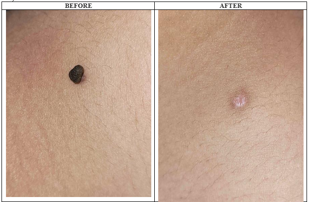
Figure 1: Acrochordon before and after CO2 laser ablation
Seborrheic Keratosis
Near-total improvement was achieved in 5/6 patients (83.3%); 1 patient showed moderate improvement. Side effects: erythema and PIH. No recurrence at 12 weeks.
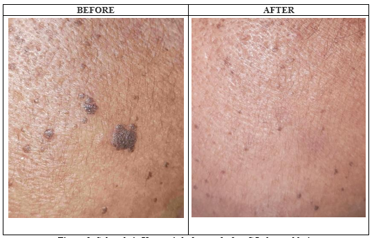
Figure 2: Seborrheic Keratosis before and after CO2 laser ablation
Dermatosis Papulosa Nigra (DPN)
All 13 patients with DPN achieved near-total improvement using pulse mode. Side effects: erythema and PIH. No recurrence at 12 weeks.
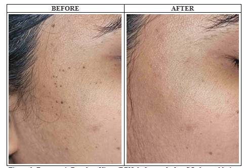
Figure 3: Dermatosis Papulosa Nigra (DPN) before and after CO2 laser ablation
Colloid Milia
Near-total improvement in 6/7 patients (85.7%); mild improvement in 1 patient. Side effects: pain, PIH, and scarring. No recurrence at 12 weeks.
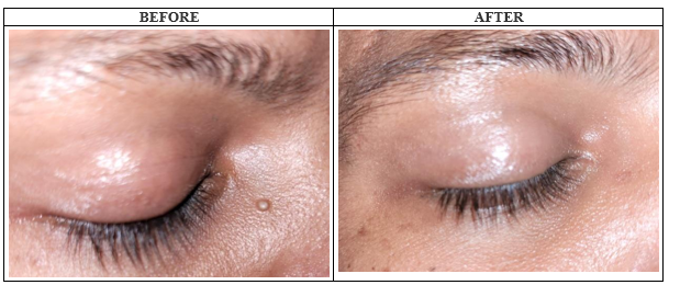
Figure 4: Colloid Milia before and after CO2 laser ablation
Syringoma
Near-total improvement in 10/11 patients (90.9%); marked improvement in 1 patient. Side effects: erythema, watery discharge, and PIH. Recurrence in 1 patient at 12 weeks.
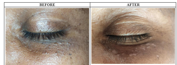
Figure 5: Periorbital Syringoma before and after CO2 laser ablation
Melanocytic Nevus
Near-total improvement achieved in all 11 patients using CW mode. Side effects: erythema, watery discharge, and PIH. No recurrence at 12 weeks.
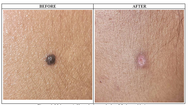
Figure 6: Melanocytic Nevus before and after CO2 laser ablation
Xanthalesma Palpebrarum
Marked improvement in 2/3 patients (66.7%); moderate improvement in 1 patient. All 3 patients had associated hyperlipidemia. Recurrence occurred in all 3 cases within 1 month, underscoring the need for concurrent lipid management.
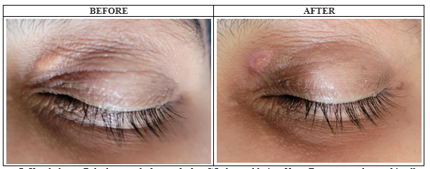
Figure 7: Xanthalesma Palpebrarum before and after CO2 laser ablation. Note: Recurrence observed in all cases at 1 month; associated hyperlipidemia was present in all patients
Linear Verrucous Epidermal Nevus (LVEN)
Near-total improvement in 2/5 patients (40%), marked improvement in 2 patients (40%), and no improvement in 1 patient (20%). CW mode used in all cases. Side effects included pain, watery discharge, PIH, and scarring.
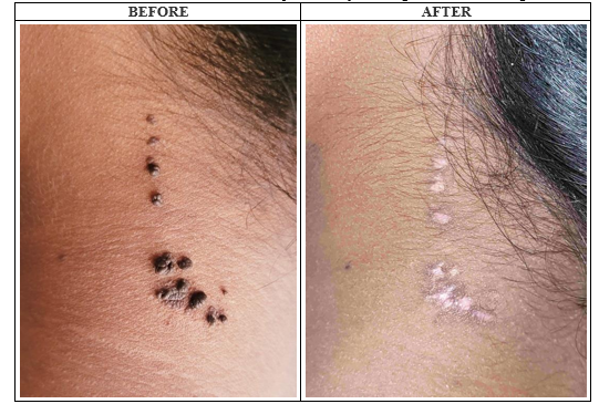
Figure 8: Linear Verrucous Epidermal Nevus (LVEN) before and after CO2 laser ablation
Pyogenic Granuloma
Near-total improvement in all 7 patients. CW mode alternating with pulse was used to control bleeding. Side effects: pain, watery discharge, and PIH. No recurrence at 12 weeks.
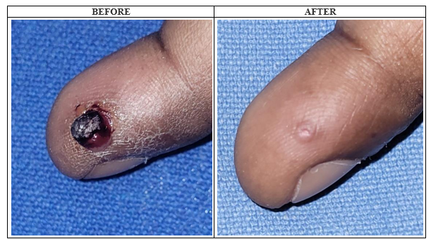
Figure 9: Pyogenic Granuloma before and after CO2 laser ablation
Fibroma
Near-total improvement in both patients with fibroma using CW mode. Side effect: PIH. No recurrence at 12 weeks.
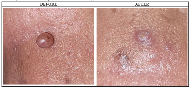
Figure 10: Fibroma before and after CO2 laser ablation
Trichoepithelioma
The single patient with trichoepithelioma achieved near-total improvement using CW mode with no side effects or recurrence at 12 weeks.
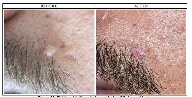
Figure 11: Trichoepithelioma before and after CO2 laser ablation
Post-Inflammatory Hyperpigmentation (Complication)
Post-inflammatory hyperpigmentation was the most common late complication, observed in 20 patients (21%). It was more prevalent in Fitzpatrick skin type IV (70% of PIH cases). The image below demonstrates PIH following CO₂ laser ablation of syringoma.
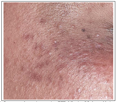
Figure 12: Post-inflammatory hyperpigmentation (PIH) following CO2 laser ablation of syringoma
Recurrence — Sebaceous Hyperplasia
The single patient with sebaceous hyperplasia showed moderate improvement but developed recurrence at 4 weeks post-procedure. This case illustrates the challenge of treating sebaceous hyperplasia, consistent with its known tendency to recur, as also reported by Kim JH et al.
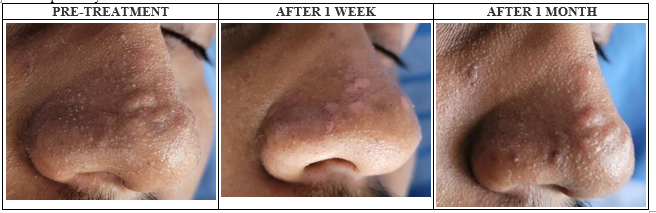
Figure 13: Recurrence after CO2 laser ablation of Sebaceous Hyperplasia (pre-treatment, 1 week, 1 month post-procedure)
DISCUSSION:
This study demonstrates that fractional CO₂ laser ablation is a highly effective modality for treating a wide spectrum of benign skin lesions. The overall near-total improvement rate of 87% and complete removal rate of 95% compare favourably with existing literature.
Sarvepalli et al. (2020) in a split-face study comparing CO₂ laser versus RFA in 70 patients reported near-total improvement in only 38.57% with CO₂ laser. The superior outcomes in our study may reflect lesion-specific laser parameter optimisation and single-session management in most cases.
Acrochordons (24%) were the most common lesion, all achieving near-total improvement. DPN (13%) showed 100% near-total improvement, consistent with Bruscino et al. (2014). For syringomas, near-total improvement in 90.9% exceeded the 8.6% reported by Cho et al. (2011), possibly reflecting differences in laser settings and lesion depth.
Xanthalesma showed 100% recurrence at 1 month, with all patients having associated hyperlipidemia — underscoring the need for concurrent lipid management alongside procedural treatment. This is consistent with findings by Esmat et al. (2014), who reported 15% recurrence in hyperlipidaemic patients.
The PIH rate (21%) exceeded that of Sarvepalli et al. (7.14%), likely reflecting our cohort's predominantly Fitzpatrick type IV skin. Of PIH cases, 70% occurred in type IV patients, highlighting the need for proactive post-procedural photoprotection and topical depigmenting agents in darker phototypes.
Limitations include a 12-week follow-up period, which may underestimate late recurrence, and 5 patients lost to follow-up due to COVID-19 pandemic restrictions.
CONCLUSION:
Fractional CO₂ laser ablation is a safe, effective, and well-tolerated modality for the management of a broad spectrum of benign skin lesions. Complete removal in a single session was achieved in 95% of patients with a low recurrence rate of 6.32%. Global improvement was near-total in 87% of cases, and 70% of patients reported very good to excellent satisfaction. Complications were predominantly mild and transient. Particular attention to post-procedural care is warranted in patients with darker skin phototypes to minimise PIH risk. Larger, multicentre studies with longer follow-up periods are recommended to validate these findings.
REFERENCES:
1. Krupa Shankar D, Chakravarthi M, Shilpakar R. Carbon dioxide laser guidelines. J Cutan Aesthet Surg. 2009;2:72-80.
2. Omi T, Numano K. The Role of the CO2 Laser and Fractional CO2 Laser in Dermatology. Laser Ther. 2014;23:49-60.
3. Cho SB, Kim HJ, Noh S, Lee SJ, Kim YK, Lee JH. Treatment of syringoma using an ablative 10,600-nm carbon dioxide fractional laser: a prospective analysis of 35 patients. Dermatol Surg. 2011;37:433-438.
4. Sarvepalli A, Sudhakar M, Rajesh V. A Split face study of CO2 fractional laser vs radiofrequency cautery for the destruction of various benign non-infectious epidermal and appendageal tumors of the skin. Ann Trop Med & Public Health. 2020;23:SP231925.
5. Zaresharifi S, Robati RM, Dadkhahfar S et al. Efficacy and safety of cryotherapy, electrodesiccation, CO2 laser, and Er:YAG laser in the treatment of seborrheic keratosis. Dermatol Ther. 2021;34:e15083.
6. Bruscino N, Conti R, Campolmi P et al. Dermatosis Papulosa Nigra and 10,600-nm CO2 laser, a good choice. J Cosmet Laser Ther. 2014;16:114-116.
7. Esmat SM, Elramly AZ, Abdel Halim DM et al. Fractional CO2 laser is an effective therapeutic modality for xanthelasma palpebrarum: a randomized clinical trial. Dermatol Surg. 2014;40:1349-1355.
8. Ozaki M, Suga H, Eto H et al. Efficacy of serial excisions of melanocytic nevi on the face using a carbon dioxide laser: a cosmetic point of view. Aesthetic Plast Surg. 2014;38:316-21.
9. Raulin C, Greve B, Hammes S. The combined continuous-wave/pulsed carbon dioxide laser for treatment of pyogenic granuloma. Arch Dermatol. 2002;138:337.
10. Kim JH, Park HY, Lee WS, Kang JS. Sebaceous hyperplasia effectively improved by the pin-hole technique with squeezing. Ann Dermatol. 2013;25:257-8.
11. Boroujeni NH, Handjani F. Cryotherapy versus CO2 laser in the treatment of plantar warts: a randomized controlled trial. Dermatol Pract Concept. 2018;8:168-173.
12. Sinha K, Mallipeddi R, Sheth N, Al-Niaimi F. Carbon dioxide laser ablation for trichoepitheliomas: The largest reported series. J Cosmet Laser Ther. 2018;20:9-11.