Cardiac Dysfunction in Patients with Chronic Kidney Disease Stages 3–5: A Study of ECG, Echocardiographic and Cardiac Biomarker Abnormalities.
- Shubhangi Verma , Professor and HOD, Dr. Panjabrao Alias Bhausaheb Deshmukh Memorial Medical College, Amravati. (Dr PDMMC AMRAVATI)
- Ajay Daphale , Professor, Dr. Panjabrao Alias Bhausaheb Deshmukh Memorial Medical College, Amravati. (Dr PDMMC AMRAVATI)
- Nikhil Badnerkar , Associate Professor, Dr. Panjabrao Alias Bhausaheb Deshmukh Memorial Medical College, Amravati.(Dr PDMMC AMRAVATI)
- Sunay Vyas , Professor, Dr. Panjabrao Alias Bhausaheb Deshmukh Memorial Medical College, Amravati. (Dr PDMMC AMRAVATI)
- Vaibhav Barad , Junior Resident, 3rd year, Dr. Panjabrao Alias Bhausaheb Deshmukh Memorial Medical College, Amravati. (Dr PDMMC AMRAVATI).
- Prasad More , Junior Resident, 3rd year, Dr. Panjabrao Alias Bhausaheb Deshmukh Memorial Medical College, Amravati. (Dr PDMMC AMRAVATI).
Article Information:
Abstract:
Background: Background: Cardiovascular abnormalities are often linked to chronic kidney disease (CKD) and significantly increase morbidity and mortality. However, clinical recognition of cardiac involvement may still be lacking, especially in patients with moderate to advanced chronic kidney disease. Objective: To use electrocardiography (ECG), two-dimensional echocardiography (2D-ECHO), cardiac biomarkers, and ejection fraction to assess cardiac dysfunction in patients with stages 3–5 of chronic kidney disease (CKD), and to analyze their correlation with CKD stage and specific clinical variables. Methods: In a tertiary care institution, 120 patients with CKD stages 3–5 participated in a long-term study. ECG, 2D-ECHO, cardiac biomarker evaluation, and ejection fraction assessment were performed on the participants. Additionally, laboratory and clinical indicators were noted. Statistical analysis was used to examine correlations between certain clinical and demographic variables and cardiac results. Results: Of the 120 individuals, 41.7% had CKD stage 3, 33.3% had stage 4, and 25.0% had stage 5. Left ventricular hypertrophy (LVH) was the most common ECG abnormality (33.3%), followed by arrhythmias and ST-T alterations (20.8% each); 25.0% had normal ECG readings. On 2D-ECHO, 37.5% of patients had left ventricular dysfunction, 20.8% had diastolic dysfunction, 12.5% had pericardial effusion, and 29.2% had normal results. 58.3% of subjects had elevated cardiac biomarkers. 37.5% had a reduced ejection fraction (less than 40%), 29.2% had an ejection fraction between 40 and 50%, and 33.3% had an ejection fraction greater than 50%. CKD stage and 2D-ECHO results were found to be significantly correlated (p=0.02). Ejection percent was strongly correlated with age (p=0.04), with older participants more likely to have a lower ejection %. Reduced ejection fraction was also substantially correlated with hypertension (p=0.01).Conclusion: Patients with CKD stages 3–5 frequently have cardiac abnormalities affecting electrical, structural, and functional cardiac parameters. Echocardiographic abnormalities increased with the severity of CKD, whereas reduced ejection fraction was linked to older age and hypertension. In patients with mild to severe CKD, routine cardiovascular evaluation using ECG, 2D-ECHO, cardiac biomarkers, and ejection fraction may help detect cardiac involvement early.
Keywords:
Article :
INTRODUCTION:
A significant worldwide health issue, chronic kidney disease (CKD) is closely linked to a higher risk of cardiovascular disease (CVD). Cardiovascular disease continues to be a major source of morbidity and mortality among people with chronic kidney disease (CKD), and the risk of cardiovascular problems rises gradually as kidney function declines.¹ Cardiovascular involvement associated with chronic kidney disease (CKD) encompasses heart failure, arrhythmias, sudden cardiac death, and structural heart damage. The burden is more noticeable in advanced stages of the disease.¹,²
Both conventional cardiovascular risk factors and CKD-specific mechanisms are involved in the complicated interaction between CKD and cardiovascular dysfunction. Myocardial remodeling and functional impairment may be caused by hypertension, diabetes mellitus, anemia, electrolyte abnormalities, vascular calcification, chronic inflammation, and uremic disturbances. Heart conduction anomalies, diastolic and systolic dysfunction, myocardial fibrosis, and left ventricular hypertrophy can all be caused by these pathways.²,³ Crucially, early diagnosis is clinically significant because structural and functional alterations in the heart may occur prior to the manifestation of overt cardiovascular symptoms.³
In patients with chronic kidney disease (CKD), electrocardiography (ECG) is a straightforward, accessible, and non-invasive test that can detect the structural and electrical effects of cardiac involvement. Cardiovascular remodeling and myocardial stress can emerge as left ventricular hypertrophy (LVH), arrhythmias, and anomalies of the ST-T segment. Complementary information about the structure and function of the heart, including as left ventricular geometry, systolic and diastolic function, valve anomalies, and pericardial changes, is provided by echocardiography. Echocardiographic anomalies are widespread in patients with chronic kidney disease (CKD), according to prior research, and they may offer crucial diagnostic and prognostic information.⁴,⁵.
An essential metric for assessing left ventricular systolic function is left ventricular ejection fraction (LVEF). CKD can be linked to early changes in heart anatomy and diastolic function, even tho some individuals may still have good systolic function. 4, 5 Therefore, even before clinically evident heart failure manifests, the existence of LV hypertrophy, poor relaxation, and other echocardiographic abnormalities may suggest early cardiac involvement.
In individuals with chronic kidney disease (CKD), cardiac biomarkers may offer further details on myocardial damage or stress. However, because decreased renal clearance may affect their circulating quantities, interpreting cardiac biomarkers in CKD can be difficult. Therefore, rather than being interpreted separately, biomarker results should ideally be interpreted in combination with clinical evaluation and cardiac procedures.¹,6
As CKD advances from mild to advanced disease, the cardiovascular burden becomes very significant. The 2024 KDIGO guideline highlights that cardiovascular risk rises as estimated glomerular filtration rate falls and that diagnosing cardiac disease in chronic kidney disease (CKD) might be difficult due to the need for careful interpretation of multiple traditional procedures.¹ As a result, a more thorough assessment of cardiac involvement may be obtained by combining an ECG, echocardiography, cardiac biomarkers, and ventricular function measurements.
ECG, two-dimensional echocardiography (2D-ECHO), cardiac biomarkers, and ejection fraction were used in the current investigation to assess cardiovascular anomalies in patients with CKD stages 3–5. The study also looked at how these cardiac findings related to certain clinical and demographic variables as well as the stage of CKD. In patients with intermediate to severe chronic kidney disease (CKD), such an integrated assessment may help identify cardiac dysfunction more accurately and support the necessity for adequate cardiovascular screening in this high-risk group.7.
MATERIALS AND METHODS:
Study Design and Setting
Over the course of eighteen months, a longitudinal research was carried out at a tertiary care hospital. Patients with stages 3–5 of chronic kidney disease (CKD) who were admitted to or visited the tertiary care hospital were included in the study.
Study Population and Sample Size
Patients with CKD stages 3–5, both male and female, made up the study population. OpenEpi version 3 was used to determine the sample size, taking into account the 9.7% reported prevalence of cardiac dysfunction in CKD patients. The estimated sample size, with a precision error of 6%, was around 120 people. As a result, the study covered 120 patients in all.
Inclusion Criteria:
Patients were eligible for inclusion if they:
• Had chronic kidney disease stage 3–5.
• Were between 18 and 85 years of age.
• Were willing to participate and provided informed consent.
Exclusion Criteria:
Patients were excluded if they
• Had CKD stage 1 or stage 2.
• Were unwilling to provide consent.
• Had acute kidney injury.
• Were younger than 18 years or older than 85 years.
Data Collection
A pre-made data collecting questionnaire was used to gather clinical and demographic data. For every participant, pertinent clinical data was documented, including the existence of concomitant conditions.
Assessment of Cardiac Dysfunction
Electrocardiography (ECG), two-dimensional echocardiography (2D-ECHO), cardiac biomarkers, and ejection fraction were used to assess cardiac dysfunction.
Electrocardiography
To find cardiac electrical anomalies, an ECG was conducted. The recorded ECG results were divided into four categories: arrhythmia, ST-T abnormalities, left ventricular hypertrophy (LVH), and normal ECG. These metrics were then examined in connection to clinical and demographic factors.
Two-Dimensional Echocardiography
To evaluate the structure and function of the heart, two-dimensional echocardiography was used. The echocardiographic results were classified as pericardial effusion, diastolic dysfunction, left ventricular dysfunction, and normal. Echocardiographic anomalies were evaluated in relation to CKD stage.
Ejection Fraction
Systolic cardiac function was measured using the left ventricular ejection fraction. Based on their ejection fraction, the participants were divided into three groups: >50%, 40–50%, and <40%. The relationship between ejection fraction, age, and hypertension was then assessed.
Cardiac Biomarkers
To find signs of myocardial damage or cardiac stress, cardiac biomarkers were evaluated. Participants were divided into groups based on whether their cardiac biomarker levels were increased or normal.
Laboratory Investigations
As part of the clinical evaluation, standard biochemical tests were carried out. Hemoglobin, serum creatinine, blood urea, salt, potassium, calcium, phosphorus, and cardiac biomarker levels were among the parameters. The study population's renal impairment and related metabolic abnormalities were described using the laboratory results.
Operational Definition of Chronic Kidney Disease
Anomalies of kidney structure or function that lasted longer than three months and had an impact on health were classified as chronic renal disease. Glomerular filtration rates below 60 mL/min/1.73 m², which correspond to G3–G5 categories, were used to designate CKD stages 3–5.
Outcome Assessment
Clinical results were classified as worsened, stable, or improved. Hospital stays were divided into three categories: fewer than five days, five to ten days, and more than ten days. Additionally, mortality was noted. These results were not the main aim for Article 1, but rather supplemental clinical characteristics.
Statistical Analysis
The Statistical Package for the Social Sciences (SPSS), version 22, was used to evaluate the data after it was input into Microsoft Excel. Appropriate statistical tests were used to evaluate associations between category variables. Statistical significance was defined as a p-value of less than 0.05.
Ethical Considerations
The relevant institutional review board granted ethical approval prior to the study's start. After outlining the goals and methods of the study, each participant provided written informed consent. Participants were made aware that they might leave the study at any time without facing any repercussions. Throughout the study, participant information was kept private and anonymous.
RESULTS:
Demographic and Clinical Characteristics
A total of 120 patients with CKD stages 3–5 were included in the study. The largest proportion of participants belonged to the 46–60-year age group (33.3%), followed by 31–45 years (29.2%), >60 years (20.8%), and 18–30 years (16.7%). Males constituted 58.3% of the study population, while females constituted 41.7%.
Regarding CKD stage, 50 (41.7%) patients had stage 3 CKD, 40 (33.3%) had stage 4, and 30 (25.0%) had stage 5. Hypertension was present in 80 (66.7%) participants, while diabetes mellitus was present in 60 (50.0%) participants.
Table 1. Demographic and clinical characteristics of the study participants
|
Characteristic |
n |
% |
|
Age group (years) |
||
|
18–30 |
20 |
16.7 |
|
31–45 |
35 |
29.2 |
|
46–60 |
40 |
33.3 |
|
>60 |
25 |
20.8 |
|
Gender |
||
|
Male |
70 |
58.3 |
|
Female |
50 |
41.7 |
|
CKD stage |
||
|
Stage 3 |
50 |
41.7 |
|
Stage 4 |
40 |
33.3 |
|
Stage 5 |
30 |
25.0 |
|
Hypertension |
||
|
Present |
80 |
66.7 |
|
Absent |
40 |
33.3 |
|
Diabetes mellitus |
||
|
Present |
60 |
50.0 |
|
Absent |
60 |
50.0 |
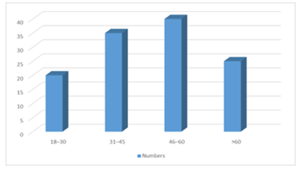
Figure 1: Distribution of study participants according to age group.
ECG Findings
ECG abnormalities were common among the study participants. Left ventricular hypertrophy (LVH) was the most frequently observed abnormality, occurring in 40 (33.3%) patients. Arrhythmia and ST–T changes were each observed in 25 (20.8%) patients, whereas 30 (25.0%) participants had a normal ECG.
Table 2. Distribution of ECG findings among study participants
|
ECG finding |
n |
% |
|
Normal |
30 |
25.0 |
|
Left ventricular hypertrophy |
40 |
33.3 |
|
Arrhythmia |
25 |
20.8 |
|
ST–T changes |
25 |
20.8 |
|
Total |
120 |
100.0 |
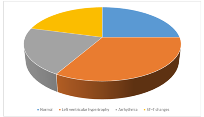
Figure 2: Distribution of ECG abnormalities among CKD patients.
Echocardiographic Findings
2D-ECHO demonstrated cardiac abnormalities in a substantial proportion of participants. Left ventricular dysfunction was the most frequent abnormality, observed in 45 (37.5%) participants, followed by diastolic dysfunction in 25 (20.8%) and pericardial effusion in 15 (12.5%). Normal echocardiographic findings were observed in 35 (29.2%) participants.
Table 3. Distribution of 2D-ECHO findings
|
2D-ECHO finding |
n |
% |
|
Normal |
35 |
29.2 |
|
Left ventricular dysfunction |
45 |
37.5 |
|
Diastolic dysfunction |
25 |
20.8 |
|
Pericardial effusion |
15 |
12.5 |
|
Total |
120 |
100.0 |
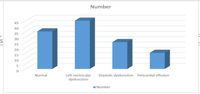
Figure 3: Distribution of 2D-ECHO findings among CKD patients.
Distribution of Ejection Fraction
Assessment of ejection fraction showed that 45 (37.5%) participants had an ejection fraction <40%, 35 (29.2%) had an ejection fraction of 40–50%, and 40 (33.3%) had an ejection fraction >50%.
Table 4. Distribution of participants according to ejection fraction
|
Ejection fraction |
n |
% |
|
>50% |
40 |
33.3 |
|
40–50% |
35 |
29.2 |
|
<40% |
45 |
37.5 |
|
Total |
120 |
100.0 |
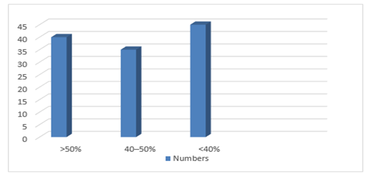
Figure 4: Distribution of participants according to ejection fraction.
Cardiac Biomarker Findings
Cardiac biomarkers were elevated in 70 (58.3%) participants, while 50 (41.7%) had normal biomarker levels.
Table 5. Distribution of cardiac biomarker levels
|
Cardiac biomarker status |
n |
% |
|
Normal |
50 |
41.7 |
|
Elevated |
70 |
58.3 |
|
Total |
120 |
100.0 |
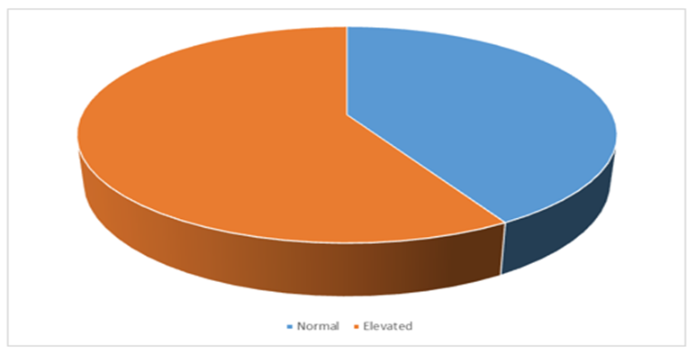
Figure 5: Distribution of participants according to cardiac biomarker status.
Association Between Age and Ejection Fraction
A statistically significant association was observed between age group and ejection fraction (p=0.04). Reduced ejection fraction (<40%) was more frequently observed in the older age groups.
Table 6. Association between age group and ejection fraction
|
Age group (years) |
>50% |
40–50% |
<40% |
Total |
|
18–30 |
10 |
6 |
4 |
20 |
|
31–45 |
12 |
13 |
10 |
35 |
|
46–60 |
10 |
10 |
20 |
40 |
|
>60 |
8 |
6 |
11 |
25 |
|
Total |
40 |
35 |
45 |
120 |
|
p-value |
0.04 |
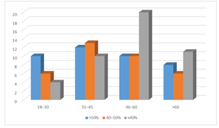
Figure 6: Association between age group and ejection fraction.
Association Between Gender and ECG Changes
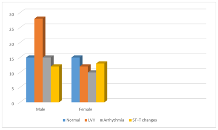
The association between gender and ECG abnormalities was not statistically significant (p=0.21). LVH was more frequently observed among males, whereas ST–T changes were relatively more frequent among females.
Table 7. Association between gender and ECG findings
|
Gender |
Normal |
LVH |
Arrhythmia |
ST–T changes |
Total |
|
Male |
15 |
28 |
15 |
12 |
70 |
|
Female |
15 |
12 |
10 |
13 |
50 |
|
Total |
30 |
40 |
25 |
25 |
120 |
|
p-value |
0.21 |
Association Between CKD Stage and 2D-ECHO Findings
A statistically significant association was observed between CKD stage and 2D-ECHO findings (p=0.02). Normal echocardiographic findings were more frequent in stage 3 CKD, whereas cardiac abnormalities were more prominent in stages 4 and 5.
Table 8. Association between CKD stage and 2D-ECHO findings
|
CKD stage |
Normal |
LV dysfunction |
Diastolic dysfunction |
Pericardial effusion |
Total |
|
Stage 3 |
20 |
15 |
10 |
5 |
50 |
|
Stage 4 |
10 |
18 |
8 |
4 |
40 |
|
Stage 5 |
5 |
12 |
7 |
6 |
30 |
|
Total |
35 |
45 |
25 |
15 |
120 |
|
p-value |
0.02 |
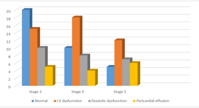
Figure 8: Association between CKD stage and 2D-ECHO findings.
Association Between Hypertension and Ejection Fraction
A statistically significant association was observed between hypertension and ejection fraction (p=0.01). Reduced EF (<40%) was more frequent among participants with hypertension
Table 9. Association between hypertension and ejection fraction
|
Hypertension |
>50% |
40–50% |
<40% |
Total |
|
Present |
20 |
25 |
35 |
80 |
|
Absent |
20 |
10 |
10 |
40 |
|
Total |
40 |
35 |
45 |
120 |
|
p-value |
0.01 |
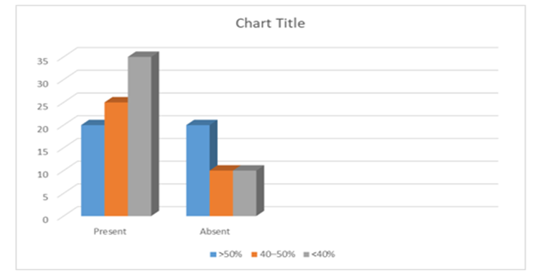
Figure 9: Association between hypertension and ejection fraction.
DISCUSSION:
The current study used electrocardiography (ECG), two-dimensional echocardiography (2D-ECHO), ejection fraction, and cardiac biomarkers to assess cardiac involvement in patients with chronic kidney disease (CKD) stages 3–5. The results show that individuals with mild to advanced chronic kidney disease (CKD) have a significant burden of cardiac abnormalities that affect the electrical, structural, and functional features of the heart. These results confirm that cardiovascular involvement and increasing renal failure are closely related.8
ECG abnormalities were found in 75.0% of patients in this study, with left ventricular hypertrophy (LVH) being the most prevalent abnormality (33.3%), followed by arrhythmias and ST-T alterations (20.8% each). LVH is a common cardiovascular symptom of chronic kidney disease (CKD) that can be brought on by anemia, volume overload, chronic hypertension, and other hemodynamic abnormalities linked to renal failure. Thus, the significant cardiovascular burden among CKD patients is reflected in the high frequency of LVH found in our study. Ventricular remodeling may have developed as a result of the coexistence of hypertension in 66.7% of the study sample.9
A high frequency of heart structural and functional abnormalities was also shown by the echocardiographic results. The most common 2D-ECHO abnormality was left ventricular dysfunction (37.5% of patients), which was followed by diastolic dysfunction (20.8%) and pericardial effusion (12.5%). The percentage of subjects with normal echocardiographic results was just 29.2%. These results suggest that cardiac involvement in chronic kidney disease (CKD) encompasses substantial structural and functional alterations in addition to electrical problems. While left ventricular systolic failure is a more severe sign of cardiac involvement, diastolic dysfunction may be the result of ventricular hypertrophy and myocardial remodeling.10
The strong correlation between CKD stage and 2D-ECHO results (p=0.02) was one of the study's key findings. Patients with stage 3 CKD were more likely to have normal echocardiographic results, but those with stages 4 and 5 disease were more likely to have abnormalities. This finding implies that increasing cardiac structural and functional defects coincide with increased renal failure. The increasing accumulation of cardiovascular risk factors and metabolic abnormalities associated with chronic kidney disease (CKD) as renal function declines may account for this connection. The results of the thesis also show that as the stage of CKD advances, cardiac involvement increases.11
Findings on ejection fraction offered more proof of compromised heart health. 37.5% of patients in the current study had an ejection fraction below 40%, 29.2% had an ejection fraction between 40 and 50%, and 33.3% had an ejection fraction above 50%. Thus, decreased systolic function was present in a significant percentage of individuals with CKD stages 3–5. Because it denotes myocardial functional impairment and may occur with other structural cardiac abnormalities, the existence of diminished ejection fraction in CKD patients is clinically significant.12
In this study, ejection fraction was substantially correlated with age (p=0.04). While younger participants showed a higher proportion of intact ejection fraction, older participants—especially those aged 46–60 and over 60—were more likely to have reduced ejection fraction. This finding implies that age-related circulatory alterations may exacerbate the effects of CKD on cardiac performance. Impaired myocardial function may result from older patients' increased cumulative exposure to hypertension, diabetes, vascular disease, and other cardiovascular risk factors.13
Additionally, there was a significant correlation (p=0.01) between hypertension and ejection fraction. Only 10 individuals without hypertension had an ejection fraction < 40%, compared to 35 individuals with hypertension. This result supports the notion that hypertension plays a significant role in the development of cardiac remodeling. Persistently high blood pressure can lead to LVH, myocardial remodeling, and eventually impaired systolic performance. It also raises left ventricular afterload. Therefore, hypertension may be a major cause of cardiovascular problems in people with chronic kidney disease (CKD) as well as a result of renal failure.14
Males were more likely than females to have LVH, although there was no statistically significant correlation between gender and ECG abnormalities (p=0.21). This implies that the distribution of ECG abnormalities in the current study sample was not significantly influenced by sex alone. Therefore, it is important to interpret the apparent difference in LVH frequency with caution. Rather than being an independent effect of gender, it may represent variations in the distribution of other cardiovascular risk factors.15
58.3% of subjects had elevated cardiac biomarkers. This result suggests that patients with intermediate to advanced chronic kidney disease have a significant burden of biochemical evidence of cardiac stress or injury. However, because renal failure may affect the circulating concentrations of cardiac biomarkers in CKD, cautious clinical interpretation is necessary Therefore, rather than being viewed as a stand-alone sign of cardiac illness, the biomarker results in this study are best evaluated in conjunction with ECG and echocardiographic abnormalities.16
Cardiovascular involvement in CKD is multifaceted, as evidenced by the combined results of ECG, echocardiography, ejection fraction, and cardiac biomarkers. Structural remodeling, reduced systolic or diastolic function, and biochemical signs of cardiac stress can all coexist with electrical anomalies. This demonstrates the value of a multimodal cardiac assessment over a single study in individuals with moderate to advanced chronic kidney disease.17
There may be clinical ramifications to the current findings. Regular cardiovascular examination may assist identify patients with clinically significant cardiac involvement because significant cardiac abnormalities were found among patients with CKD stages 3–5. While echocardiography can detect structural and functional issues that might not be seen clinically, ECG can offer an easily accessible initial assessment. While cardiac biomarkers may offer supplementary biochemical information, ejection fraction assessment can further describe systolic dysfunction.18
However, the limitations of the study should be considered while interpreting it. The study was carried out in a single location and had a somewhat limited sample size, which would restrict how broadly the results can be applied. Additionally, the follow-up period was somewhat brief, which made it difficult to evaluate the long-term course of renal and cardiac disease. Furthermore, confounding variables including lifestyle traits and treatment differences were not thoroughly assessed.19
Overall, the current study shows that individuals with CKD stages 3–5 have a substantial burden of cardiac anomalies. While age and hypertension were strongly linked to a lower ejection fraction, the considerable correlation between CKD stage and echocardiographic abnormalities suggests that growing cardiac involvement is related with worsening renal disease. These results highlight how crucial it is for individuals with mild to severe CKD to have an early and thorough cardiovascular evaluation.
CONCLUSION:
The current study shows that patients with stages 3–5 of chronic kidney disease (CKD) have a high prevalence of cardiac anomalies. While 2D-ECHO showed a significant burden of left ventricular and diastolic dysfunction, ECG abnormalities were frequent, with left ventricular hypertrophy being the most prevalent observation. Elevated cardiac biomarkers and decreased ejection fraction were also often noted.
Echocardiographic observations and CKD stage were shown to be significantly correlated, suggesting that increasing renal failure is associated with increased cardiac involvement. Ejection fraction was substantially correlated with age and hypertension, indicating that these variables may exacerbate heart dysfunction in CKD patients.
These results emphasize the significance of thorough cardiovascular evaluation in individuals with intermediate to advanced chronic kidney disease. Combining ECG, 2D-ECHO, ejection fraction, and cardiac biomarkers may help identify cardiac involvement earlier and enable suitable therapeutic treatment.
Limitations
There are a number of restrictions on the study. First, the study was carried out at a single tertiary care facility and had a rather limited sample size, which would restrict how broadly the results can be applied. Second, because the follow-up period was brief, it might not accurately represent the long-term development of heart dysfunction. Third, a thorough assessment of potential confounding variables, such as treatment differences and lifestyle traits, was lacking. To confirm these results, more multicentric research with bigger sample sizes and longer follow-up is needed.
Recommendations:
Cardiovascular evaluation should be regarded as a crucial part of the evaluation of patients with CKD stages 3–5, according to the results of this study. While echocardiography and ejection fraction measurement can provide information about structural and functional cardiac problems, ECG can be employed as a first accessible examination. Interpreting cardiac biomarkers in conjunction with clinical and cardiac imaging results may yield further information
To better understand the evolution of cardiac dysfunction across stages of chronic kidney disease (CKD) and to identify individuals who are most at risk for cardiovascular disease, prospective multicenter studies with bigger patient populations and longer follow-up are advised.
REFERENCES:
1. Vaidya SR, Aeddula NR. Chronic Kidney Disease. [Updated 2022 Oct 24]. In: StatPearls. Treasure Island (FL): StatPearls Publishing; 2024.
2. Lv JC, Zhang LX. Prevalence and disease burden of chronic kidney disease. Adv Exp Med Biol. 2019;1165:3-15.
3. Ene-Iordache B, Perico N, Bikbov B, Carminati S, Remuzzi A, Perna A, et al. Chronic kidney disease and cardiovascular risk in six regions of the world (ISN-KDDC): a cross-sectional study. Lancet Glob Health. 2016;4:e307-e319.
4. Jankowski J, Floege J, Fliser D, Böhm M, Marx N. Cardiovascular disease in chronic kidney disease: pathophysiological insights and therapeutic options. Circulation. 2021;143(11):1157-1172.
5. Zoccali C, Mark PB, Sarafidis P, Agarwal R, Adamczak M, Bueno de Oliveira R, et al. Diagnosis of cardiovascular disease in patients with chronic kidney disease. Nat Rev Nephrol. 2023;19(11):733-746.
6. Xanthopoulos A, Papamichail A, Briasoulis A, Loritis K, Bourazana A, Magouliotis DE, et al. Heart failure in patients with chronic kidney disease. J Clin Med. 2023;12(18):6105.
7. Guaricci AI, Sturda F, Russo R, Basile P, Baggiano A, Mushtaq S, et al. Assessment and management of heart failure in patients with chronic kidney disease. Heart Fail Rev. 2024;29(2):379-394.
8. Chinnappa S, White E, Lewis N, Baldo O, Tu YK, Glorieux G, et al. Early and asymptomatic cardiac dysfunction in chronic kidney disease. Nephrol Dial Transplant. 2018;33(3):450-458.
9. He J, Shlipak M, Anderson A, Roy JA, Feldman HI, Kallem RR, et al. Risk factors for heart failure in patients with chronic kidney disease: the CRIC study. J Am Heart Assoc. 2017;6(5):e005336.
10. Segall L, Nistor I, Covic A. Heart failure in patients with chronic kidney disease: a systematic integrative review. Biomed Res Int. 2014;2014:937398.
11. McIntyre CW, Odudu A, Eldehni MT. Cardiac assessment in chronic kidney disease. Curr Opin Nephrol Hypertens. 2009;18(6):501-506.
12. Agarwal S, Dangri P, Kalra OP, Rajpal S. Echocardiographic assessment of cardiac dysfunction in patients of chronic renal failure. J Indian Acad Clin Med. 2003;4(4):297.
13. Veeramani Kartheek AS, Srinivas Reddy VC. Assessment of cardiac dysfunction in patients with end stage renal disease in a tertiary care hospital. J Evid Based Med Healthc. 2019;6(24):1668-1672.
14. Dobre M, Yang W, Pan Q, Appel L, Bellovich K, Chen J, et al. Persistent high serum bicarbonate and the risk of heart failure in patients with chronic kidney disease: a report from the CRIC study. J Am Heart Assoc. 2015;4(4):e001599.
15. Murthy VL, Naya M, Foster CR, Hainer J, Gaber M, Dorbala S, et al. Coronary vascular dysfunction and prognosis in patients with chronic kidney disease. JACC Cardiovasc Imaging. 2012;5(10):1025-1034.
16. Triposkiadis F, Giamouzis G, Parissis J, Starling RC, Boudoulas H, Skoularigis J, et al. Reframing the association and significance of co-morbidities in heart failure. Eur J Heart Fail. 2016;18:744-758.
17. Szlagor M, Dybiec J, Mlynarska E, Rysz J, Franczyk B. Chronic kidney disease as a comorbidity in heart failure. Int J Mol Sci. 2023;24:2988.
18. Beldhuis IE, Lam CSP, Testani JM, Voors AA, Van Spall HGC, Ter Maaten JM, et al. Evidence-based medical therapy in patients with heart failure with reduced ejection fraction and chronic kidney disease. Circulation. 2022;145:693-712.
19. Ortiz A, Navarro-Gonzalez JF, Nunez J, de la Espriella R, Cobo M, Santamaria R, et al. The unmet need of evidence-based therapy for patients with advanced chronic kidney disease and heart failure: position paper from the Cardiorenal Working Groups of the Spanish Society of Nephrology and the Spanish Society of Cardiology. Clin Kidney J. 2022;15:865-872.
20. Bagshaw SM, Cruz DN, Aspromonte N, et al. Epidemiology of cardio-renal syndromes: workgroup statements from the 7th ADQI Consensus Conference. Nephrol Dial Transplant. 2010;25:1406-1416.