The Adult Skull as a Forensic Timepiece: Analyzing the Coronal Suture after the Third Decade.
- Dipak B. Chotaliya , Assistant Professor, Department of Forensic Medicine & Toxicology, JNU Institute for Medical Sciences and Research Centre, Jaipur National University, Jaipur, Rajasthan, India
- Deepak J. Sangle , Associate Professor, Department of Forensic Medicine & Toxicology, JNU Institute for Medical Sciences and Research Centre, Jaipur National University, Jaipur, Rajasthan, India
- Mahesh Devadas , Associate Professor, Department of Forensic Medicine & Toxicology, JNU Institute for Medical Sciences and Research Centre, Jaipur National University, Jaipur, Rajasthan, India
- Abhijit Rudra , Professor and Head of the Department, Department of Forensic Medicine & Toxicology, JNU Institute for Medical Sciences and Research Centre, Jaipur National University, Jaipur, Rajasthan, India
- Krunal Pipaliya , Professor and Head of the Department, Department of Forensic Medicine & Toxicology, Bhagyoday Medical College, Kadi, Gujarat, India.
Article Information:
Abstract:
Background: Adult age estimation is important in forensic identification when conventional indicators are unavailable or unreliable. This study evaluated the progression and forensic utility of coronal suture fusion on endocranial and ectocranial surfaces for adult age estimation in the Rajkot region. Materials and Methods: A postmortem study was conducted on 100 individuals (68 males and 32 females) of known age at P.D.U. Medical College & Hospital, Rajkot. The coronal suture was divided into three bilateral segments (C1, C2, and C3) and assessed using the Acsadi-Nemeskeri five-point scoring system (0–4). The association between suture fusion and chronological age was evaluated using Spearman's rank correlation coefficient. Results: Endocranial fusion commenced earlier and showed a more consistent progression than ectocranial fusion. In males, endocranial fusion commenced at 31.29 ± 1.94 years and reached complete obliteration at 64.04 ± 1.07 years. In females, fusion commenced at 28.37 ± 3.20 years and reached complete obliteration at approximately 75 years. No significant bilateral differences were observed. Conclusion: Endocranial coronal suture fusion appears to be a more consistent indicator for adult age estimation than ectocranial fusion. However, due to biological variability, it should be used to provide an age range rather than an exact age and interpreted alongside other age indicators.
Keywords:
Article :
INTRODUCTION:
Age estimation is an important component of forensic identification, particularly when chronological age cannot be established from documentary evidence. In cases involving decomposed, mutilated, burned, or skeletal remains, biological indicators can help estimate an approximate age range. Adult age estimation is challenging because skeletal changes show considerable inter-individual variation. Cranial suture closure is therefore used as an adjunctive skeletal indicator of adult age. [1,2]
Cranial sutures undergo progressive fusion with advancing age. The coronal suture, located between the frontal and parietal bones, extends bilaterally from the bregma toward the pterion. Its fusion can be assessed macroscopically using established scoring systems. The Acsadi-Nemeskeri method grades suture closure on a five-point scale, ranging from an open suture to complete obliteration, and has been widely used in forensic and anthropological studies. [2]
The use of cranial suture closure for age estimation remains controversial because the timing and extent of fusion show considerable variation, limiting its accuracy as an independent age marker. However, cranial sutures may provide useful adjunctive information when combined with other skeletal and dental indicators. The relationship between age and suture closure may also vary according to the suture, anatomical surface, sex, population, and assessment method. [3,4]
Differences in fusion between the endocranial and ectocranial surfaces may influence their forensic utility. Population-specific studies are therefore important, as the chronology of cranial suture closure may vary among populations. Although studies have been conducted in Indian populations, including north-western India, regional data remain limited. [1]
The present study evaluated the chronological progression of coronal suture fusion on the endocranial and ectocranial surfaces in adult postmortem cases from the Rajkot region. Bilateral coronal suture segments were assessed using the Acsadi-Nemeskeri scoring system, and the relationship between fusion and chronological age was examined. Fusion patterns were also compared between sexes and between endocranial and ectocranial surfaces to assess the utility of coronal suture fusion for adult age estimation.
MATERIALS AND METHODS:
Study Design and Study Population: A postmortem observational study was conducted on 100 cases received for medico-legal autopsy at P.D.U. Medical College & Hospital, Rajkot, from December 2010 to September 2012. The study included individuals aged ≥20 years whose chronological age was verified from birth certificates, identity documents, or school records.
Inclusion and Exclusion Criteria: Cases aged ≥20 years with documented chronological age were included. Unknown bodies and skulls with pathological deformities, fractures, or severe thermal damage that could interfere with sutural assessment were excluded.
Assessment of Cranial Sutures: Routine autopsy procedures were followed. Following a mastoid-to-mastoid incision and reflection of the scalp, the major cranial vault sutures, including the sagittal, coronal, and lambdoid sutures, were examined. The calvarium was cleared of soft tissue bilaterally using hydrogen peroxide in fresh bodies; in decomposed bodies, soft tissues were removed by stripping. The coronal suture was divided into three segments: C1 (pars bregmatica), C2 (pars complicata), and C3 (pars pterica). The degree of sutural fusion was assessed separately on the ectocranial and endocranial surfaces using the five-point Acsadi-Nemeskeri scoring system.2
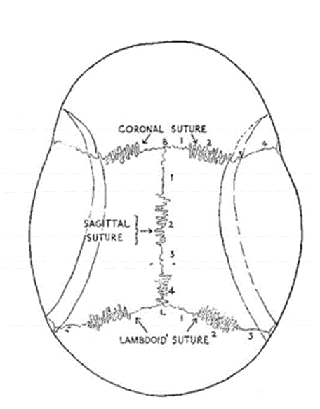
Figure 1: Skull Sutures and their Patterns in Segments. [4,5,6] SEGMENTS
Coronal:
1= Pars bregmatica (C1)
2= Pars complicata (C2)
3= Pars pterica (C3)
Scoring System (Acsadi-Nemeskeri Method):
Score 0: Open; fusion has not started.
Score 1: Incipient fusion; less than half the length is fused.
Score 2: Fusion in process; approximately half the length is fused.
Score 3: Advanced fusion; more than half the length is fused.
Score 4: Obliterated; complete fusion.
For endocranial assessment, the skull vault was removed to expose the internal surface of the coronal suture, and the same scoring system was applied.
Statistical Analysis: Data were analyzed using SPSS version 17 and Microsoft Excel. Spearman's rank correlation coefficient (rho) was used to assess the relationship between chronological age and the degree of sutural fusion. A p-value <0.05 was considered statistically significant.
RESULTS:
A total of 100 cases were included in the study, comprising 68 males and 32 females.
Table 1. Score-wise distribution of coronal suture fusion on the endocranial surface among males.
|
Suture |
Score |
Left Total Cases |
Left Mean Age |
Left SD |
Left Min Age |
Left Max Age |
Right Total Cases |
Right Mean Age |
Right SD |
Right Min Age |
Right Max Age |
|
C1 |
0 |
00 |
- |
- |
- |
- |
00 |
- |
- |
- |
- |
|
C1 |
1 |
08 |
32.13 |
8.82 |
22 |
50 |
09 |
31.89 |
8.28 |
22 |
50 |
|
C1 |
2 |
07 |
39.71 |
12.05 |
25 |
59 |
12 |
45.25 |
10.73 |
25 |
60 |
|
C1 |
3 |
33 |
52.24 |
12.62 |
21 |
75 |
27 |
52.93 |
13.45 |
21 |
75 |
|
C1 |
4 |
20 |
64.70 |
11.38 |
45 |
80 |
20 |
64.70 |
11.38 |
45 |
80 |
|
C2 |
0 |
00 |
- |
- |
- |
- |
00 |
- |
- |
- |
- |
|
C2 |
1 |
07 |
28.86 |
4.63 |
22 |
35 |
07 |
28.86 |
4.63 |
22 |
35 |
|
C2 |
2 |
07 |
41.71 |
7.27 |
32 |
50 |
09 |
41.89 |
6.43 |
32 |
50 |
|
C2 |
3 |
31 |
50.06 |
12.58 |
21 |
75 |
26 |
51.27 |
12.95 |
21 |
75 |
|
C2 |
4 |
23 |
65.52 |
10.47 |
38 |
80 |
26 |
63.12 |
12.44 |
32 |
80 |
|
C3 |
0 |
00 |
- |
- |
- |
- |
00 |
- |
- |
- |
- |
|
C3 |
1 |
01 |
33.00 |
- |
33 |
33 |
01 |
33.00 |
- |
33 |
33 |
|
C3 |
2 |
14 |
36.71 |
9.37 |
22 |
50 |
11 |
35.82 |
9.48 |
22 |
50 |
|
C3 |
3 |
23 |
48.13 |
12.88 |
21 |
67 |
23 |
45.65 |
12.44 |
21 |
60 |
|
C3 |
4 |
30 |
63.30 |
11.48 |
35 |
80 |
23 |
62.91 |
11.20 |
35 |
80 |
Endocranial Coronal Suture Fusion: The score-wise distribution of coronal suture fusion on the endocranial surface among males and females is presented in Tables 1 and 2, respectively. In males, endocranial fusion (Score 1) was observed at a mean age ranging from 28.86 to 33.00 years across the three coronal suture segments. The C2 segment (pars complicata) showed the earliest mean age of fusion onset at 28.86 years, followed by C1 (pars bregmatica) at 32.13 years and C3 (pars pterica) at 33.00 years. Complete obliteration (Score 4) was observed at a mean age ranging from 63.30 to 65.52 years across the three segments. In females, endocranial fusion (Score 1) was observed at a mean age ranging from 26.00 to 33.00 years. The C2 segment showed the earliest mean age of fusion onset at 26.33 years, followed by C3 at 26.00 years and C1 at 33.00 years, with variation between sides. Complete obliteration (Score 4) was observed at a mean age of 75 years in C1, C2, and C3. Overall, endocranial fusion showed a progressive increase in mean age with increasing fusion score. The standard deviation varied across segments and fusion stages.
Table 2. Score-wise distribution of coronal suture fusion on the endocranial surface among females.
|
Suture |
Score |
Left Total Cases |
Left Mean Age |
Left SD |
Left Min Age |
Left Max Age |
Right Total Cases |
Right Mean Age |
Right SD |
Right Min Age |
Right Max Age |
|
C1 |
0 |
02 |
25.00 |
7.07 |
20 |
30 |
02 |
30.00 |
14.14 |
20 |
40 |
|
C1 |
1 |
09 |
33.00 |
9.43 |
25 |
55 |
09 |
31.89 |
9.09 |
25 |
55 |
|
C1 |
2 |
09 |
40.56 |
9.08 |
30 |
60 |
07 |
40.29 |
10.44 |
30 |
60 |
|
C1 |
3 |
09 |
51.11 |
8.04 |
40 |
60 |
11 |
49.36 |
8.20 |
40 |
60 |
|
C1 |
4 |
03 |
75.00 |
5.00 |
70 |
80 |
03 |
75.00 |
5.00 |
70 |
80 |
|
C2 |
0 |
02 |
26.50 |
9.19 |
20 |
33 |
02 |
36.50 |
4.95 |
33 |
40 |
|
C2 |
1 |
03 |
26.33 |
1.53 |
25 |
28 |
05 |
26.00 |
4.06 |
20 |
31 |
|
C2 |
2 |
06 |
32.33 |
4.59 |
27 |
40 |
06 |
34.00 |
6.96 |
27 |
45 |
|
C2 |
3 |
18 |
47.00 |
9.67 |
30 |
60 |
16 |
47.81 |
9.71 |
30 |
60 |
|
C2 |
4 |
03 |
75.00 |
5.00 |
70 |
80 |
03 |
75.00 |
5.00 |
70 |
80 |
|
C3 |
0 |
02 |
26.50 |
9.19 |
20 |
33 |
01 |
33.00 |
- |
33 |
33 |
|
C3 |
1 |
01 |
26.00 |
- |
26 |
26 |
02 |
27.00 |
1.41 |
26 |
28 |
|
C3 |
2 |
07 |
31.00 |
6.58 |
25 |
45 |
06 |
27.50 |
4.51 |
20 |
32 |
|
C3 |
3 |
19 |
46.11 |
9.90 |
30 |
60 |
20 |
46.00 |
9.72 |
30 |
60 |
|
C3 |
4 |
03 |
75.00 |
5.00 |
70 |
80 |
03 |
75.00 |
5.00 |
70 |
80 |
Ectocranial Coronal Suture Fusion: The score-wise distribution of coronal suture fusion on the ectocranial surface among males and females is presented in Tables 3 and 4, respectively. In males, ectocranial fusion (Score 1) occurred at a mean age ranging from 39.75 to 52.38 years across the three segments. The C3 segment (pars pterica) showed the earliest mean age of fusion onset at 39.75 years, followed by C2 at 52.38 years and C1 at 51.31 years. Complete obliteration (Score 4) was not observed in the C1 segment. In C2 and C3, complete obliteration was observed at mean ages of 77.50 and 61.11 years, respectively, with variation between sides. In females, ectocranial fusion (Score 1) occurred at a mean age ranging from 32.63 to 47.71 years across the three segments. The C3 segment showed the earliest mean age of fusion onset, at 32.63 years on the left side and 31.14 years on the right side. Complete obliteration (Score 4) was not observed on the left side of C3, whereas it was observed on the right side at a mean age of 77.50 years. In C1 and C2, complete obliteration was observed at later ages, with considerable variation between segments and sides. Compared with the endocranial surface, ectocranial fusion demonstrated greater variability in the age associated with individual fusion scores. The highest standard deviation was 22.89 years, observed for C3 Score 4 on the left side among males.
Table 3. Score-wise distribution of coronal suture fusion on the ectocranial surface among males.
|
Suture |
Score |
Left Total Cases |
Left Mean Age |
Left Standard Deviation |
Left Min Age |
Left Max Age |
Right Total Cases |
Right Mean Age |
Right Standard Deviation |
Right Min Age |
Right Max Age |
|
C1 |
0 |
16 |
41.44 |
17.85 |
21 |
75 |
17 |
42.94 |
18.37 |
21 |
75 |
|
C1 |
1 |
29 |
51.31 |
10.60 |
30 |
70 |
28 |
51.29 |
10.34 |
30 |
65 |
|
C1 |
2 |
14 |
54.43 |
14.39 |
25 |
70 |
15 |
54.13 |
14.60 |
25 |
70 |
|
C1 |
3 |
09 |
71.11 |
8.34 |
55 |
80 |
08 |
71.88 |
8.58 |
55 |
80 |
|
C1 |
4 |
00 |
- |
- |
- |
- |
00 |
- |
- |
- |
- |
|
C2 |
0 |
11 |
31.91 |
6.58 |
21 |
45 |
14 |
32.50 |
8.10 |
21 |
50 |
|
C2 |
1 |
21 |
52.38 |
15.55 |
22 |
78 |
22 |
54.91 |
12.97 |
34 |
78 |
|
C2 |
2 |
20 |
56.25 |
7.17 |
43 |
67 |
16 |
57.69 |
7.74 |
43 |
70 |
|
C2 |
3 |
14 |
58.71 |
16.22 |
25 |
80 |
14 |
58.00 |
15.90 |
25 |
80 |
|
C2 |
4 |
02 |
77.50 |
3.54 |
75 |
80 |
02 |
77.50 |
3.54 |
75 |
80 |
|
C3 |
0 |
03 |
33.00 |
10.82 |
24 |
45 |
04 |
30.25 |
10.40 |
22 |
45 |
|
C3 |
1 |
16 |
39.75 |
15.52 |
21 |
78 |
16 |
41.50 |
14.95 |
21 |
78 |
|
C3 |
2 |
18 |
52.17 |
10.32 |
32 |
70 |
20 |
54.30 |
10.92 |
32 |
70 |
|
C3 |
3 |
22 |
60.41 |
7.04 |
50 |
72 |
18 |
60.11 |
7.03 |
50 |
72 |
|
C3 |
4 |
09 |
61.11 |
22.89 |
25 |
80 |
10 |
60.00 |
21.21 |
25 |
80 |
Table 4. Score-wise distribution of coronal suture fusion on the ectocranial surface among females.
|
Suture |
Score |
Left Total Cases |
Left Mean Age |
Left SD |
Left Min Age |
Left Max Age |
Right Total Cases |
Right Mean Age |
Right Standard Deviation |
Right Min Age |
Right Max Age |
|
C1 |
0 |
13 |
31.08 |
6.25 |
20 |
40 |
13 |
31.08 |
6.25 |
20 |
40 |
|
C1 |
1 |
14 |
47.71 |
9.64 |
30 |
60 |
14 |
47.71 |
9.64 |
30 |
60 |
|
C1 |
2 |
03 |
56.67 |
15.28 |
40 |
70 |
03 |
56.67 |
15.28 |
40 |
70 |
|
C1 |
3 |
01 |
80.00 |
- |
80 |
80 |
01 |
80.00 |
- |
80 |
80 |
|
C1 |
4 |
01 |
75.00 |
- |
75 |
75 |
01 |
75.00 |
- |
75 |
75 |
|
C2 |
0 |
10 |
29.73 |
5.41 |
20 |
40 |
12 |
29.75 |
5.15 |
20 |
40 |
|
C2 |
1 |
10 |
43.30 |
9.53 |
30 |
60 |
11 |
46.82 |
9.11 |
30 |
60 |
|
C2 |
2 |
08 |
51.50 |
8.50 |
40 |
60 |
04 |
46.25 |
9.46 |
40 |
60 |
|
C2 |
3 |
02 |
72.50 |
3.54 |
70 |
75 |
04 |
65.00 |
9.13 |
55 |
75 |
|
C2 |
4 |
02 |
77.50 |
3.54 |
75 |
80 |
01 |
80.00 |
- |
80 |
80 |
|
C3 |
0 |
06 |
29.83 |
6.85 |
20 |
40 |
06 |
29.83 |
6.85 |
20 |
40 |
|
C3 |
1 |
10 |
32.63 |
6.23 |
26 |
43 |
07 |
31.14 |
4.98 |
26 |
40 |
|
C3 |
2 |
08 |
45.00 |
9.40 |
30 |
60 |
08 |
43.13 |
9.32 |
30 |
60 |
|
C3 |
3 |
08 |
55.25 |
9.29 |
40 |
70 |
09 |
55.56 |
8.29 |
40 |
70 |
|
C3 |
4 |
00 |
- |
- |
- |
- |
02 |
77.50 |
3.54 |
75 |
80 |
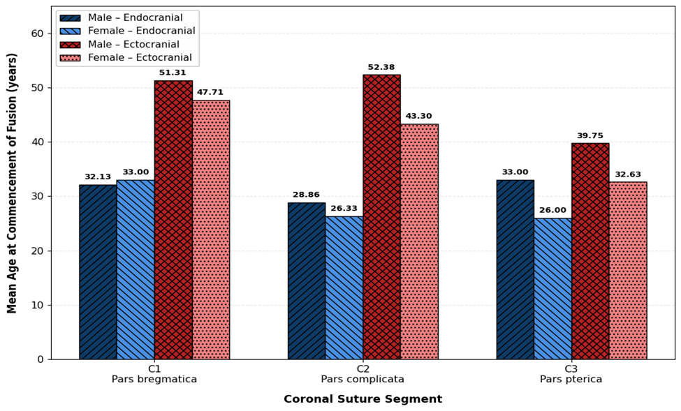
Figure 2. Mean age at commencement of coronal suture fusion (Score 1) according to anatomical segment, sex, and cranial surface.
Correlation Between Ectocranial and Endocranial Fusion: The age-group-wise correlation between the ectocranial-to-endocranial fusion score ratio and age, based on Spearman's rank correlation coefficient, is presented in Table 5. In males, none of the assessed age groups showed a statistically significant correlation between age and the ectocranial-to-endocranial fusion score ratio (p > 0.05). Among females, no statistically significant correlation was observed in the 20–29, 30–39, 40–49, or 50–59-year age groups. A statistically significant correlation was observed in females aged 70–79 years (rho = −1.000, p = 0.010). Correlation analysis was not estimable in some age groups because of insufficient observations or data availability. Overall, the findings indicate that the ectocranial-to-endocranial fusion score ratio did not show a consistent statistically significant association with age across the assessed age groups.
Table 5. Age-group-wise correlation between the ectocranial-to-endocranial fusion score ratio and age using Spearman's rank correlation coefficient (rho) among males and females.
|
Age Group |
Parameter (C/P) |
ECTLC/ENDLC (Male) – C |
ECTLC/ENDLC (Male) – P |
ECTLC/ENDLC (Female) – C |
ECTLC/ENDLC (Female) – P |
|
20–29 |
C |
0.406 |
0.618 |
0.359 |
0.616 |
|
20–29 |
P |
0.425 |
0.191 |
0.553 |
0.269 |
|
30–39 |
C |
-0.057 |
-0.006 |
0.075 |
0.187 |
|
30–39 |
P |
0.876 |
0.986 |
0.873 |
0.688 |
|
40–49 |
C |
-0.509 |
-0.509 |
0.185 |
0.401 |
|
40–49 |
P |
0.161 |
0.161 |
0.608 |
0.251 |
|
50–59 |
C |
-0.192 |
-0.005 |
0.000 |
-0.544 |
|
50–59 |
P |
0.445 |
0.985 |
1.000 |
0.456 |
|
60–69 |
C |
0.259 |
-0.499 |
XXX |
XXX |
|
60–69 |
P |
0.392 |
0.083 |
XXX |
XXX |
|
70–79 |
C |
0.026 |
0.026 |
-1.000 |
-1.000 |
|
70–79 |
P |
0.942 |
0.942 |
0.010 |
0.010 |
|
80–>80 |
C |
XXX |
XXX |
XXX |
XXX |
|
80–>80 |
P |
XXX |
XXX |
XXX |
XXX |
Note: rho = Spearman's rank correlation coefficient; p = p-value.
DISCUSSION:
The present study evaluated coronal suture fusion on endocranial and ectocranial surfaces in 100 adult postmortem cases from Rajkot. Fusion increased with age but varied by sex, segment, and surface, supporting its use as an adjunctive rather than independent age indicator.
Endocranial fusion commenced earlier than ectocranial fusion. In males, Score 1 occurred at mean ages of 32.13, 28.86, and 33.00 years in C1, C2, and C3, respectively; in females, the corresponding ages were 33.00, 26.33, and 26.00 years. Complete endocranial obliteration occurred at approximately 64 years in males and 75 years in females. These findings are consistent with Sahni et al., who studied 665 adults from north-western India and reported earlier endocranial than ectocranial obliteration, with generally earlier closure in males than females. [1] This similarity suggests that endocranial assessment may provide useful chronological information in Indian populations.
However, our findings differ from Meindl and Lovejoy, who found ectocranial assessment more reliable for age estimation in 236 crania. [4] This difference may reflect variation in population, sample characteristics, anatomical sites, scoring methods, and age distribution. Thus, the greater utility of endocranial assessment in our study should be considered specific to the Rajkot population.
Segmental variation was also observed. C2 (pars complicata) showed the earliest endocranial fusion in males, while C2 and C3 showed earlier fusion in females. On the ectocranial surface, C3 showed the earliest Score 1 in both sexes. Similar segmental variation was reported by Sahni et al., supporting assessment of individual suture segments rather than treating the coronal suture as a single unit. [4,6]
Ectocranial fusion showed greater variability than endocranial fusion, with SD reaching 22.89 years in males (C3, Score 4) and incomplete obliteration in some segments. Ruengdit et al. reported similar limitations, with cranial suture methods showing errors of approximately 13–22 years in a Thai population, highlighting the influence of population differences on age estimation. [7]
The ectocranial-to-endocranial fusion ratio showed no consistent association with age. Most age groups had non-significant correlations, with significance observed only in females aged 70–79 years (rho = −1.000, p = 0.010). This isolated finding should therefore be interpreted cautiously and does not establish the ratio as a reliable age-estimation parameter.
Recent Indian evidence supports a cautious interpretation. Akbar et al., using 3D CT in 263 individuals, found an association between ectocranial suture obliteration and age but emphasized its limitations for precise age estimation. [8] A recent systematic review similarly concluded that cranial sutures should not be used as sole age markers because of their limited accuracy. [9]
Overall, the present study demonstrates an age-related but variable pattern of coronal suture fusion. Endocranial fusion was earlier and more orderly than ectocranial fusion in the Rajkot population, while the ectocranial-to-endocranial ratio showed limited correlation with age. Thus, coronal suture fusion should be considered a supplementary forensic indicator, with age reported as an approximate range and interpreted alongside other skeletal and dental parameters.
CONCLUSION:
The present study demonstrates a progressive, but variable, age-related pattern of coronal suture fusion in the Rajkot population. Endocranial fusion commenced earlier and showed a comparatively more consistent progression than ectocranial fusion, suggesting greater utility for adult age estimation in this population. Fusion generally commenced earlier in females but complete obliteration occurred at a later age than in males. No significant bilateral variation was observed in the progression of coronal suture closure.
However, the considerable inter-individual and segmental variation limits the use of coronal suture closure as an independent indicator of chronological age. The ectocranial-to-endocranial fusion ratio also showed no consistent association with age. Therefore, coronal suture assessment should be used as an adjunctive forensic parameter, with age preferably reported as an approximate range rather than an exact value and interpreted in conjunction with other established skeletal and dental indicators.
REFERENCES:
1. Sahni D, Jit I. Time of closure of cranial sutures in northwest Indian adults. Forensic science international. 2005 Mar 10;148(2-3):199-205.
2. Acsádi G, Nemeskéri J, Balás K. History of human life span and mortality. Akademiai kiado; 1970.
3. Singer R. Estimation of age from cranial suture closure. J Forensic Med. 1953;1:52-9.
4. Meindl RS, Lovejoy CO. Ectocranial suture closure: A revised method for the determination of skeletal age at death based on the lateral‐anterior sutures. American journal of physical anthropology. 1985 Sep;68(1):57-66.
5. Erenko O, Kilhberg J. Closure of cranial sutures and age. A.V.medica- anthropologica.1995;43:1-31
6. Oppenheim S. Die suturren des menschlichen Schadels in ihrer anthropologieschen Bedeutung. 1907;38:128-135
7. Ruengdit S, Prasitwattanaseree S, Mekjaidee K, Sinthubua A, Mahakkanukrauh P. Age estimation approaches using cranial suture closure: A validation study on a Thai population. Journal of forensic and legal medicine. 2018 Jan 1;53:79-86.
8. Akbar NJM, Shekhawat RS, Kanchan T, Yadav T, Meshram VP, Shedge R, Vempalli SR, Setia P, Meshram V. Computed tomographic evaluation of cranial suture obliteration for age estimation in an Indian population. Cureus. 2023 Mar 14;15(3).
9. Macedo F, Stefanel ME, Sakurada A, Moreira D, Junqueira JL, Franco A. Skull joints assessed via CT for age estimation—a systematic review. Dentomaxillofacial Radiology. 2025 Jul;54(5):332-41.