Ultrasound Assessment Of Diaphragmatic Function As A Predictor Of Extubation Outcome In Mechanically Ventilated Patients: A Prospective Observational Study.
- Vishwakiran , Resident Department of anesthesiology MR medical College, Kalaburagi – corresponding author
- Satish sharanappa , Assistant professor Department of anesthesiology MR medical College, Kalaburagi
- Varsha S Patil , Senior resident Department of anesthesiology MR medical College, Kalaburagi
- Pratima S Kamareddy , Professor Department of anesthesiology MR medical College, Kalaburagi.
Article Information:
Abstract:
Background: Successful weaning from mechanical ventilation is a critical step in intensive care management, and extubation failure is associated with increased morbidity and mortality. Conventional weaning indices have limited predictive accuracy, highlighting the need for reliable bedside assessment tools. Diaphragmatic ultrasonography has emerged as a promising non-invasive method for evaluating diaphragmatic function. Objectives: To evaluate the role of diaphragmatic ultrasound parameters in predicting extubation outcome, correlate these parameters with conventional weaning indices, and determine their diagnostic accuracy. Methods: This prospective observational study included 50 adult mechanically ventilated patients planned for elective extubation. Diaphragmatic excursion and diaphragmatic thickness fraction (DTF) were measured using bedside ultrasonography before extubation. Patients were followed for 48 hours and classified into successful extubation and extubation failure groups. Ultrasound findings were compared with conventional weaning parameters, and receiver operating characteristic (ROC) curve analysis was performed. Results: Extubation was successful in 40 (80%) patients and failed in 10 (20%). Patients with successful extubation had significantly higher diaphragmatic excursion (1.72 ± 0.29 cm vs. 1.12 ± 0.21 cm) and DTF (49.6 ± 10.8% vs. 23.8 ± 7.2%; p<0.001). Both parameters correlated significantly with rapid shallow breathing index and oxygenation indices. ROC analysis demonstrated excellent diagnostic performance, with an AUC of 0.90 for diaphragmatic excursion and 0.93 for DTF. Conclusion: Diaphragmatic ultrasonography is an accurate, non-invasive bedside tool for predicting extubation outcome and may serve as a valuable adjunct to conventional weaning assessment in mechanically ventilated patients.
Keywords:
Article :
INTRODUCTION:
Mechanical ventilation (MV) is an indispensable life-support intervention for critically ill patients with acute respiratory failure admitted to intensive care units (ICUs)[1]. Although MV improves oxygenation and reduces the work of breathing, prolonged ventilatory support is associated with complications such as ventilator-associated pneumonia (VAP), ventilator-induced lung injury (VILI), ventilator-induced diaphragmatic dysfunction (VIDD), ICU-acquired weakness, prolonged ICU stay, increased healthcare costs, and higher mortality. Timely liberation from mechanical ventilation through successful extubation is therefore a major goal of critical care[2]. However, determining the optimal timing for extubation remains challenging, as both premature and delayed extubation are associated with adverse outcomes. Extubation failure, usually defined as the need for reintubation within 48–72 hours, occurs in approximately 10–20% of patients and is associated with increased morbidity, mortality, and healthcare utilization [3,4].
Current weaning strategies combine clinical assessment, arterial blood gas analysis, spontaneous breathing trials (SBTs), and physiological indices such as the rapid shallow breathing index (RSBI), tidal volume, and maximal inspiratory pressure[5]. Although RSBI is widely used because of its simplicity, its predictive accuracy is only moderate, and many patients who successfully complete an SBT subsequently require reintubation. These limitations have highlighted the need for additional objective bedside tools to improve prediction of extubation readiness [8].The diaphragm is the principal inspiratory muscle, contributing nearly 70–80% of tidal ventilation during spontaneous breathing. Mechanical ventilation, particularly when prolonged, can rapidly lead to VIDD through diaphragmatic atrophy, oxidative stress, and impaired contractility. Diaphragmatic dysfunction may develop within 24–48 hours of controlled ventilation and has emerged as a major reversible cause of weaning failure, emphasizing the importance of assessing diaphragmatic function before extubation [9].
Bedside diaphragm ultrasonography has gained increasing acceptance as a non-invasive, portable, radiation-free, and repeatable technique for evaluating diaphragmatic function in critically ill patients. It allows real-time assessment without transporting unstable patients outside the ICU. The two most commonly used parameters are diaphragmatic excursion (DE), measured by M-mode ultrasonography, and diaphragmatic thickening fraction (DTF), measured by B-mode ultrasonography. These indices objectively assess diaphragmatic movement and contractility and have shown promising utility in predicting successful weaning and extubation [9].
Several prospective studies and systematic reviews have demonstrated that reduced DE and lower DTF are significantly associated with extubation failure. Proposed cutoff values of DE (approximately 1.0–1.5 cm) and DTF (20–30%) have shown good diagnostic performance. However, variability in patient selection, ventilator settings, spontaneous breathing trial protocols, patient positioning, and ultrasound methodology has resulted in heterogeneous findings. Ventilator parameters, particularly positive end-expiratory pressure (PEEP), and neurological status can influence diaphragm performance and extubation outcomes, highlighting the need for standardized assessment protocols [10,11].Recent international recommendations increasingly support integrating point-of-care ultrasound into routine ICU practice.
When combined with conventional weaning parameters, diaphragm ultrasonography may improve identification of patients ready for extubation, reduce reintubation rates, shorten ICU stay, and improve clinical outcomes. Nevertheless, further standardized prospective studies are needed to establish optimal diagnostic thresholds and confirm its routine clinical utility [12].Therefore, the present prospective observational study was designed to evaluate the role of ultrasound assessment of diaphragmatic function as a predictor of extubation outcome in mechanically ventilated patients and to determine its usefulness as a simple, non-invasive adjunct to conventional weaning parameters.
MATERIALS AND METHODS:
This study was conducted as a prospective observational study to evaluate the role of diaphragmatic ultrasonography in predicting extubation outcomes among mechanically ventilated patients.The study was carried out in the Intensive Care Unit (ICU) of Basaveshwara Teaching and General Hospital attached to Mahadevappa Rampure Medical College, Kalaburagi.
Study Duration
The study was conducted over a period of four months, from March 2026 to June 2026.
Study Population
The study population consisted of adult patients receiving invasive mechanical ventilation in the ICU who were planned for elective extubation after successful completion of a spontaneous breathing trial (SBT).
Sample Size
A total of 50 patients fulfilling the eligibility criteria were enrolled in the study.
Sampling Technique
Patients were recruited using a consecutive sampling technique, whereby all eligible patients admitted during the study period were included until the required sample size was achieved.
Inclusion Criteria
The following patients were included in the study:
• Adult patients aged 18 years or older.
• Patients who had received invasive mechanical ventilation for 48 hours or more.
• Patients who had successfully completed a spontaneous breathing trial and were considered suitable for elective extubation.
Exclusion Criteria
Patients were excluded if they had any of the following:
• Known diaphragmatic paralysis.
• Neuromuscular disorders affecting respiratory muscles.
• Cervical spinal cord injury or phrenic nerve injury.
• Chest wall deformities interfering with ultrasonographic assessment.
• Poor ultrasonographic window.
• Refusal to provide informed consent by the patient or legally authorized representative.
Ethical Considerations
Ethical approval for the study was obtained from the Institutional Ethics Committee of Mahadevappa Rampure Medical College, Kalaburagi, before commencement of the study. Written informed consent was obtained from the patient or the legally authorized representative in their vernacular language after explaining the purpose, nature, benefits, and potential risks of the study. Confidentiality of patient information was maintained throughout the study.
Study Procedure
Eligible patients were enrolled after obtaining written informed consent. Baseline demographic and clinical data, including age, sex, primary diagnosis, duration of mechanical ventilation, duration of ICU stay, and relevant comorbidities, were recorded using a predesigned data collection proforma. Conventional weaning parameters, including respiratory rate, tidal volume, rapid shallow breathing index (RSBI), arterial blood gas findings, and oxygenation status, were also documented before extubation.Diaphragmatic ultrasonography was performed immediately before planned extubation by an experienced investigator using a portable ultrasound machine equipped with both a low-frequency curvilinear transducer (2–5 MHz) and a high-frequency linear transducer (7–12 MHz).Diaphragmatic excursion was assessed using M-mode ultrasonography through the right subcostal approach with the patient in a semi-recumbent position. Three consecutive measurements were obtained during quiet spontaneous breathing, and the average value was recorded in centimeters.Diaphragmatic thickness was measured at the zone of apposition using the linear probe at both end-expiration and end-inspiration. The diaphragmatic thickness fraction (DTF) was calculated using the following formula:
DTF (%) = [(Thickness at end inspiration − Thickness at end expiration) / Thickness at end expiration] × 100
Following ultrasonographic assessment, all patients underwent extubation according to the standard ICU weaning and extubation protocol. Patients were monitored for 48 hours after extubation.
Extubation outcome was classified into two groups:
• Extubation Success: Patients who did not require reintubation within 48 hours following extubation.
• Extubation Failure: Patients who required reintubation within 48 hours after extubation due to respiratory failure.
The diaphragmatic ultrasound parameters were subsequently compared between patients with successful extubation and those who experienced extubation failure.
Statistical Analysis
The collected data were entered into Microsoft Excel and analyzed using IBM SPSS Statistics version 29.0. Continuous variables were expressed as mean ± standard deviation (SD) or median with interquartile range (IQR), depending on data distribution, while categorical variables were expressed as frequencies and percentages. Normality of data distribution was assessed using the Shapiro–Wilk test. Comparisons between the extubation success and failure groups were performed using the independent Student's t-test for normally distributed continuous variables and the Mann–Whitney U test for non-normally distributed variables. Categorical variables were compared using the Chi-square test or Fisher's exact test, as appropriate.
Receiver operating characteristic (ROC) curve analysis was performed to evaluate the diagnostic accuracy of diaphragmatic excursion and diaphragmatic thickness fraction for predicting extubation outcome. The area under the curve (AUC), optimal cut-off values, sensitivity, specificity, positive predictive value, negative predictive value, and overall diagnostic accuracy were calculated. A p-value of less than 0.05 was considered statistically significant.
RESULTS:
A total of 50 mechanically ventilated patients who fulfilled the inclusion criteria were enrolled in the study. Among them, 40 patients (80.0%) had successful extubation, whereas 10 patients (20.0%) experienced extubation failure. The overall demographic profile, clinical characteristics, diaphragmatic ultrasound findings, conventional weaning parameters, and extubation outcomes are summarized in Tables 1–6 and Figures 1–3.The baseline demographic and clinical characteristics of the study participants are presented in Table 1.
The mean age of the study population was 56.8 ± 14.2 years, with the majority of patients belonging to the 41–60 years age group (44.0%). Males constituted 64.0% of the study population. The mean body mass index was 24.9 ± 3.8 kg/m², while the mean duration of mechanical ventilation and ICU stay were 5.9 ± 2.6 days and 8.3 ± 3.4 days, respectively. Pneumonia was the most common indication for mechanical ventilation (40.0%), followed by sepsis (22.0%), COPD exacerbation (16.0%), postoperative respiratory failure (14.0%), and other causes (8.0%). Diabetes mellitus and hypertension were present in 32.0% and 40.0% of patients, respectively (Table 1).
Table 1. Baseline Demographic and Clinical Characteristics of Study Participants (n = 50)
|
Parameter |
Value |
|
Age (years), Mean ± SD |
56.8 ± 14.2 |
|
Age group, n (%) |
|
|
18–40 years |
10 (20.0) |
|
41–60 years |
22 (44.0) |
|
>60 years |
18 (36.0) |
|
Male sex, n (%) |
32 (64.0) |
|
Female sex, n (%) |
18 (36.0) |
|
BMI (kg/m²), Mean ± SD |
24.9 ± 3.8 |
|
Duration of mechanical ventilation (days), Mean ± SD |
5.9 ± 2.6 |
|
Duration of ICU stay (days), Mean ± SD |
8.3 ± 3.4 |
|
Primary diagnosis, n (%) |
|
|
Pneumonia |
20 (40.0) |
|
Sepsis |
11 (22.0) |
|
COPD exacerbation |
8 (16.0) |
|
Postoperative respiratory failure |
7 (14.0) |
|
Other causes |
4 (8.0) |
|
Diabetes mellitus |
16 (32.0) |
|
Hypertension |
20 (40.0) |
Comparison of diaphragmatic ultrasound parameters between patients with successful extubation and those with extubation failure is shown in Table 2 and Figure 1. Patients with successful extubation demonstrated significantly greater diaphragmatic excursion (1.72 ± 0.29 cm vs. 1.12 ± 0.21 cm; p<0.001) and higher diaphragmatic thickness fraction (49.6 ± 10.8% vs. 23.8 ± 7.2%; p<0.001) compared with patients who experienced extubation failure. End-inspiratory diaphragmatic thickness was also significantly greater in the success group (0.34 ± 0.05 cm vs. 0.27 ± 0.04 cm; p<0.001). However, end-expiratory diaphragmatic thickness did not differ significantly between the two groups (p=0.468) (Table 2, Figure 1).
Table 2. Diaphragmatic Ultrasound Parameters According to Extubation Outcome
|
Parameter |
Extubation Success (n=40) |
Extubation Failure (n=10) |
p-value |
|
Diaphragmatic excursion (cm) |
1.72 ± 0.29 |
1.12 ± 0.21 |
<0.001 |
|
End-expiratory thickness (cm) |
0.23 ± 0.04 |
0.22 ± 0.03 |
0.468 |
|
End-inspiratory thickness (cm) |
0.34 ± 0.05 |
0.27 ± 0.04 |
<0.001 |
|
Diaphragmatic thickness fraction (%) |
49.6 ± 10.8 |
23.8 ± 7.2 |
<0.001 |
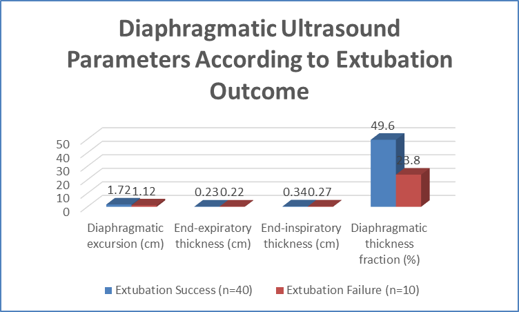
Figure 1 Diaphragmatic Ultrasound Parameters According to Extubation Outcome
The comparison of conventional weaning parameters between the two groups is presented in Table 3 and Figure 2. Patients with successful extubation had significantly lower respiratory rates (20.4 ± 2.8 vs. 24.1 ± 3.2 breaths/min; p=0.002) and Rapid Shallow Breathing Index (RSBI) values (58.7 ± 12.6 vs. 82.4 ± 14.8; p<0.001). In contrast, tidal volume (470 ± 62 vs. 401 ± 55 mL; p=0.004), PaO₂/FiO₂ ratio (252.8 ± 48.2 vs. 211.4 ± 39.5; p=0.011), and SpO₂ (97.3 ± 1.4% vs. 95.8 ± 1.8%; p=0.018) were significantly higher among patients with successful extubation (Table 3, Figure 2).
Table 3. Comparison of Conventional Weaning Parameters Between Extubation Success and Failure Groups
|
Parameter |
Success (n=40) |
Failure (n=10) |
p-value |
|
Respiratory rate (breaths/min) |
20.4 ± 2.8 |
24.1 ± 3.2 |
0.002 |
|
Tidal volume (mL) |
470 ± 62 |
401 ± 55 |
0.004 |
|
Rapid Shallow Breathing Index (RSBI) |
58.7 ± 12.6 |
82.4 ± 14.8 |
<0.001 |
|
PaO₂/FiO₂ ratio |
252.8 ± 48.2 |
211.4 ± 39.5 |
0.011 |
|
SpO₂ (%) |
97.3 ± 1.4 |
95.8 ± 1.8 |
0.018 |
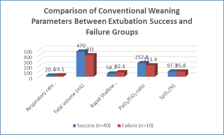
Figure 2 Comparison of Conventional Weaning Parameters Between Extubation Success and Failure Groups
Correlation analysis between diaphragmatic ultrasound parameters and conventional weaning indices is summarized in Table 4 and illustrated in Figure 3. Diaphragmatic excursion demonstrated a significant negative correlation with RSBI (r = −0.69, p<0.001) and a moderate positive correlation with PaO₂/FiO₂ ratio (r = 0.55, p<0.001). Similarly, diaphragmatic thickness fraction showed a strong negative correlation with RSBI (r = −0.74, p<0.001) and significant positive correlations with tidal volume (r = 0.67, p<0.001) and PaO₂/FiO₂ ratio (r = 0.59, p<0.001) (Table 4, Figure 3).
Table 4. Correlation Between Diaphragmatic Ultrasound Parameters and Conventional Weaning Indices
|
Variables |
Correlation coefficient (r) |
p-value |
|
Diaphragmatic excursion vs RSBI |
−0.69 |
<0.001 |
|
Diaphragmatic excursion vs PaO₂/FiO₂ |
0.55 |
<0.001 |
|
Diaphragmatic thickness fraction vs RSBI |
−0.74 |
<0.001 |
|
Diaphragmatic thickness fraction vs Tidal volume |
0.67 |
<0.001 |
|
Diaphragmatic thickness fraction vs PaO₂/FiO₂ |
0.59 |
<0.001 |
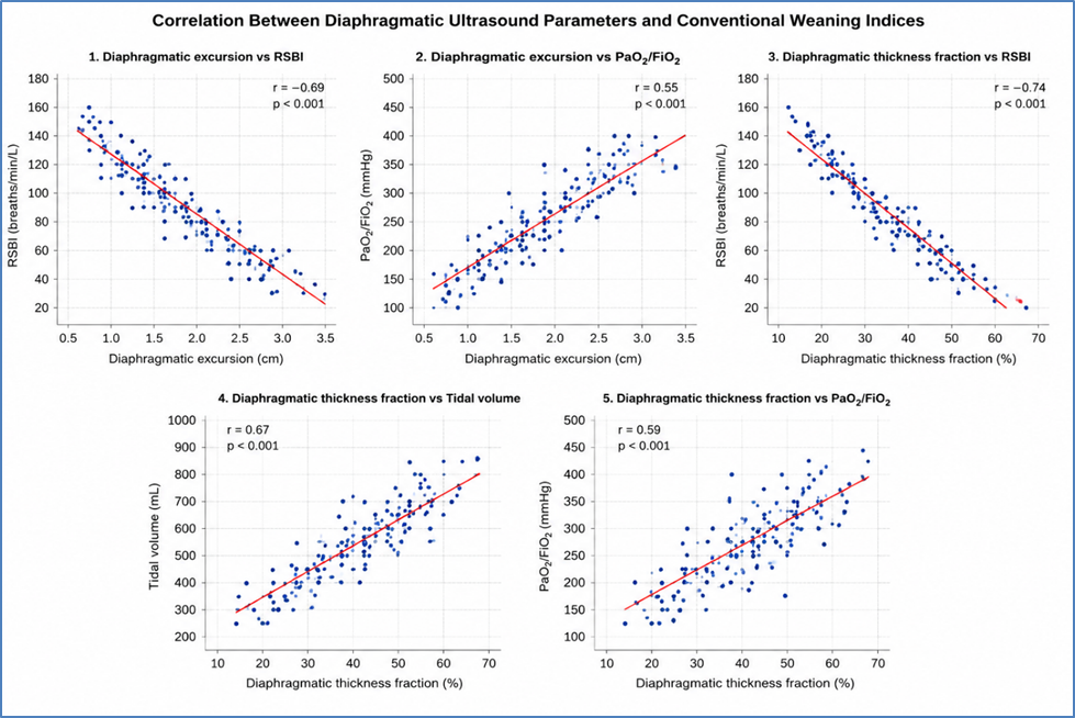
Figure 3 Correlation Between Diaphragmatic Ultrasound Parameters and Conventional Weaning Indices
Receiver operating characteristic (ROC) curve analysis demonstrated excellent diagnostic performance of diaphragmatic ultrasound parameters in predicting extubation outcome (Table 5). A diaphragmatic excursion cut-off value of ≥1.45 cm predicted successful extubation with a sensitivity of 87.5%, specificity of 80.0%, and an area under the curve (AUC) of 0.90 (95% CI: 0.81–0.99; p<0.001). Similarly, a diaphragmatic thickness fraction cut-off value of ≥32% demonstrated a sensitivity of 90.0%, specificity of 85.0%, and an AUC of 0.93 (95% CI: 0.86–1.00; p<0.001), indicating superior predictive accuracy (Table 5).
Table 5. Diagnostic Performance of Diaphragmatic Ultrasound Parameters in Predicting Extubation Outcome
|
Parameter |
Cut-off value |
Sensitivity (%) |
Specificity (%) |
AUC (95% CI) |
p-value |
|
Diaphragmatic excursion |
≥1.45 cm |
87.5 |
80.0 |
0.90 (0.81–0.99) |
<0.001 |
|
Diaphragmatic thickness fraction |
≥32% |
90.0 |
85.0 |
0.93 (0.86–1.00) |
<0.001 |
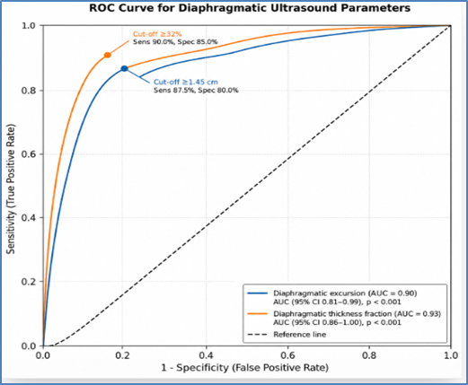
The overall extubation outcomes are presented in Table 6. Successful extubation was achieved in 40 patients (80.0%), whereas 10 patients (20.0%) experienced extubation failure. Among the failure group, 8 patients (16.0%) required reintubation within 48 hours, while 2 patients (4.0%) required rescue non-invasive ventilation. The mean ICU stay following extubation was 4.6 ± 2.3 days. Overall ICU mortality was 6.0%, and 47 patients (94.0%) were discharged from the hospital (Table 6).
Table 6. Extubation Outcomes and Clinical Characteristics
|
Outcome |
n (%) |
|
Successful extubation |
40 (80.0) |
|
Extubation failure |
10 (20.0) |
|
Reintubation within 48 hours |
8 (16.0) |
|
Rescue non-invasive ventilation |
2 (4.0) |
|
ICU mortality |
3 (6.0) |
|
Mean ICU stay after extubation (days), Mean ± SD |
4.6 ± 2.3 |
|
Hospital discharge |
47 (94.0) |
DISCUSSION:
The present prospective observational study evaluated the usefulness of diaphragmatic ultrasonography as a bedside tool for predicting extubation outcomes in mechanically ventilated patients. Patients who were successfully extubated demonstrated significantly greater diaphragmatic excursion (DE) and diaphragmatic thickness fraction (DTF) than those with extubation failure. Diaphragmatic ultrasound parameters also showed significant correlations with conventional weaning indices and excellent diagnostic accuracy, supporting their role as reliable adjuncts to routine weaning assessment.The mean age of the study population was 56.8 ± 14.2 years, with males accounting for 64% of participants. Pneumonia (40%) was the leading indication for mechanical ventilation, followed by sepsis (22%) and COPD exacerbation (16%). The mean duration of mechanical ventilation was 5.9 ± 2.6 days, and the overall extubation success rate was 80%.
These findings are comparable with those reported by Eltrabili et al. [13], who observed a similar patient profile with an extubation success rate of 75%, and by Khilnani et al. [14], who reported successful extubation in approximately 77% of mechanically ventilated ICU patients.Patients with successful extubation had significantly higher diaphragmatic excursion (1.72 ± 0.29 vs. 1.12 ± 0.21 cm; p<0.001) and DTF (49.6 ± 10.8% vs. 23.8 ± 7.2%; p<0.001) than those with extubation failure. End-inspiratory diaphragmatic thickness was also significantly greater, whereas end-expiratory thickness showed no significant difference. Similar observations have been reported by Eltrabili et al. [13], who demonstrated significantly higher DE, inspiratory thickness, and DTF among successfully extubated patients. Khilnani et al. [14] also found diaphragmatic excursion to be strongly associated with successful extubation, while Zhang et al. [15] further confirmed that preserved diaphragmatic contractility, reflected by higher DE and DTF, is closely associated with successful liberation from mechanical ventilation.Conventional weaning indices also differed significantly between the two groups. Successfully extubated patients had lower respiratory rate and rapid shallow breathing index (RSBI), while tidal volume and PaO₂/FiO₂ ratio were significantly higher.
These findings are consistent with previous studies showing that diaphragmatic ultrasound parameters outperform RSBI alone in predicting extubation success [13,14].Correlation analysis demonstrated significant negative relationships between diaphragmatic excursion and RSBI (r = −0.69) and between DTF and RSBI (r = −0.74), whereas positive correlations were observed with tidal volume and PaO₂/FiO₂ ratio. Similar associations between diaphragmatic function, respiratory mechanics, and successful spontaneous breathing have been reported by Zhang et al. [15] and Eltrabili et al. [13].Receiver operating characteristic analysis further demonstrated excellent diagnostic performance. A diaphragmatic excursion cut-off of ≥1.45 cm yielded 87.5% sensitivity, 80% specificity, and an AUC of 0.90, while a DTF cut-off of ≥32% achieved 90% sensitivity, 85% specificity, and an AUC of 0.93. These results closely align with the meta-analysis by Mahmoodpoor et al. [16], which reported pooled DTF sensitivity of 89%, specificity of 81%, and an AUC of 0.93. Similarly, Li et al. [17] demonstrated superior pooled diagnostic performance of DTF compared with diaphragmatic excursion, while the recent 2025 meta-analysis published in the Indian Journal of Anaesthesia further confirmed the high sensitivity and specificity of diaphragmatic ultrasonography for predicting successful extubation.
CONCLUSION:
The present study demonstrated that diaphragmatic ultrasonography is a reliable, non-invasive bedside tool for predicting extubation outcomes in mechanically ventilated patients. Patients with successful extubation had significantly greater diaphragmatic excursion and diaphragmatic thickness fraction than those who experienced extubation failure. These ultrasound parameters showed excellent diagnostic accuracy and correlated well with conventional weaning indices. Incorporating diaphragmatic ultrasound into routine weaning assessment may improve the prediction of extubation success, facilitate timely decision-making, and reduce the incidence of extubation failure in critically ill patients.
Limitations
The present study was conducted at a single tertiary care center with a relatively small sample size, which may limit the generalizability of the findings. Diaphragmatic ultrasonography is an operator-dependent technique, and inter-observer variability was not assessed. Additionally, patients were followed only for 48 hours after extubation; therefore, late extubation failures and long-term clinical outcomes could not be evaluated.
REFERENCES:
1. Santana PV, Cardenas LZ, de Albuquerque ALP. Diaphragm ultrasound in critically ill patients on mechanical ventilation: evolving concepts. Diagnostics (Basel). 2023;13(6):1116.
2. Gagliardi V, Butturini A, Gioconda F, Gagliardi G. Diaphragm ultrasound: a valuable predictor of the outcome of extubation. An observational pilot study in COVID-19-related ARDS. Arch Clin Biomed Res. 2022;6(5):771-780.
3. Parada-Gereda HM, Tibaduiza AL, Rico-Mendoza A, Molano-Franco D, Nieto VH, Arias-Ortiz WA, et al. Effectiveness of diaphragmatic ultrasound as a predictor of successful weaning from mechanical ventilation: a systematic review and meta-analysis. Crit Care. 2023;27(1):174.
4. Farghaly S, Hasan AA. Diaphragm ultrasound as a new method to predict extubation outcome in mechanically ventilated patients. Aust Crit Care. 2017;30(1):37-43.
5. Eksombatchai D, Sukkratok C, Sutherasan Y, Junhasavasdikul D, Theerawit P. The ratio of respiratory rate to diaphragm thickening fraction for predicting extubation success. BMC Pulm Med. 2023;23:109.
6. Sferrazza Papa GF, Michela PG, Di Marco F, Imeri G, Laurent B, Goligher EC, et al. A review of the ultrasound assessment of diaphragmatic function in clinical practice. Respiration. 2016;91(5):403-411.
7. Goligher EC, Laghi F, Detsky ME, Farias P, Murray A, Brace D, et al. Measuring diaphragm thickness with ultrasound in mechanically ventilated patients: feasibility, reproducibility and validity. Intensive Care Med. 2015;41(4):734-741.
8. Llamas-Álvarez AM, Tenza-Lozano EM, Latour-Pérez J. Diaphragm and lung ultrasound to predict weaning outcome: systematic review and meta-analysis. Chest. 2017;152(6):1140-1150.
9. Ge H, Zhang A, Teng Y, Hu L. Evaluation of the combined predictive value of multiple indicators based on diaphragmatic ultrasound using logistic regression and ROC curve in weaning from mechanical ventilation in pediatric patients. Front Pediatr. 2024;12:1344709.
10. Jansen D, Jonkman AH, de Vries HJ, Wennen M, Elshof J, Hoofs MA, et al. Positive end-expiratory pressure affects geometry and function of the human diaphragm. J Appl Physiol (1985). 2021;131(4):1328-1339.
11. Soilemezi E, Koco E, Tsimpos C, Tsagourias M, Savvidou S, Matamis D, et al. Effects of continuous positive airway pressure on diaphragmatic kinetics and breathing pattern in healthy individuals. Respirology. 2016;21(7):1262-1269.
12. Zambon M, Greco M, Speranza B, Cabrini L, Beccaria PF, Zangrillo A, et al. Assessment of diaphragmatic dysfunction in the critically ill patient with ultrasound: a systematic review. Intensive Care Med. 2017;43(1):29-38.
13. Eltrabili HM, Hassan AA, Abdelazeem RM. Diaphragm ultrasound as a new method to predict extubation outcome in mechanically ventilated patients. Aust Crit Care. 2017;30(1):37-43.
14. Khilnani GC, Galle AD, Hadda V, Sharma SK. Non-invasive ventilation after extubation in patients with chronic obstructive airways disease: a randomised controlled trial. Anaesth Intensive Care. 2011;39(2):217-223.
15. Zhang T, Wang X, Zhang Y. Diaphragmatic dynamics assessed by bedside ultrasound for predicting successful extubation in mechanically ventilated patients: a prospective observational study. Crit Care. 2024;28:343.
16. Mahmoodpoor A, Fouladi DF, Sanaie S, Shadvar K, Saghaleini SH, Nader ND, et al. Diaphragm ultrasound to predict weaning outcome: systematic review and meta-analysis. Anestezjol Intens Ter. 2022;54(3):165-176.
17. Li C, Li X, Han H, Cui H, Wang G, Wang Z, et al. Diaphragmatic ultrasonography for predicting ventilator weaning: a meta-analysis. Medicine (Baltimore). 2018;97(22):e10968.