Comparison of Video Laryngoscopy and Direct Laryngoscopy for Tracheal Intubation in Patients with Anticipated Difficult Airways.
- Safoora Siddiqui , Assistant Professor, Dept of Anaesthesia, Shri Atal Bihari Vajpayee Medical College Bangalore (SABVMCRI).
- Shruti , Anaesthesiologist, Charaka Superspeciality Hospital, SABVMCRI.
Article Information:
Abstract:
Background: Tracheal intubation in patients with anticipated difficult airways remains a major challenge in anaesthetic practice. Video laryngoscopy may improve glottic visualization and facilitate successful intubation compared with conventional direct laryngoscopy. This study aimed to compare video laryngoscopy and direct laryngoscopy for tracheal intubation in patients with anticipated difficult airways. Materials and Methods: This prospective, randomized, comparative study was conducted at Rajarajeshwari Medical College and Hospital, Bangalore, over one year from 2024 to 2025. A total of 30 adult patients with anticipated difficult airways undergoing elective surgery under general anaesthesia were included. Patients were randomized into two equal groups of 15 patients each. Group VL underwent tracheal intubation using video laryngoscopy, while Group DL underwent conventional direct laryngoscopy. The primary outcome was first-attempt intubation success. Secondary outcomes included overall intubation success, intubation time, Cormack-Lehane grade, requirement for optimization manoeuvres, haemodynamic changes, oxygen saturation, and airway-related complications. Results: First-attempt intubation success was significantly higher in the video laryngoscopy group compared with the direct laryngoscopy group (100% vs 66.7%, P=0.042), while overall intubation success was 100% in both groups. Mean intubation time was significantly shorter with video laryngoscopy (31.8 ± 6.9 seconds) than with direct laryngoscopy (45.7 ± 10.8 seconds, P<0.001). Cormack-Lehane grade I visualization was achieved in 80.0% of patients in Group VL compared with 33.3% in Group DL. Conclusion: Video laryngoscopy provided a higher first-attempt intubation success rate, better glottic visualization, shorter intubation time, fewer optimization manoeuvres, and a lower haemodynamic response compared with conventional direct laryngoscopy in patients with anticipated difficult airways.
Keywords:
Article :
INTRODUCTION:
Securing the airway by tracheal intubation is one of the most important responsibilities of an anaesthesiologist during general anaesthesia. Successful intubation ensures adequate oxygenation and ventilation and protects the airway from aspiration. Although conventional direct laryngoscopy remains a widely used technique for tracheal intubation, the procedure may become challenging in patients with anatomical or clinical features suggesting a difficult airway. Difficult or failed tracheal intubation can lead to serious complications, including hypoxaemia, airway trauma, oesophageal intubation, aspiration, haemodynamic instability, and, in severe cases, cardiac arrest. The risk of complications increases when multiple attempts at intubation are required, making first-attempt success an important goal of safe airway management [1]. In a large observational study, the need for more than one intubation attempt was associated with a markedly increased frequency of intubation-related complications.
An anticipated difficult airway may be suspected during preoperative airway assessment based on findings such as reduced mouth opening, limited neck movement, high Mallampati grade, short thyromental distance, restricted mandibular movement, obesity, abnormal dentition, or structural abnormalities involving the face, neck, or upper airway. Recognition of these factors before induction of anaesthesia allows the anaesthesiologist to select an appropriate airway management strategy and prepare alternative techniques if initial intubation is unsuccessful. However, no individual bedside test can predict every difficult intubation, and the choice of an intubation device remains an important component of airway management.
Direct laryngoscopy using the Macintosh laryngoscope has traditionally been considered the standard method for tracheal intubation. With this technique, the operator must obtain a direct line of sight from the mouth to the glottic opening. This generally requires appropriate positioning of the head and neck and displacement of the tongue and other upper airway structures. These requirements may make visualization of the vocal cords difficult in patients with restricted cervical movement, limited mouth opening, abnormal airway anatomy, or other predictors of difficult laryngoscopy. Jungbauer et al. compared direct and video laryngoscopy in 200 patients with expected difficult intubation and demonstrated that video laryngoscopy provided better glottic visualization, a higher overall intubation success rate, a shorter intubation time, and less need for additional optimization manoeuvres [2].
Video laryngoscopes were developed to overcome some of the limitations associated with direct laryngoscopy. These devices incorporate a miniature camera near the distal portion of the laryngoscope blade and display an enlarged image of the laryngeal structures on an external screen. Consequently, alignment of the oral, pharyngeal, and laryngeal axes is less dependent on obtaining a direct line of sight. This feature may provide a particular advantage in patients with anticipated difficult airways. In a randomized controlled trial involving patients with predictors of difficult intubation, Aziz et al. reported a first-attempt intubation success rate of 93% with the C-MAC video laryngoscope compared with 84% with conventional direct laryngoscopy. Video laryngoscopy also produced better Cormack-Lehane views and reduced the need for adjunctive airway manoeuvres [3].
Further evidence has demonstrated the potential value of video-assisted devices in difficult airway management. Serocki et al. evaluated the C-MAC D-Blade, GlideScope, and conventional direct laryngoscopy in patients with suspected difficult airways. Both video laryngoscopes produced significantly better laryngeal views than direct laryngoscopy, while successful tracheal intubation was achieved in all patients using the video devices [4]. Similarly, Raja et al. reported that a channeled King Vision video laryngoscope produced a first-pass success rate of 88.6%, compared with 76.5% using a Macintosh laryngoscope in patients with anticipated difficult intubation. The video laryngoscope also resulted in significantly better Cormack-Lehane grades, although intubation required slightly more time [5].
Video laryngoscopy may also have a role in awake management of anticipated difficult airways. Rosenstock et al. compared awake fibreoptic intubation with awake McGrath video laryngoscopy in patients with anticipated difficult intubation and demonstrated that both techniques could be successfully used by experienced anaesthesiologists, with no significant difference in the time required for intubation [6]. These findings indicate that video laryngoscopy may expand the available options for difficult airway management, although patient selection, operator experience, airway anatomy, and device characteristics remain important determinants of success.
More recent evidence has further strengthened interest in video laryngoscopy. A large multicentre randomized trial involving 2,092 elective surgical patients demonstrated significantly greater first-pass success with McGrath video laryngoscopy than with conventional direct laryngoscopy, with rates of 94% and 82%, respectively [7]. Moreover, advances in blade design may further improve performance in difficult airways. In a randomized trial involving adults with anticipated difficult airway management, Köhl et al. found that hyperangulated video laryngoscopy provided substantially better visualization of the glottis and a first-attempt success rate of 97%, compared with 67% using a Macintosh-shaped video laryngoscope blade [8].
Despite these advantages, video laryngoscopy is not without limitations. A good view of the glottis does not always guarantee easy passage of the tracheal tube, particularly when highly angulated blades are used. Intubation may require a preshaped stylet or other adjunct, and performance may vary according to the type of video laryngoscope and the experience of the operator. Furthermore, the additional equipment cost and requirement for appropriate training may influence its routine use in some healthcare settings. Direct laryngoscopy, in contrast, remains inexpensive, familiar, widely available, and effective in many patients.
Therefore, direct comparison of video laryngoscopy and direct laryngoscopy in patients with anticipated difficult airways is clinically important. Evaluating parameters such as first-attempt intubation success, overall success, glottic visualization, number of attempts, time required for intubation, need for optimization manoeuvres, and airway-related complications can help determine which technique provides safer and more effective airway management. The present study was therefore undertaken to compare video laryngoscopy with conventional direct laryngoscopy for tracheal intubation in patients with anticipated difficult airways.
MATERIALS AND METHODS:
Study Design and Setting
This prospective, randomized, comparative clinical study was conducted in the Department of Anaesthesiology at Rajarajeshwari Medical College and Hospital, Bangalore, India, over a period of one year from 2024 to 2025. The study was designed to compare the efficacy and safety of video laryngoscopy with conventional direct laryngoscopy for tracheal intubation in adult patients with an anticipated difficult airway.
Study Population
A total of 30 adult patients scheduled to undergo elective surgical procedures under general anaesthesia requiring orotracheal intubation were enrolled. All included patients had one or more clinical predictors suggesting a potentially difficult airway during preoperative airway assessment.
Inclusion Criteria
Patients were eligible for inclusion if they fulfilled the following criteria:
1. Age between 18 and 65 years.
2. American Society of Anesthesiologists physical status I to III.
3. Scheduled for elective surgery under general anaesthesia requiring endotracheal intubation.
4. Presence of an anticipated difficult airway based on preoperative airway assessment.
5. Ability to provide written informed consent.
An anticipated difficult airway was considered when the patient demonstrated at least two of the following predictors: modified Mallampati class III or IV, inter-incisor distance less than 3.5 cm, thyromental distance less than 6.5 cm, restricted cervical spine movement, limited mandibular protrusion, short or thick neck, or previous documented history of difficult laryngoscopy or intubation.
Exclusion Criteria
Patients were excluded in the presence of:
1. Requirement for emergency surgery.
2. Full-stomach status or increased risk of pulmonary aspiration requiring rapid-sequence induction.
3. Severe restriction of mouth opening that prevented insertion of the study laryngoscope.
4. Upper airway tumour, major facial trauma, or gross anatomical distortion requiring an alternative airway strategy.
5. Need for awake fibreoptic intubation as the primary airway management technique.
6. Severe cardiorespiratory instability.
7. Pregnancy.
8. Refusal to participate in the study.
Sample Size and Group Allocation
The final sample comprised 30 patients, who were allocated equally into two groups of 15 patients each.
Group VL (n = 15): Tracheal intubation was performed using a video laryngoscope.
Group DL (n = 15): Tracheal intubation was performed using a conventional Macintosh direct laryngoscope.
Patients were randomized in a 1:1 ratio using a computer-generated random allocation sequence. Allocation was concealed using sequentially numbered, opaque, sealed envelopes, which were opened immediately before induction of anaesthesia. Because of the nature of the airway devices, blinding of the anaesthesiologist performing tracheal intubation was not feasible. Whenever possible, postoperative outcome assessment and data analysis were performed by investigators who were not involved in the intubation procedure.
Preoperative Assessment
All patients underwent a detailed pre-anaesthetic evaluation on the day before surgery. Demographic parameters including age, sex, body weight, height, body mass index, ASA physical status, and relevant comorbidities were recorded.
A structured airway examination was performed and included modified Mallampati classification, inter-incisor distance, thyromental distance, neck circumference, cervical spine mobility, mandibular protrusion, upper lip bite test where applicable, and previous history of difficult airway management. The presence and number of predictors of difficult intubation were documented before randomization.
Patients were kept fasting according to institutional preoperative fasting guidelines and received premedication according to the standard departmental protocol.
Anaesthetic Technique
On arrival in the operating room, standard monitoring was established, including continuous electrocardiography, non-invasive blood pressure, pulse oximetry, and capnography after tracheal intubation. Baseline heart rate, systolic blood pressure, diastolic blood pressure, mean arterial pressure, and peripheral oxygen saturation were recorded.
All patients were positioned in an appropriate sniffing position unless cervical movement was clinically restricted. Preoxygenation was performed with 100% oxygen for approximately 3 minutes or until adequate end-tidal oxygen concentration was achieved.
General anaesthesia was induced according to a standardized institutional protocol using an intravenous opioid followed by an induction agent. Neuromuscular blockade was achieved with an appropriate non-depolarizing muscle relaxant. As an example of the standardized protocol, fentanyl approximately 2 µg/kg, propofol approximately 2 mg/kg, and rocuronium approximately 0.6 mg/kg could be administered unless clinically contraindicated. The final drug doses were individualized according to patient characteristics and institutional practice.
Adequate neuromuscular relaxation was ensured before laryngoscopy. All tracheal intubations were performed by an anaesthesiologist with experience in both conventional direct laryngoscopy and the video-laryngoscopy device used in the study.
Intubation Procedure
In Group VL, the video laryngoscope blade was introduced according to the manufacturer's recommended technique. The glottic opening was visualized on the video monitor, and an appropriately sized cuffed endotracheal tube, with a stylet when required, was advanced through the vocal cords.
In Group DL, conventional direct laryngoscopy was performed using an appropriately sized Macintosh blade. The laryngeal structures were visualized directly, and a cuffed endotracheal tube was advanced into the trachea.
Correct placement of the endotracheal tube was confirmed by bilateral chest expansion, auscultation, and the presence of a sustained end-tidal carbon dioxide waveform.
Definition of an Intubation Attempt
An intubation attempt was defined as insertion of the laryngoscope blade into the oral cavity with the intention of placing an endotracheal tube. Removal of the laryngoscope from the mouth before successful tube placement was considered the end of an attempt.
First-pass success was defined as successful placement of the endotracheal tube into the trachea during the first laryngoscope insertion without complete withdrawal and reinsertion of the device.
If intubation was unsuccessful, the patient was ventilated with 100% oxygen before subsequent attempts. Optimization techniques including external laryngeal manipulation, change in head position, use of a stylet or bougie, or alteration of blade size were permitted and documented.
If tracheal intubation could not be achieved after the predefined maximum number of attempts, the study procedure was discontinued and airway management proceeded according to the institutional difficult-airway protocol, with patient safety taking priority over continuation in the study.
Outcome Measures
The primary outcome measure was:
· First-attempt tracheal intubation success rate.
The secondary outcome measures included:
· Overall successful tracheal intubation rate.
· Number of intubation attempts.
· Time required for successful tracheal intubation.
· Laryngeal view obtained during laryngoscopy.
· Need for optimization manoeuvres or airway adjuncts.
· Difficulty of endotracheal tube advancement.
· Changes in heart rate and blood pressure associated with laryngoscopy and intubation.
· Lowest oxygen saturation during airway manipulation.
· Incidence of airway-related complications.
Intubation Time
Intubation time was measured using a stopwatch. It was defined as the time from insertion of the laryngoscope blade between the teeth until confirmation of successful tracheal intubation by the appearance of a sustained end-tidal carbon dioxide waveform.
In patients requiring more than one attempt, the duration of individual attempts and the total time required to achieve successful tracheal intubation were recorded separately.
Assessment of Laryngeal View
The laryngeal view obtained during laryngoscopy was graded using the Cormack-Lehane classification:
· Grade I: Full view of the glottis.
· Grade II: Partial view of the glottis.
· Grade III: Only the epiglottis visible.
· Grade IV: Neither the epiglottis nor the glottis visible.
For analytical purposes, Cormack-Lehane grades I and II were considered favourable laryngeal views, whereas grades III and IV were considered poor views.
Haemodynamic Monitoring
Heart rate, systolic blood pressure, diastolic blood pressure, mean arterial pressure, and oxygen saturation were recorded at the following predefined time points:
· Baseline before induction.
· Immediately before laryngoscopy.
· Immediately after successful intubation.
· 1 minute after intubation.
· 3 minutes after intubation.
· 5 minutes after intubation.
Any clinically significant haemodynamic disturbance requiring pharmacological intervention was documented.
Airway-Related Complications
Patients were evaluated for complications occurring during or immediately after airway management. These included oxygen desaturation, defined as SpO₂ below 90%, oesophageal intubation, visible blood on the laryngoscope blade or endotracheal tube, dental or oral mucosal injury, bronchospasm, laryngospasm, and other adverse airway events.
Postoperatively, patients were assessed for sore throat, hoarseness, dysphagia, and other symptoms suggestive of airway trauma during the early postoperative period.
Standardization and Safety Measures
The same general anaesthetic principles were followed in both study groups to minimize potential confounding. Appropriate difficult-airway rescue equipment, including supraglottic airway devices, bougies, stylets, alternative video-laryngoscopes, flexible fibreoptic bronchoscope where available, and equipment for emergency invasive airway access, was immediately accessible throughout the procedure.
Patient safety was considered the primary priority. The study protocol permitted termination of the assigned intubation technique at any stage if continuation was considered unsafe by the attending anaesthesiologist.
Statistical Analysis
Data were entered into Microsoft Excel and analysed using IBM SPSS Statistics version 26.0. Continuous variables were assessed for distribution and expressed as mean ± standard deviation for normally distributed data or median with interquartile range for non-normally distributed data. Categorical variables were presented as frequencies and percentages.
Continuous variables such as intubation time, heart rate, and blood pressure were compared between the two groups using the independent-samples Student's t-test when normally distributed. The Mann-Whitney U test was used for non-normally distributed continuous variables. Categorical variables, including first-pass success, Cormack-Lehane grade, requirement for optimization manoeuvres, and complications, were analysed using the chi-square test or Fisher's exact test as appropriate.
Repeated haemodynamic measurements were analysed using an appropriate repeated-measures model or repeated-measures analysis of variance after assessment of the assumptions of the statistical test. A two-tailed P value <0.05 was considered statistically significant. Effect estimates with 95% confidence intervals were reported wherever applicable.
RESULTS:
A total of 30 patients with anticipated difficult airways undergoing elective surgery under general anaesthesia were included in the study. Patients were equally allocated to the video laryngoscopy group (Group VL, n = 15) and the direct laryngoscopy group (Group DL, n = 15). All enrolled patients completed the study and were included in the final analysis.
The two groups were comparable with respect to demographic characteristics, ASA physical status, and baseline airway characteristics. Video laryngoscopy demonstrated a significantly higher first-attempt intubation success rate, shorter intubation time, improved glottic visualization, and reduced requirement for optimization manoeuvres compared with direct laryngoscopy.
Table 1. Demographic and baseline characteristics of the study groups
|
Characteristic |
Group VL (n=15) |
Group DL (n=15) |
P value |
|
Age, years |
46.1 ± 11.8 |
47.7 ± 10.6 |
0.699 |
|
Male, n (%) |
8 (53.3) |
9 (60.0) |
1.000 |
|
Female, n (%) |
7 (46.7) |
6 (40.0) |
|
|
Body mass index, kg/m² |
26.8 ± 3.4 |
27.1 ± 3.6 |
0.816 |
|
ASA I, n (%) |
6 (40.0) |
5 (33.3) |
0.924 |
|
ASA II, n (%) |
7 (46.7) |
8 (53.3) |
|
|
ASA III, n (%) |
2 (13.3) |
2 (13.3) |
There were no statistically significant differences between the groups with respect to age, sex distribution, body mass index, or ASA physical status, indicating satisfactory baseline comparability.
Table 2. Preoperative airway assessment in the two groups
|
Airway parameter |
Group VL (n=15) |
Group DL (n=15) |
P value |
|
Mallampati class III, n (%) |
11 (73.3) |
10 (66.7) |
1.000 |
|
Mallampati class IV, n (%) |
4 (26.7) |
5 (33.3) |
|
|
Inter-incisor distance, cm |
3.12 ± 0.31 |
3.06 ± 0.34 |
0.617 |
|
Thyromental distance, cm |
6.02 ± 0.42 |
6.08 ± 0.46 |
0.712 |
|
Restricted neck movement, n (%) |
5 (33.3) |
6 (40.0) |
1.000 |
|
Short/thick neck, n (%) |
7 (46.7) |
8 (53.3) |
1.000 |
|
Limited mandibular protrusion, n (%) |
4 (26.7) |
5 (33.3) |
1.000 |
|
Previous difficult intubation, n (%) |
3 (20.0) |
4 (26.7) |
1.000 |
The distribution of predictors of difficult airway was comparable between the study groups. Mallampati class III or IV was present in all patients, together with one or more additional predictors of difficult airway. None of the assessed airway characteristics showed a statistically significant intergroup difference.
Table 3. Comparison of tracheal intubation outcomes
|
Outcome |
Group VL (n=15) |
Group DL (n=15) |
P value |
|
First-attempt success, n (%) |
15 (100.0) |
10 (66.7) |
0.042 |
|
Overall intubation success, n (%) |
15 (100.0) |
15 (100.0) |
1.000 |
|
Successful in one attempt, n (%) |
15 (100.0) |
10 (66.7) |
0.041 |
|
Successful in two attempts, n (%) |
0 |
4 (26.7) |
|
|
Successful in three attempts, n (%) |
0 |
1 (6.7) |
|
|
Intubation time, seconds |
31.8 ± 6.9 |
45.7 ± 10.8 |
<0.001 |
|
Lowest SpO₂ during intubation, % |
97.2 ± 1.5 |
95.8 ± 2.6 |
0.082 |
The first-attempt success rate was significantly higher with video laryngoscopy, with successful tracheal intubation achieved on the first attempt in all 15 patients compared with 10 of 15 patients in the direct laryngoscopy group (100% vs 66.7%, P=0.042). All patients were ultimately successfully intubated in both groups. However, five patients in Group DL required more than one attempt, including four requiring two attempts and one requiring three attempts. Mean intubation time was significantly shorter in Group VL compared with Group DL (31.8 ± 6.9 seconds vs 45.7 ± 10.8 seconds, P<0.001). The lowest oxygen saturation observed during airway manipulation was slightly higher with video laryngoscopy, although this difference did not reach statistical significance.
Table 4. Glottic visualization and requirement for optimization manoeuvres
|
Parameter |
Group VL (n=15) |
Group DL (n=15) |
P value |
|
Cormack-Lehane grade I |
12 (80.0) |
5 (33.3) |
0.019 |
|
Cormack-Lehane grade II |
3 (20.0) |
6 (40.0) |
|
|
Cormack-Lehane grade III |
0 |
4 (26.7) |
|
|
Cormack-Lehane grade IV |
0 |
0 |
|
|
Any optimization manoeuvre |
2 (13.3) |
9 (60.0) |
0.021 |
|
External laryngeal manipulation |
1 (6.7) |
6 (40.0) |
0.080 |
|
Stylet/bougie required |
1 (6.7) |
7 (46.7) |
0.035 |
|
Repositioning required |
1 (6.7) |
5 (33.3) |
0.169 |
Video laryngoscopy provided significantly better glottic visualization. A Cormack-Lehane grade I view was obtained in 80.0% of patients in Group VL compared with 33.3% in Group DL. No patient in the video laryngoscopy group had a grade III or IV view, whereas four patients in the direct laryngoscopy group demonstrated grade III visualization.
The overall requirement for optimization manoeuvres was significantly lower with video laryngoscopy (13.3% vs 60.0%, P=0.021). The need for a stylet or bougie was also significantly reduced in Group VL.
Table 5. Haemodynamic changes during laryngoscopy and tracheal intubation
|
Time point |
Group VL |
Group DL |
P value |
|
Heart rate, beats/min |
|||
|
Baseline |
78.5 ± 9.2 |
79.1 ± 8.7 |
0.856 |
|
Before laryngoscopy |
76.8 ± 8.8 |
77.4 ± 8.5 |
0.851 |
|
Immediately after intubation |
88.6 ± 9.8 |
97.8 ± 10.9 |
0.022 |
|
1 min after intubation |
86.2 ± 9.4 |
95.5 ± 10.2 |
0.015 |
|
3 min after intubation |
82.4 ± 8.7 |
89.7 ± 9.3 |
0.035 |
|
5 min after intubation |
79.8 ± 8.1 |
84.2 ± 8.6 |
0.160 |
|
Mean arterial pressure, mmHg |
|||
|
Baseline |
92.4 ± 8.6 |
91.8 ± 8.9 |
0.852 |
|
Before laryngoscopy |
89.7 ± 8.1 |
90.1 ± 8.4 |
0.895 |
|
Immediately after intubation |
101.5 ± 9.7 |
111.8 ± 10.6 |
0.010 |
|
1 min after intubation |
99.2 ± 9.2 |
108.7 ± 9.9 |
0.011 |
|
3 min after intubation |
95.6 ± 8.7 |
101.9 ± 9.1 |
0.063 |
|
5 min after intubation |
93.1 ± 8.3 |
96.4 ± 8.5 |
0.291 |
Baseline haemodynamic parameters were comparable between the groups. Both groups showed an increase in heart rate and mean arterial pressure following laryngoscopy and intubation. However, the magnitude of the haemodynamic response was greater in the direct laryngoscopy group.
Heart rate was significantly lower in Group VL immediately after intubation and at 1 and 3 minutes following intubation. Similarly, mean arterial pressure was significantly lower with video laryngoscopy immediately after and at 1 minute after intubation. By 5 minutes, haemodynamic parameters had approached baseline values in both groups.
Table 6. Airway-related and postoperative complications
|
Complication |
Group VL (n=15) |
Group DL (n=15) |
P value |
|
Desaturation SpO₂ <90%, n (%) |
0 |
3 (20.0) |
0.224 |
|
Visible blood on device/tube |
1 (6.7) |
4 (26.7) |
0.330 |
|
Sore throat |
2 (13.3) |
6 (40.0) |
0.215 |
|
Hoarseness |
1 (6.7) |
4 (26.7) |
0.330 |
|
Dental/oral injury |
0 |
1 (6.7) |
1.000 |
|
Oesophageal intubation |
0 |
1 (6.7) |
1.000 |
|
Any airway-related complication |
2 (13.3) |
7 (46.7) |
0.109 |
Airway-related complications occurred less frequently in the video laryngoscopy group. No episode of oxygen desaturation below 90% occurred in Group VL, whereas three patients in Group DL developed transient desaturation. Similarly, sore throat, hoarseness, visible airway trauma, and other complications were numerically more frequent following direct laryngoscopy. However, owing to the small sample size, these differences did not reach statistical significance.
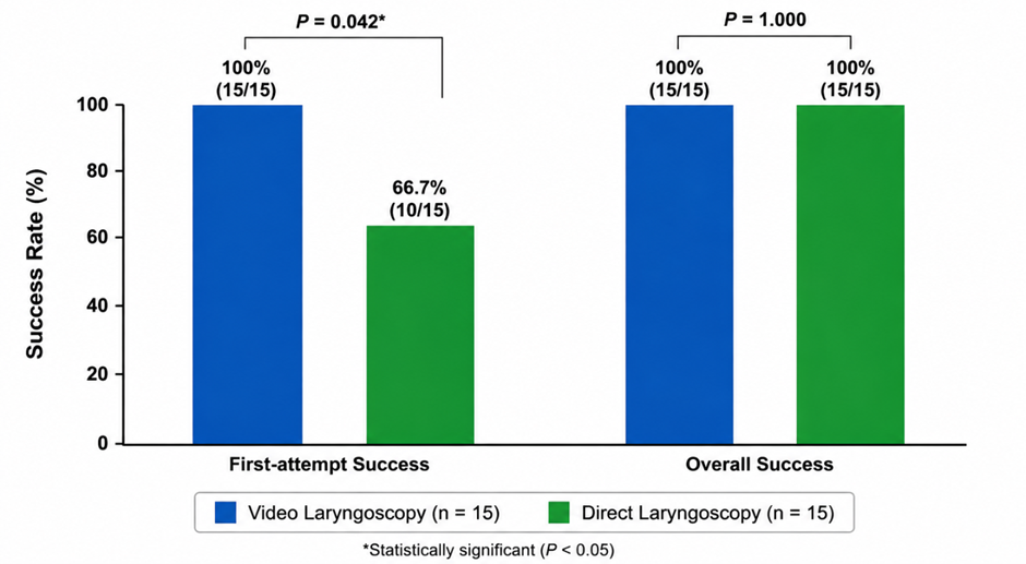
Figure 1. First-attempt and overall tracheal intubation success rates with video laryngoscopy and direct laryngoscopy in patients with anticipated difficult airways.
Figure 1 demonstrates the comparison of first-attempt and overall tracheal intubation success between video laryngoscopy and direct laryngoscopy in patients with anticipated difficult airways. First-attempt success was achieved in 100% (15/15) of patients in the video laryngoscopy group compared with 66.7% (10/15) in the direct laryngoscopy group, showing a statistically significant difference (P = 0.042). Overall intubation success was 100% in both groups. These findings indicate that video laryngoscopy improved first-pass intubation success while maintaining complete overall intubation success.
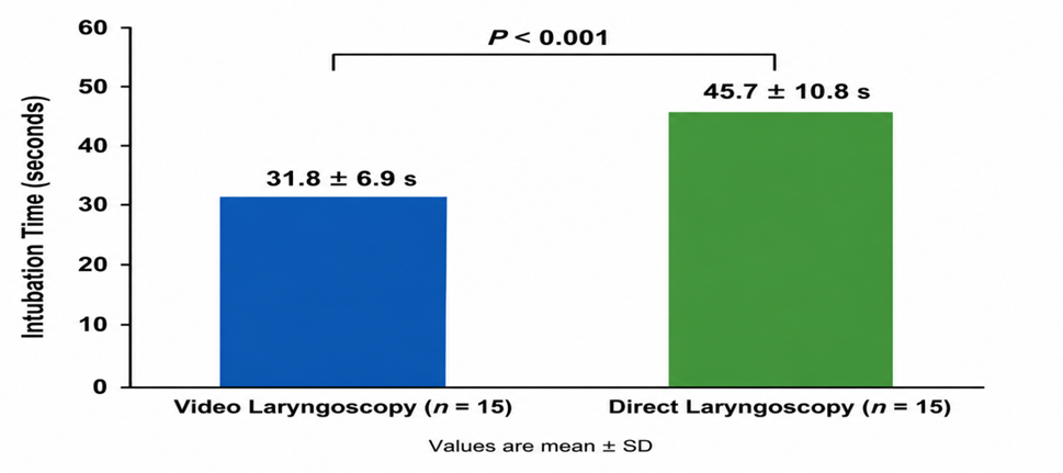
Figure 2. Comparison of mean tracheal intubation time between video laryngoscopy and direct laryngoscopy groups.
Figure 2 shows that the mean tracheal intubation time was significantly shorter with video laryngoscopy compared with direct laryngoscopy. The mean intubation time was 31.8 ± 6.9 seconds in the video laryngoscopy group and 45.7 ± 10.8 seconds in the direct laryngoscopy group. This difference was statistically significant (P < 0.001), indicating that video laryngoscopy facilitated faster successful tracheal intubation in patients with anticipated difficult airways.
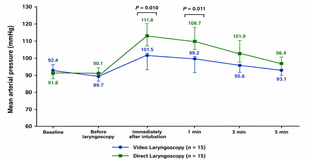
Figure 3. Serial changes in mean arterial pressure before and following tracheal intubation with video laryngoscopy and direct laryngoscopy.
Figure 3 demonstrates the serial changes in mean arterial pressure (MAP) before and after tracheal intubation in the two groups. Baseline MAP was comparable between video laryngoscopy and direct laryngoscopy groups (92.4 ± 8.6 vs 91.8 ± 8.9 mmHg). Following intubation, MAP increased in both groups; however, the rise was greater with direct laryngoscopy. Immediately after intubation, MAP was 101.5 ± 9.7 mmHg in the video laryngoscopy group compared with 111.8 ± 10.6 mmHg in the direct laryngoscopy group (P = 0.010). At 1 minute, the corresponding values were 99.2 ± 9.2 and 108.7 ± 9.9 mmHg, respectively (P = 0.011). Thereafter, MAP gradually declined toward baseline in both groups. These findings indicate a lower haemodynamic response with video laryngoscopy compared with conventional direct laryngoscopy.
DISCUSSION:
The present study compared video laryngoscopy with conventional direct laryngoscopy for tracheal intubation in patients with anticipated difficult airways. The principal findings were that video laryngoscopy was associated with a significantly higher first-attempt intubation success rate, shorter intubation time, superior visualization of the glottis, and a lower requirement for optimization manoeuvres. In addition, video laryngoscopy produced a comparatively attenuated haemodynamic response following tracheal intubation. Although airway-related complications were numerically less frequent with video laryngoscopy, the difference between the groups was not statistically significant. Collectively, these observations suggest that video laryngoscopy may provide important procedural advantages when difficulty with conventional laryngoscopy is anticipated.
An important finding of the present study was the significantly higher first-attempt success rate observed with video laryngoscopy. All 15 patients in the video laryngoscopy group were successfully intubated on the first attempt compared with 10 of 15 patients in the direct laryngoscopy group, corresponding to rates of 100% and 66.7%, respectively. This finding is consistent with the accumulating evidence supporting video-assisted laryngoscopy for difficult airway management. Hansel et al., in an updated Cochrane systematic review and meta-analysis, reported that video laryngoscopy was associated with fewer failed intubations and improved glottic visualization compared with direct laryngoscopy and also reduced certain complications, including hypoxaemia [9].
The clinical importance of improving first-pass success is particularly relevant in patients with anticipated difficult airways because repeated instrumentation increases the duration and complexity of airway management. Ruderman et al. evaluated more than 12,000 patients with predicted or anatomically difficult airways from the National Emergency Airway Registry and reported a first-pass success rate of 89.1% with video laryngoscopy compared with 77.7% using direct laryngoscopy [10]. They also observed lower rates of oesophageal intubation with video laryngoscopy. Although their investigation was conducted in an emergency department setting and involved a substantially larger population than the present study, the direction of effect is consistent with our findings.
More recent evidence further supports the benefit of video laryngoscopy in reducing repeated attempts. Ruetzler et al. conducted a large cluster-randomized clinical trial involving 8,429 surgical procedures and found that more than one attempt was required in only 1.7% of procedures assigned to hyperangulated video laryngoscopy compared with 7.6% with direct laryngoscopy. First-attempt success was therefore achieved in 98.3% and 92.4%, respectively, and intubation failure was also substantially less frequent with video laryngoscopy [12]. These data strengthen the clinical relevance of the present finding that none of the patients assigned to video laryngoscopy required a second attempt, whereas five patients undergoing direct laryngoscopy required multiple attempts.
A second important observation was the superior laryngeal view obtained with video laryngoscopy. Cormack-Lehane grade I visualization was achieved in 80.0% of patients in the video laryngoscopy group compared with only 33.3% in the direct laryngoscopy group. Furthermore, no patient undergoing video laryngoscopy demonstrated a grade III or IV view, whereas 26.7% of patients undergoing direct laryngoscopy had a grade III view. This advantage is anatomically plausible because video laryngoscopy permits visualization of the glottis without requiring the same degree of alignment of the oral, pharyngeal, and laryngeal axes necessary during conventional direct laryngoscopy. A network meta-analysis by de Carvalho et al., incorporating 179 randomized studies, demonstrated important differences in difficult laryngoscopy and glottic visualization among laryngoscope designs, with several video-laryngoscope configurations performing favourably compared with conventional Macintosh laryngoscopy [11].
Improved visualization probably contributed to the reduced requirement for optimization manoeuvres observed in the present study. Only 13.3% of patients in the video laryngoscopy group required an additional optimization manoeuvre compared with 60.0% in the direct laryngoscopy group. Similarly, the need for a stylet or bougie was significantly lower in the video laryngoscopy group. In an anticipated difficult airway, minimizing repositioning, external laryngeal manipulation, repeated blade insertion, and rescue adjuncts may simplify airway management and potentially reduce airway trauma. Nevertheless, the requirement for adjuncts is influenced by the specific video-laryngoscope design. Hyperangulated blades, for example, may provide an excellent view while still requiring a preshaped stylet to facilitate passage of the endotracheal tube through the glottis. This distinction is important when comparing studies involving different video-laryngoscope platforms.
The mean time required for tracheal intubation was also significantly shorter with video laryngoscopy in the present study, being 31.8 ± 6.9 seconds compared with 45.7 ± 10.8 seconds using direct laryngoscopy. The approximately 14-second reduction may be clinically relevant in a difficult airway because prolongation of airway manipulation can increase the risk of oxygen desaturation, particularly in patients with reduced physiological reserve. However, the literature regarding intubation time is less uniform than that regarding glottic visualization or first-pass success. De Carvalho et al. found statistically detectable differences between different laryngoscopes but concluded that differences in intubation time were generally not clinically important [11].
Some investigators have even reported longer intubation times with particular video-laryngoscope designs. Colak et al., while demonstrating reduced haemodynamic fluctuations with the McGRATH MAC video laryngoscope in elderly patients, found that intubation duration was longer with the video device [13]. Differences between these findings and the present study may be attributable to variation in video-laryngoscope geometry, airway difficulty, operator familiarity, use of stylets, definitions of intubation time, and the clinical environment. In the present study, the improved visualization and reduced requirement for optimization manoeuvres may have contributed to the shorter time to successful placement.
The present study also demonstrated differences in the haemodynamic response to laryngoscopy and intubation. Both techniques resulted in increases in heart rate and mean arterial pressure after intubation, which is an expected consequence of sympathetic stimulation during airway instrumentation. However, the increase was more pronounced following direct laryngoscopy. Heart rate was significantly lower in the video laryngoscopy group immediately after intubation and at 1 and 3 minutes, while mean arterial pressure was significantly lower immediately after intubation and at 1 minute. These differences subsequently diminished, with values approaching baseline in both groups by 5 minutes.
Similar findings have been reported in previous investigations. Colak et al. compared the McGRATH MAC video laryngoscope with the Macintosh direct laryngoscope in elderly patients and observed greater increases in heart rate and arterial pressure following direct laryngoscopy, concluding that video laryngoscopy reduced haemodynamic fluctuations associated with tracheal intubation [13]. Yokose et al. also reported that use of the McGRATH MAC video laryngoscope may reduce the incidence of hypertension following tracheal intubation compared with conventional Macintosh laryngoscopy [14].
The attenuated cardiovascular response observed with video laryngoscopy in the present study may potentially be explained by the reduced lifting force and reduced requirement for manipulation of upper-airway structures to establish a direct line of sight. This could decrease stimulation of the tongue base, epiglottis, and pharyngolaryngeal structures. However, haemodynamic responses to tracheal intubation are multifactorial and are influenced by the depth of anaesthesia, opioid administration, duration of airway manipulation, patient characteristics, and the operator's technique. Consequently, the apparent haemodynamic benefit should not be attributed exclusively to the device itself.
With regard to airway-related complications, adverse events were less frequent after video laryngoscopy, although statistical significance was not achieved. Any airway-related complication occurred in 13.3% of patients in the video laryngoscopy group compared with 46.7% in the direct laryngoscopy group. No patient in the video group developed an oxygen saturation below 90%, whereas transient desaturation was observed in three patients undergoing direct laryngoscopy. Visible airway trauma, postoperative sore throat, hoarseness, oesophageal intubation, and dental or oral injury were also numerically more frequent with direct laryngoscopy.
The absence of statistical significance for these outcomes should be interpreted cautiously because the study included only 15 patients in each group and was therefore inadequately powered to reliably detect differences in relatively infrequent complications. A large randomized trial by Ruetzler et al. similarly found no significant difference in airway or dental injury despite a clear reduction in repeated intubation attempts with video laryngoscopy [12]. Conversely, the updated systematic review by Hansel et al. suggested that video laryngoscopy reduces some complications, particularly hypoxaemia, while also reducing failed intubation and improving glottic visualization [9].
The lower number of complications observed in the video laryngoscopy group may also relate to the greater first-pass success rate. Repeated laryngoscopic attempts expose patients to repeated mechanical airway stimulation and prolong the period during which ventilation may be interrupted. Hasegawa et al. demonstrated an association between repeated intubation attempts and an increased frequency of adverse events during emergency airway management [15]. Thus, techniques that maximize the likelihood of successful placement during the initial attempt may have clinical benefits extending beyond procedural convenience.
The findings of this study therefore suggest that the principal advantage of video laryngoscopy in anticipated difficult airways lies not merely in obtaining a better image of the vocal cords but in translating improved visualization into clinically relevant outcomes, including greater first-pass success, fewer repeated attempts, reduced use of optimization manoeuvres, and potentially less physiological disturbance. Nevertheless, an excellent video view does not invariably guarantee uncomplicated passage of the endotracheal tube. Device geometry, stylet configuration, operator experience, secretions or blood obscuring the camera, and restricted mouth opening can all influence the performance of video laryngoscopy. Therefore, video laryngoscopy should be regarded as an important component of a comprehensive difficult-airway strategy rather than a replacement for appropriate preparation, alternative airway devices, and established rescue techniques.
Strengths and Limitations
An important strength of the present study was the direct comparison of two commonly used laryngoscopic approaches specifically in patients with predictors of difficult airway management. Standardized assessment of first-attempt success, intubation time, Cormack-Lehane grade, optimization requirements, haemodynamic variables, and complications allowed evaluation of both technical and patient-centred outcomes.
However, several limitations should be acknowledged. First, the small sample size of 30 patients limits statistical power, particularly for uncommon complications, and may produce imprecise estimates of treatment effect. Second, this was a single-centre study, which may limit generalizability to other institutions and operators with different levels of experience. Third, blinding of the anaesthesiologist performing intubation was impossible because the devices were visibly different. Fourth, the results may depend on the particular model and blade geometry of the video laryngoscope used and therefore should not automatically be generalized to all video-laryngoscopy systems. Fifth, individual variations in airway anatomy and anaesthetic depth could have influenced intubation conditions and haemodynamic responses. Finally, the study evaluated short-term procedural outcomes and was not designed to assess rare major airway complications or longer-term patient outcomes. Larger multicentre randomized studies would therefore be valuable to confirm these findings and better characterize clinically important adverse events.
Overall, the findings indicate that video laryngoscopy provides favourable intubating conditions in patients with anticipated difficult airways. The higher first-pass success rate, improved glottic visualization, shorter intubation time, reduced need for optimization manoeuvres, and attenuated immediate haemodynamic response observed in this study support the use of video laryngoscopy as an effective initial technique when difficult tracheal intubation is anticipated.
CONCLUSION:
Video laryngoscopy was more effective than conventional direct laryngoscopy for tracheal intubation in patients with anticipated difficult airways. It provided a higher first-attempt success rate, better glottic visualization, shorter intubation time, and reduced requirement for optimization manoeuvres. Video laryngoscopy was also associated with a lower haemodynamic response following intubation and fewer airway-related complications, although the difference in complications was not statistically significant. These findings support the use of video laryngoscopy as a preferred initial technique in patients with anticipated difficult airways. However, considering the small sample size and single-centre design, larger multicentre studies are required to confirm these findings and establish their broader clinical applicability.
REFERENCES:
1. Hypes C, Sakles J, Joshi R, Greenberg J, Natt B, Malo J, et al. Failure to achieve first attempt success at intubation using video laryngoscopy is associated with increased complications. Intern Emerg Med. 2017;12(8):1235-1243. doi:10.1007/s11739-016-1549-9.
2. Jungbauer A, Schumann M, Brunkhorst V, Börgers A, Groeben H. Expected difficult tracheal intubation: a prospective comparison of direct laryngoscopy and video laryngoscopy in 200 patients. Br J Anaesth. 2009;102(4):546-550. doi:10.1093/bja/aep013.
3. Aziz MF, Dillman D, Fu R, Brambrink AM. Comparative effectiveness of the C-MAC video laryngoscope versus direct laryngoscopy in the setting of the predicted difficult airway. Anesthesiology. 2012;116(3):629-636. doi:10.1097/ALN.0b013e318246ea34.
4. Serocki G, Neumann T, Scharf E, Dörges V, Cavus E. Indirect videolaryngoscopy with C-MAC D-Blade and GlideScope: a randomized, controlled comparison in patients with suspected difficult airways. Minerva Anestesiol. 2013;79(2):121-129.
5. Raja R, Gupta S, Mehta N, Attal P. First-pass success rate and number of attempts required for intubation in anticipated difficult airway: comparison between Macintosh and channeled King Vision video laryngoscopes. Anesth Essays Res. 2022;16(3):340-344. doi:10.4103/aer.aer_68_22.
6. Rosenstock CV, Thøgersen B, Afshari A, Christensen AL, Eriksen C, Gätke MR. Awake fiberoptic or awake video laryngoscopic tracheal intubation in patients with anticipated difficult airway management: a randomized clinical trial. Anesthesiology. 2012;116(6):1210-1216. doi:10.1097/ALN.0b013e318254d085.
7. Kriege M, Noppens RR, Turkstra T, Payne S, Kunitz O, Tzanova I, et al. A multicentre randomised controlled trial of the McGrath Mac videolaryngoscope versus conventional laryngoscopy. Anaesthesia. 2023. doi:10.1111/anae.15985.
8. Köhl V, Wünsch VA, Müller MC, Sasu PB, Dohrmann T, Peters T, et al. Hyperangulated vs. Macintosh videolaryngoscopy in adults with anticipated difficult airway management: a randomised controlled trial. Anaesthesia. 2024;79(9):957-966. doi:10.1111/anae.16326.
9. Hansel J, Rogers AM, Lewis SR, Cook TM, Smith AF. Videolaryngoscopy versus direct laryngoscopy for adults undergoing tracheal intubation: a Cochrane systematic review and meta-analysis update. Br J Anaesth. 2022;129(4):612-623. doi:10.1016/j.bja.2022.05.027.
10. Ruderman BT, Mali M, Kaji AH, Kilgo R, Watts S, Wells R, et al. Direct vs video laryngoscopy for difficult airway patients in the emergency department: a National Emergency Airway Registry study. West J Emerg Med. 2022;23(5):706-715. doi:10.5811/westjem.2022.6.55551.
11. de Carvalho CC, da Silva DM, Lemos VM, Dos Santos TGB, Agra IC, Pinto GM, et al. Videolaryngoscopy vs. direct Macintosh laryngoscopy in tracheal intubation in adults: a ranking systematic review and network meta-analysis. Anaesthesia. 2022;77(3):326-338. doi:10.1111/anae.15626.
12. Ruetzler K, Bustamante S, Schmidt MT, Almonacid-Cardenas F, Duncan A, Bauer A, et al. Video laryngoscopy vs direct laryngoscopy for endotracheal intubation in the operating room: a cluster randomized clinical trial. JAMA. 2024;331(15):1279-1286. doi:10.1001/jama.2024.0762.
13. Colak F, Ozgul U, Erdogan MA, Kayhan GE, Erdil FA, Colak C, et al. Comparison of hemodynamic responses and QTc intervals to tracheal intubation with the McGRATH MAC videolaryngoscope and the Macintosh direct laryngoscope in elderly patients. Kaohsiung J Med Sci. 2019;35(2):116-122. doi:10.1002/kjm2.12017.
14. Yokose M, Mihara T, Kuwahara S, Goto T. Effect of the McGRATH MAC video laryngoscope on hemodynamic response during tracheal intubation: a retrospective study. PLoS One. 2016;11(5). doi:10.1371/journal.pone.0155566.
15. Hasegawa K, Shigemitsu K, Hagiwara Y, Chiba T, Watase H, Brown CA 3rd, et al. Association between repeated intubation attempts and adverse events in emergency departments: an analysis of a multicenter prospective observational study. Ann Emerg Med. 2012;60(6):749-754.e2. doi:10.1016/j.annemergmed.2012.04.005.