Diagnostic Accuracy of Point-of-Care Ultrasound in the Rapid Assessment of Undifferentiated Shock in the Emergency Department
- Ram Gulhane , Resident, Department of Emergency Medicine, K. D. Medical College Hospital & Research Center, Mathura, Uttar Pradesh, India
- Ajay Kumar Agrawal , Professor, Department of Emergency Medicine, K. D. Medical College Hospital & Research Center, Mathura, Uttar Pradesh, India
- Naziya Hamid , Assistant Professor, Department of Emergency Medicine, K. D. Medical College Hospital & Research Center, Mathura, Uttar Pradesh, India
Article Information:
Abstract:
Background: Rapid identification of the cause of shock is essential for timely treatment in the emergency department. Point-of-care ultrasound (POCUS) provides immediate bedside information regarding cardiac function, intravascular volume, pulmonary abnormalities, and obstructive causes. Objective: To assess the diagnostic accuracy of POCUS in identifying the underlying etiology of undifferentiated shock in emergency department patients. Materials and Methods: A prospective observational study was conducted among 150 adults presenting with undifferentiated shock. A structured POCUS examination of the heart, lungs, inferior vena cava, and abdomen was performed during initial assessment. POCUS findings were compared with the final clinical diagnosis. Results: POCUS correctly classified 132 patients (88.0%). Sensitivity was highest for cardiogenic shock (100.0%), followed by hypovolemic shock (94.2%), while specificity was highest for hypovolemic shock (100.0%). Overall agreement with the final diagnosis was strong (κ=0.84, p<0.001). Conclusion: POCUS demonstrated high diagnostic accuracy and may facilitate rapid differentiation of shock etiologies during emergency assessment.
Keywords:
Article :
INTRODUCTION:
Shock is a life-threatening clinical syndrome characterized by inadequate tissue perfusion and impaired oxygen delivery. In the emergency department, patients with shock may initially present with nonspecific hypotension and signs of hypoperfusion, while the underlying cause may remain uncertain. Early differentiation of hypovolemic, cardiogenic, distributive, and obstructive shock is important because each category requires a different therapeutic approach [1].
Point-of-care ultrasound (POCUS) has increasingly become an important component of emergency assessment because it allows rapid bedside evaluation without requiring transfer of an unstable patient. A structured examination can assess ventricular function, right ventricular size, pericardial fluid, pulmonary abnormalities, inferior vena cava characteristics, abdominal free fluid, and other findings relevant to shock [2,3].
Several ultrasound-based protocols have been developed to improve the evaluation of undifferentiated hypotension. The SEARCH 8Es and RUSH approaches integrate cardiac, pulmonary, vascular, and abdominal findings to narrow the differential diagnosis [4,5]. Prospective studies have demonstrated that combining POCUS with clinical assessment can substantially improve diagnostic accuracy compared with clinical assessment alone [6].
However, diagnostic performance differs among shock subtypes, and distributive shock may be more difficult to identify because its ultrasound findings are often less specific. Therefore, the present study was undertaken to evaluate the diagnostic accuracy of structured POCUS in rapidly identifying the etiology of undifferentiated shock in patients presenting to the emergency department.
MATERIALS AND METHODS:
A prospective observational study was conducted in the Emergency Department of a tertiary care hospital over a period of 18 months. Adult patients presenting with undifferentiated shock requiring immediate hemodynamic assessment were evaluated. A total of 150 adult patients presenting with undifferentiated shock were included in the study.
Inclusion Criteria
· Adults aged ≥18 years.
· Patients presenting with undifferentiated shock or significant hypotension.
· Systolic blood pressure <90 mmHg or clinical evidence of tissue hypoperfusion.
· Patients requiring immediate hemodynamic assessment.
· Patients in whom POCUS could be performed during the initial emergency evaluation.
Exclusion Criteria
· Traumatic shock.
· Pregnancy.
· Patients with an established cause of shock before emergency assessment.
· Patients transferred after definitive resuscitation and diagnosis at another hospital.
· Patients in whom adequate ultrasound images could not be obtained.
· Patients unwilling to participate.
POCUS Examination
A structured bedside POCUS examination was performed during the initial assessment, including focused cardiac, lung, inferior vena cava, and abdominal evaluation. Findings were used to categorize shock as hypovolemic, cardiogenic, distributive, obstructive, or mixed.
Reference Standard
The final diagnosis was established from clinical assessment, relevant investigations, imaging, and clinical course and was used as the reference standard for comparison with POCUS findings.
Statistical Analysis
Categorical variables were expressed as frequency and percentage. Diagnostic accuracy was assessed using sensitivity, specificity, positive predictive value, and negative predictive value, while agreement was evaluated using Cohen’s kappa coefficient. A p-value <0.05 was considered statistically significant.
RESULTS:
A total of 150 patients with undifferentiated shock were included in the study. The majority were male, and tachycardia was the most frequent clinical finding. Hypovolemic shock was the most common final shock category, followed by distributive shock. POCUS demonstrated good diagnostic performance across the major shock subtypes, with particularly high sensitivity for cardiogenic and hypovolemic shock and high specificity for hypovolemic and cardiogenic shock.
Table 1: Demographic and Clinical Characteristics of the Study Population
|
Characteristic |
Number (n=150) |
Percentage |
|
Age <60 years |
92 |
61.3% |
|
Age ≥60 years |
58 |
38.7% |
|
Male |
94 |
62.7% |
|
Female |
56 |
37.3% |
|
SBP <70 mmHg |
43 |
28.7% |
|
SBP 70–89 mmHg |
107 |
71.3% |
|
Tachycardia |
119 |
79.3% |
|
Altered sensorium |
46 |
30.7% |
|
Raised lactate |
103 |
68.7% |
Most patients were younger than 60 years, and males constituted 62.7% of the study population. Tachycardia was observed in 79.3% of patients, while raised lactate was present in 68.7%, indicating significant circulatory compromise in a substantial proportion of patients.
Table 2: Distribution of Patients According to Final Etiology of Shock
|
Shock category |
Number (n=150) |
Percentage |
|
Hypovolemic |
52 |
34.7% |
|
Distributive |
48 |
32% |
|
Cardiogenic |
28 |
18.7% |
|
Obstructive |
12 |
8% |
|
Mixed |
10 |
6.7% |
Hypovolemic shock was the most common category, accounting for 34.7% of cases, followed by distributive shock in 32.0%. Cardiogenic shock represented 18.7% of cases, whereas obstructive and mixed shock were less frequent.
Table 3: Diagnostic Performance of POCUS According to Shock Etiology
|
Shock etiology |
Sensitivity (%) |
Specificity (%) |
PPV (%) |
NPV (%) |
|
Hypovolemic |
94.2% |
100% |
100% |
97% |
|
Distributive |
85.4% |
96.1% |
91.1% |
93.3% |
|
Cardiogenic |
100% |
98.4% |
93.3% |
100% |
|
Obstructive |
66.7% |
95.7% |
57.1% |
97.1% |
|
Mixed |
60% |
95.7% |
50% |
97.1% |
POCUS demonstrated the highest sensitivity for cardiogenic shock (100.0%), followed by hypovolemic shock (94.2%) and distributive shock (85.4%). Specificity was highest for hypovolemic shock (100.0%), followed by cardiogenic shock (98.4%). The negative predictive value was high across all shock categories, whereas positive predictive values were comparatively lower for obstructive and mixed shock.
Table 4: Agreement Between POCUS Assessment and Final Diagnosis
|
Diagnostic parameter |
Result |
|
Correctly classified by POCUS |
132 (88%) |
|
Incorrectly classified |
18 (12%) |
|
Cohen's kappa |
0.84 |
|
Overall agreement |
88% |
|
p-value |
<0.001 |
POCUS correctly classified 132 of 150 patients (88%). The Cohen's kappa coefficient of 0.84 indicated strong agreement between the initial POCUS assessment and the final diagnosis, with statistically significant agreement.
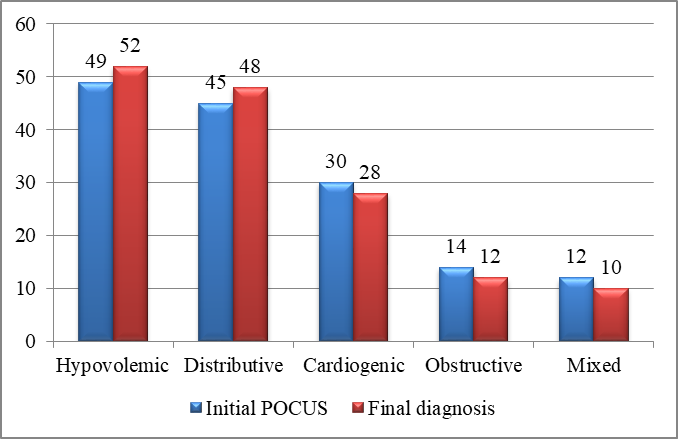
Figure 1: Comparison of initial POCUS impression with final diagnosis across different shock categories
Figure 1 shows a close correspondence between the initial POCUS impression and the final diagnosis across the different shock categories. Hypovolemic and distributive shock were the most frequent categories. The distribution of cardiogenic shock was also closely comparable between POCUS assessment and final diagnosis. Overall, the findings demonstrate good concordance between the initial POCUS impression and the final clinical diagnosis.
DISCUSSION:
The present study demonstrated that structured POCUS provided a high level of diagnostic agreement with the final diagnosis in patients presenting with undifferentiated shock. The overall correct classification rate was 88%, with a Cohen's kappa of 0.84. These findings support the value of bedside ultrasound as an adjunct to conventional clinical assessment in the early evaluation of shock.
In a prospective Indian study, Humbal et al. reported a kappa value of 0.86 for the RUSH protocol, with particularly high diagnostic accuracy for obstructive, cardiogenic, and hypovolemic shock [7]. Their findings are comparable with the present results, particularly the good diagnostic performance observed for hypovolemic, distributive, and cardiogenic shock.
The SHOC-ED randomized trial demonstrated that POCUS performed well in diagnosing shock subtypes, particularly as a rule-in tool, although its diagnostic performance was not significantly different from standard clinical assessment [8]. This suggests that POCUS should be considered an extension of clinical evaluation rather than a replacement for bedside examination.
Berg et al. reported in a systematic review that diagnostic accuracy improved when POCUS findings were combined with clinical information, with overall diagnostic accuracy increasing from approximately 45–60% with standard assessment to 80–89% when clinical assessment and POCUS were integrated [9]. This supports the combined approach used in the present study.
A meta-analysis by Yoshida et al. found good diagnostic performance of POCUS for different shock subtypes, with particularly high specificity and positive likelihood ratios for obstructive shock [10]. More recent pooled evidence involving 2,088 patients similarly showed high sensitivity and specificity for hypovolemic, cardiogenic, and obstructive shock, while performance was lower for distributive shock [11]. This pattern closely corresponds to the present findings.
Ultrasound-based assessment has also demonstrated good agreement with final clinical diagnosis in patients with undifferentiated shock. A prospective study evaluating ultrasound in critically ill patients reported good overall agreement between ultrasound-based shock classification and the final clinical diagnosis, with particularly strong diagnostic performance for obstructive shock [12]. These findings further support the use of bedside ultrasound as a rapid adjunct for identifying the underlying mechanism of shock.
Overall, the present findings indicate that structured POCUS can provide rapid and clinically relevant information during the initial assessment of undifferentiated shock. Its strongest diagnostic performance in the present study was observed for cardiogenic and hypovolemic shock, while distributive shock also showed good diagnostic accuracy. Obstructive and mixed shock demonstrated comparatively lower sensitivity and may require greater integration with clinical and laboratory findings.
CONCLUSION:
Structured point-of-care ultrasound demonstrated good diagnostic accuracy in the rapid assessment of undifferentiated shock. POCUS correctly classified 88.0% of patients and showed strong agreement with the final diagnosis. Diagnostic performance was particularly high for hypovolemic and cardiogenic shock, whereas distributive shock also showed good sensitivity. Obstructive and mixed shock demonstrated comparatively lower sensitivity. POCUS can therefore serve as a valuable bedside adjunct to clinical assessment, helping emergency physicians rapidly narrow the differential diagnosis and initiate appropriate management in patients with shock.
REFERENCES:
1. Atkinson P, Bowra J, Milne J, Lewis D, Lambert M, Jarman B, et al. International Federation for Emergency Medicine Consensus Statement: Sonography in hypotension and cardiac arrest (SHoC): An international consensus on the use of point of care ultrasound for undifferentiated hypotension and during cardiac arrest. CJEM. 2017 Nov;19(6):459-470. doi: 10.1017/cem.2016.394.
2. Sasmaz MI, Gungor F, Guven R, Akyol KC, Kozaci N, Kesapli M. Effect of Focused Bedside Ultrasonography in Hypotensive Patients on the Clinical Decision of Emergency Physicians. Emerg Med Int. 2017;2017:6248687. doi: 10.1155/2017/6248687.
3. Shokoohi H, Boniface KS, Zaragoza M, Pourmand A, Earls JP. Point-of-care ultrasound leads to diagnostic shifts in patients with undifferentiated hypotension. Am J Emerg Med. 2017 Dec;35(12):1984.e3-1984.e7. doi: 10.1016/j.ajem.2017.08.054.
4. Ahn JH, Jeon J, Toh HC, Noble VE, Kim JS, Kim YS, et al. SEARCH 8Es: A novel point of care ultrasound protocol for patients with chest pain, dyspnea or symptomatic hypotension in the emergency department. PLoS One. 2017 Mar 29;12(3):e0174581. doi: 10.1371/journal.pone.0174581.
5. Stickles SP, Carpenter CR, Gekle R, Kraus CK, Scoville C, Theodoro D, et al. The diagnostic accuracy of a point-of-care ultrasound protocol for shock etiology: A systematic review and meta-analysis. CJEM. 2019 May;21(3):406-417. doi: 10.1017/cem.2018.498.
6. Javali RH, Loganathan A, Srinivasarangan M, Patil A, Siddappa GB, Satyanarayana N, et al. Reliability of Emergency Department Diagnosis in Identifying the Etiology of Nontraumatic Undifferentiated Hypotension. Indian J Crit Care Med. 2020 May;24(5):313-320. doi: 10.5005/jp-journals-10071-23429.
7. Rahulkumar HH, Bhavin PR, Shreyas KP, Krunalkumar HP, Atulkumar S, Bansari C. Utility of Point-of-Care Ultrasound in Differentiating Causes of Shock in Resource-Limited Setup. J Emerg Trauma Shock. 2019 Jan-Mar;12(1):10-17. doi: 10.4103/JETS.JETS_61_18.
8. Peach M, Milne J, Diegelmann L, Lamprecht H, Stander M, Lussier D, et al. Does point-of-care ultrasonography improve diagnostic accuracy in emergency department patients with undifferentiated hypotension? An international randomized controlled trial from the SHOC-ED investigators. CJEM. 2023 Jan;25(1):48-56. doi: 10.1007/s43678-022-00431-9.
9. Berg I, Walpot K, Lamprecht H, Valois M, Lanctôt JF, Srour N, et al. A Systemic Review on the Diagnostic Accuracy of Point-of-Care Ultrasound in Patients With Undifferentiated Shock in the Emergency Department. Cureus. 2022 Mar 15;14(3):e23188. doi: 10.7759/cureus.23188.
10. Yoshida T, Yoshida T, Noma H, Nomura T, Suzuki A, Mihara T. Diagnostic accuracy of point-of-care ultrasound for shock: a systematic review and meta-analysis. Crit Care. 2023 May 25;27(1):200. doi: 10.1186/s13054-023-04495-6.
11. Basmaji J, Tang JE, Arntfield R, Desai K, Ball IM, Fiorini K, et al. The diagnostic accuracy of point-of-care ultrasound in shock: a systematic review and meta-analysis. Can J Anaesth. 2025 Jul;72(7):1118-1129. English. doi: 10.1007/s12630-025-02997-1.
12. Vaidya T, D'costa P, Pande S. Role of Ultrasound in Evaluation of Undifferentiated Shock in ICU Settings. J Assoc Physicians India. 2018 Aug;66(8):13-17.