Clinical Spectrum, Etiological Patterns, Management Strategies, and Factors Associated with Complications in Liver Abscess: A Cross-Sectional Study from Central India.
- Arpita Chouhan , MBBS, MS General Surgery Resident ,Sri Aurobindo Institute of Medical Sciences Indore MP.
- Harshita Gupta , MBBS, MS General Surgery Resident, Sri Aurobindo Institute of Medical Sciences Indore MP.
- Manoj Kela , MS, Professor, HOD and Head of Unit, Department of General Surgery, Sri Aurobindo Institute of Medical Sciences Indore..
Article Information:
Abstract:
Background: Liver abscess remains an important cause of morbidity in tropical regions. Amoebic and pyogenic liver abscesses may have overlapping clinical features but differ in their etiological and therapeutic implications. This study evaluated the clinical spectrum, etiological patterns, management strategies, and factors associated with complications among patients with liver abscess in Central India. Methods: This hospital-based cross-sectional study included 100 adults with clinically and radiologically confirmed liver abscess treated at a tertiary-care centre in Indore between June 2023 and November 2024. Demographic, clinical, laboratory, microbiological, radiological, management, and outcome data were recorded. Associations between selected variables and complications were assessed using Pearson’s chi-square test, with P<0.05 considered statistically significant. Results: Most patients were male (84%), and the largest age group was 41–50 years (28%). Abdominal pain (100%), fever (82%), and jaundice (37%) were the principal presenting features. Amoebic, pyogenic, and tubercular liver abscesses accounted for 63%, 33%, and 4% of cases, respectively. Leukocytosis occurred in 75%, hyperbilirubinaemia in 38%, elevated INR in 41%, and hypoalbuminaemia in 89%. Most cultures were sterile (71%); Klebsiella species and Escherichia coli were the commonest isolates. Right-lobe involvement (61%) and solitary abscesses (65%) predominated. Percutaneous catheter drainage was the most frequently reported intervention. Complications occurred in 37%, with recurrence being the most common. Diabetes mellitus (P=0.006), abscess etiology (P=0.028), and hypoalbuminaemia (P=0.042) were significantly associated with complications. Conclusion: Liver abscess predominantly affected middle-aged men and was most frequently amoebic. Early diagnosis, image-guided drainage, and closer monitoring of patients with diabetes, hypoalbuminaemia, or atypical etiology may reduce complications and recurrence.
Keywords:
Article :
INTRODUCTION:
Liver abscess is a potentially life-threatening infection characterized by a localized collection of pus within the hepatic parenchyma. Despite substantial improvements in diagnostic imaging, antimicrobial therapy, interventional radiology, and critical care, liver abscess continues to cause considerable morbidity, prolonged hospitalization, and occasional mortality, particularly when diagnosis or drainage is delayed. The condition encompasses a heterogeneous group of infections that differ in their causative organisms, routes of hepatic invasion, clinical manifestations, management requirements, and outcomes. Amoebic liver abscess and pyogenic liver abscess are the two major forms encountered in clinical practice, although fungal, tubercular, and other parasitic abscesses may occur in selected immunocompromised or high-risk populations [1,2].
Amoebic liver abscess is the most common extraintestinal manifestation of infection with Entamoebahistolytica. The organism is transmitted through ingestion of food or water contaminated with mature cysts. Following intestinal colonization, invasive trophozoites may penetrate the colonic mucosa and reach the liver through the portal circulation. Amoebic liver abscess remains particularly important in tropical and subtropical regions where inadequate sanitation, unsafe drinking water, overcrowding, malnutrition, and poor socioeconomic conditions facilitate transmission [1]. In contrast, pyogenic liver abscess develops because of bacterial infection through the biliary tract, portal venous circulation, hepatic arterial circulation, direct extension from a nearby infective focus, penetrating trauma, or secondary infection of an existing hepatic lesion. Biliary tract disease and obstruction are now among the leading causes of pyogenic abscess, while Escherichia coli, Klebsiellapneumoniae, enterococci, streptococci, and anaerobic bacteria are frequently identified pathogens [2].
The epidemiological pattern of liver abscess in India differs from that reported in many developed countries. Large Indian cohorts have demonstrated that amoebic liver abscess remains the predominant etiological type, especially among middle-aged men. Chronic alcohol consumption, diabetes mellitus, poor nutritional status, and delayed access to medical services are commonly observed associated factors. In a large Indian series of 1,630 adult patients, amoebic liver abscess accounted for most cases, whereas pyogenic abscess was more frequently associated with older age, biliary disease, multiple lesions, bilobar involvement, and systemic complications [3]. A study from Central India similarly found a marked male predominance, frequent chronic alcohol use, and a high occurrence of solitary right-lobe abscesses. Abdominal pain, fever, tachycardia, hepatomegaly, leukocytosis, anemia, hypoalbuminemia, and electrolyte disturbances were common clinical and laboratory findings [4]. These observations emphasize the influence of regional socioeconomic, behavioural, environmental, and healthcare-related factors on the presentation of liver abscess.
The clinical spectrum of liver abscess is broad and often nonspecific. Fever and right upper-quadrant abdominal pain are the most frequent symptoms, but patients may also present with anorexia, weight loss, nausea, vomiting, cough, dyspnoea, diarrhea, jaundice, abdominal distension, or altered sensorium. Physical examination may reveal hepatic tenderness, hepatomegaly, pallor, icterus, pleural effusion, or features of sepsis. Laboratory findings commonly include neutrophilicleukocytosis, elevated alkaline phosphatase, hypoalbuminemia, hyperbilirubinemia, anemia, and deranged coagulation parameters. However, none of these findings independently distinguishes amoebic from pyogenic disease [5,6]. Ultrasonography is generally the initial imaging investigation because it is widely available, inexpensive, and useful for identifying the number, size, location, and internal characteristics of an abscess. Contrast-enhanced computed tomography offers greater sensitivity for small, multiple, multiloculated, or deeply situated lesions and may demonstrate rupture, vascular thrombosis, biliary communication, and adjacent organ involvement.
Complications may develop through local extension, rupture, vascular involvement, biliary communication, secondary bacterial infection, or systemic dissemination. Recognized complications include intraperitoneal rupture, pleuropulmonary rupture, pericardial extension, biliary rupture, portal or hepatic venous thrombosis, inferior vena cava compression, acute kidney injury, septic shock, encephalopathy, disseminated infection, and multiorgan dysfunction. Larger abscesses, bilobar or multiple lesions, left-lobe location, pyogenic etiology, jaundice, diabetes mellitus, liver cirrhosis, hypoalbuminemia, elevated bilirubin, acute kidney injury, and delayed treatment have been associated with complicated disease or poor outcomes [3]. A prospective study of amoebic liver abscess identified hyperbilirubinemia, severe hypoalbuminemia, multiple abscesses, large abscess volume, and encephalopathy as important indicators of an adverse prognosis [7]. Early recognition of these factors can support timely escalation of monitoring, drainage, and intensive care.
Management depends on the suspected etiology, abscess size and location, clinical severity, microbiological findings, response to initial therapy, and presence of complications. Amoebic liver abscess is primarily treated with a tissue-active nitroimidazole, followed by a luminal amoebicide to eradicate intestinal colonization. Uncomplicated, small amoebic abscesses often respond to medical therapy alone. Pyogenic liver abscess generally requires broad-spectrum intravenous antibiotics covering enteric Gram-negative organisms, streptococci, enterococci, and anaerobes, followed by modification according to blood or pus culture results [8]. Image-guided percutaneous needle aspiration or catheter drainage is indicated for large abscesses, poor clinical response, diagnostic uncertainty, impending rupture, left-lobe lesions, multiloculated collections, or pyogenic abscesses requiring source control [9]. Percutaneous catheter drainage may provide more effective continuous evacuation than repeated needle aspiration, particularly in large or thick-walled collections [10]. Nevertheless, routine aspiration does not appear necessary for every uncomplicated amoebic abscess, and intervention should be individualized according to clinical and radiological risk features [11]. Surgical drainage is now reserved mainly for freely ruptured abscesses, failed percutaneous drainage, complex multiloculated disease, associated intra-abdominal pathology, or situations requiring definitive surgical source control [12].
Although several studies have described liver abscess in different parts of India, variations in etiology, comorbidities, microbiological profiles, referral practices, and access to image-guided procedures may influence outcomes across regions. Contemporary information from Central India regarding the combined clinical spectrum, etiological distribution, management practices, and factors associated with complications remains comparatively limited. Therefore, the present cross-sectional study was undertaken to describe the demographic and clinical characteristics of patients with liver abscess, determine the principal etiological patterns, evaluate the management strategies employed, and identify clinical, biochemical, and radiological factors associated with the development of complications. Such evidence may help improve early risk stratification, guide appropriate use of drainage procedures, and reduce preventable morbidity and mortality.
MATERIALS AND METHODS:
Study design and setting
This hospital-based, observational cross-sectional study was conducted in the Department of General Surgery, Sri Aurobindo Medical College and Postgraduate Institute, Indore, Madhya Pradesh, India. The study was performed over 18 months, from June 2023 to November 2024. Patients were recruited from the General Surgery outpatient department, casualty and emergency services, inpatient wards, and referrals received from other healthcare facilities.
Study population and sampling
The study population comprised adult patients diagnosed with liver abscess during the study period. A consecutive sampling approach was followed, in which all eligible patients presenting to the study centre and providing written informed consent were considered for enrolment. A total of 100 patients were included. The target sample was determined from the expected institutional caseload of approximately seven to eight eligible liver abscess cases per month during the planned study period.
Patients aged 18 years or older with a clinically and radiologically confirmed diagnosis of liver abscess were eligible. Both bacterial and parasitic liver abscesses were included. Patients were enrolled irrespective of whether the abscess was evolving, liquefied or ruptured and whether peritonitis was present. Patients with a history of a previously treated liver abscess, those with a traumatic liver abscess, and those who declined voluntary written informed consent were excluded.
Diagnostic assessment
The diagnosis of liver abscess was established using a combination of clinical presentation and imaging findings on abdominal ultrasonography or contrast-enhanced computed tomography. At enrolment, each patient underwent a detailed clinical assessment using a structured study proforma.
The demographic variables recorded included age, sex and occupation. Presenting complaints, duration and pattern of symptoms, previous surgical history and any previous episode of a similar illness were documented. Particular attention was given to potential predisposing factors and comorbidities, including alcohol consumption, diabetes mellitus, immunodeficiency states, biliary tract disease, previous amoebic dysentery and jaundice.
A complete general and systemic examination was performed. The clinical assessment included evaluation of vital status and signs of systemic illness. Respiratory examination was undertaken to identify possible pleuropulmonary involvement. Abdominal examination included inspection, palpation and percussion, with assessment for tenderness, guarding, hepatomegaly, abdominal distension and signs of peritonitis. A per-rectal examination was performed when clinically indicated.
Laboratory and radiological investigations
Baseline investigations included a complete haemogram, total leukocyte count, liver-function tests, serum bilirubin, serum albumin, prothrombin time and international normalized ratio. Serological testing for amoebic antigen was performed at presentation. These parameters were used to characterize the systemic inflammatory response, hepatic dysfunction, nutritional status and coagulation abnormalities.
Patients undergoing aspiration or drainage had the aspirated material evaluated microbiologically, including culture and antimicrobial-susceptibility testing. The final etiological categories recorded in the study were amoebic, pyogenic and tubercular liver abscess. Microbiological findings were documented as sterile culture or according to the isolated organism.
Radiological evaluation included chest radiography and ultrasonography of the abdomen and pelvis. Contrast-enhanced computed tomography of the abdomen was performed as part of the diagnostic assessment, particularly to define the anatomical extent of disease. Imaging findings were used to document the affected hepatic lobe, number of abscess cavities, degree of liquefaction and evidence of rupture or involvement of adjacent structures. Abdominal ultrasonography was repeated on the third day after initiation of treatment to assess the residual abscess cavity and early treatment response.
Management protocol
Treatment was individualized according to the clinical condition of the patient and the site, size, number and radiological characteristics of the abscess. The management modalities were classified as medical management, percutaneous needle aspiration, percutaneous catheter drainage and surgical drainage.
Patients considered suitable for conservative treatment received medical management and supportive care according to the treating surgical team’s institutional practice. Image-guided intervention was performed in patients requiring drainage. Percutaneous needle aspiration was used for selected drainable collections, whereas percutaneous catheter drainage was undertaken when continuous evacuation of the abscess cavity was considered necessary.
Open surgical drainage was reserved for patients in whom operative management was clinically required. Patients who developed rupture into a serosal cavity, peritonitis or another complication requiring urgent source control were taken for surgery. Separate written informed consent was obtained before every invasive or operative procedure. The thesis did not specify uniform antibiotic regimens, catheter sizes or predetermined abscess-size thresholds; therefore, these were not introduced into the manuscript methodology.
Follow-up and outcome assessment
All enrolled patients were monitored daily during hospitalization. Clinical response was assessed through changes in symptoms, physical findings and the development or resolution of systemic or abdominal complications. Liver-function tests were monitored during follow-up, and repeat ultrasonography was used to assess the residual collection. The principal study outcomes were the clinical spectrum, etiological distribution, radiological pattern, microbiological profile, management modality and final clinical outcome. Treatment effectiveness was evaluated according to the patient’s clinical response following medical, image-guided or surgical management. For outcome analysis, patients were categorized as having no complications or having developed a complication. The complications documented in the study included recurrence, typhlitis, pancreatitis and sinus-tract formation. Rupture of the liver abscess and extension into a serosal cavity were additionally monitored as clinically important complications requiring urgent intervention. Associations between the final outcome and age, sex, alcohol use, diabetes mellitus, abscess type, leukocyte count, bilirubin, international normalized ratio and serum albumin were evaluated.
Statistical analysis
Data were entered and organized using Microsoft Excel 2016 and analysed using IBM SPSS Statistics, version 22. Categorical variables were summarized as frequencies and percentages. The clinical characteristics, etiological patterns, laboratory parameters, radiological findings, management modalities and outcomes were presented using tables and graphical representations. Associations between categorical independent variables and the occurrence of complications were examined using Pearson’s chi-square test. A two-sided P value of less than 0.05 was considered statistically significant. The analysis was complete-case based, as the thesis did not describe data imputation or another method for handling missing observations.
RESULTS:
A total of 100 patients diagnosed with liver abscess were included. The largest proportion belonged to the 41–50-year age group, accounting for 28 (28.0%) patients, followed by 21 (21.0%) aged 31–40 years and 20 (20.0%) aged 51–60 years. There was a marked male predominance, with 84 (84.0%) male and 16 (16.0%) female patients. Abdominal pain was reported by all 100 patients. Fever was the second most frequent presenting feature, occurring in 82 (82.0%) patients, followed by jaundice in 37 (37.0%). Vomiting, breathlessness, loss of appetite, diarrhoea and abdominal distension were comparatively uncommon. Alcohol consumption was documented in 28 (28.0%) patients, while 25 (25.0%) had diabetes mellitus.
Table 1. Demographic characteristics, presenting complaints and personal history of the study population
|
Characteristic |
Category |
Number, n |
Percentage |
|
Age group |
21–30 years |
13 |
13.0 |
|
31–40 years |
21 |
21.0 |
|
|
41–50 years |
28 |
28.0 |
|
|
51–60 years |
20 |
20.0 |
|
|
61–70 years |
12 |
12.0 |
|
|
>70 years |
6 |
6.0 |
|
|
Sex |
Male |
84 |
84.0 |
|
Female |
16 |
16.0 |
|
|
Presenting complaints |
Abdominal pain |
100 |
100.0 |
|
Fever |
82 |
82.0 |
|
|
Jaundice |
37 |
37.0 |
|
|
Vomiting |
9 |
9.0 |
|
|
Breathlessness |
5 |
5.0 |
|
|
Loss of appetite |
4 |
4.0 |
|
|
Diarrhoea |
2 |
2.0 |
|
|
Abdominal distension |
1 |
1.0 |
|
|
Personal history |
Alcohol consumption |
28 |
28.0 |
|
Diabetes mellitus |
25 |
25.0 |
|
|
Other history |
1 |
1.0 |
Amoebic liver abscess was the most common etiological category, identified in 63 (63.0%) patients. Pyogenic abscess was diagnosed in 33 (33.0%), while 4 (4.0%) patients were classified as having tubercular liver abscess.
Leukocytosis was present in 75 (75.0%) patients. Hyperbilirubinaemia was observed in 38 (38.0%), and an elevated international normalized ratio was found in 41 (41.0%). Hypoalbuminaemia was particularly frequent and was recorded in 89 (89.0%) patients.
Table 2. Etiological classification and laboratory profile
|
Variable |
Category |
Number, n |
Percentage |
|
Type of liver abscess |
Amoebic |
63 |
63.0 |
|
Pyogenic |
33 |
33.0 |
|
|
Tubercular |
4 |
4.0 |
|
|
White blood cell count |
Elevated |
75 |
75.0 |
|
Normal |
25 |
25.0 |
|
|
Low |
0 |
0.0 |
|
|
Serum bilirubin |
Elevated |
38 |
38.0 |
|
Normal |
61 |
61.0 |
|
|
Low |
1 |
1.0 |
|
|
International normalized ratio |
Elevated |
41 |
41.0 |
|
Normal |
58 |
58.0 |
|
|
Low |
1 |
1.0 |
|
|
Serum albumin |
Low |
89 |
89.0 |
|
Normal |
11 |
11.0 |
|
|
Elevated |
0 |
0.0 |
Culture examination was sterile in 71 (71.0%) patients. Among the reported positive findings, Klebsiella species and Escherichia coli were each isolated in 8 (8.0%) patients. Mycobacterium tuberculosis was reported in 5 (5.0%), gram-positive cocci in 3 (3.0%) and gram-negative bacilli in 2 (2.0%). Enterococcus and Acinetobacter were each identified in one patient.
The right hepatic lobe was the most frequently affected anatomical site, being involved alone in 61 (61.0%) patients. Both hepatic lobes were affected in 23 (23.0%), and isolated left-lobe involvement was recorded in 14 (14.0%). A single abscess cavity was present in 65 (65.0%) patients, whereas 35 (35.0%) had multiple cavities.
Table 3. Microbiological findings and radiological characteristics
|
Variable |
Category |
Number, n |
Percentage |
|
Culture finding |
Sterile culture |
71 |
71.0 |
|
Klebsiella species |
8 |
8.0 |
|
|
Escherichia coli |
8 |
8.0 |
|
|
Mycobacterium tuberculosis |
5 |
5.0 |
|
|
Gram-positive cocci |
3 |
3.0 |
|
|
Gram-negative bacilli |
2 |
2.0 |
|
|
Enterococcus species |
1 |
1.0 |
|
|
Acinetobacter species |
1 |
1.0 |
|
|
Occasional pus cells |
1 |
1.0 |
|
|
Anatomical location |
Right lobe |
61 |
61.0 |
|
Both lobes |
23 |
23.0 |
|
|
Left lobe |
14 |
14.0 |
|
|
Caudate lobe |
1 |
1.0 |
|
|
Left and caudate lobes |
1 |
1.0 |
|
|
Number of cavities |
Single |
65 |
65.0 |
|
Multiple |
35 |
35.0 |
Percutaneous catheter drainage was the most frequently reported treatment modality and was used in 41 (41.0%) patients. Medical management alone was reported in 21 (21.0%), while percutaneous needle aspiration and surgical intervention were each reported in 20 (20.0%) patients.
Overall, 63 (63.0%) patients experienced no complication. Recurrence was the most frequent adverse outcome, occurring in 24 (24.0%) patients. Typhlitis developed in 11 (11.0%), while pancreatitis and sinus-tract formation were each reported in one patient.
Table 4. Management strategies and final clinical outcomes
|
Variable |
Category |
Number, n |
Percentage |
|
Management modality |
Percutaneous catheter drainage |
41 |
41.0 |
|
Medical management |
21 |
21.0 |
|
|
Percutaneous needle aspiration |
20 |
20.0 |
|
|
Surgical intervention |
20 |
20.0 |
|
|
Final outcome |
No complication |
63 |
63.0 |
|
Recurrence |
24 |
24.0 |
|
|
Typhlitis |
11 |
11.0 |
|
|
Pancreatitis |
1 |
1.0 |
|
|
Sinus-tract formation |
1 |
1.0 |
Abdominal pain was universally present across amoebic, pyogenic and tubercular abscess groups. Fever occurred in 50 of 63 (79.4%) patients with amoebic abscess, 29 of 33 (87.9%) with pyogenic abscess and 3 of 4 (75.0%) with tubercular abscess. The association between fever and etiological category was not statistically significant (χ² = 1.202, df = 2, P = 0.548).
Jaundice was more frequent among patients with pyogenic liver abscess, occurring in 17 of 33 (51.5%), compared with 19 of 63 (30.2%) amoebic and 1 of 4 (25.0%) tubercular cases. However, this difference was also not statistically significant (χ² = 4.495, df = 2, P = 0.106).
Table 5. Association between major clinical features and type of liver abscess
|
Clinical feature |
Amoebic, n = 63 |
Pyogenic, n = 33 |
Tubercular, n = 4 |
Total, n = 100 |
χ², df |
P value |
|
Pain present |
63 (100.0) |
33 (100.0) |
4 (100.0) |
100 (100.0) |
Not applicable |
Not applicable |
|
Fever present |
50 (79.4) |
29 (87.9) |
3 (75.0) |
82 (82.0) |
1.202, 2 |
0.548 |
|
Fever absent |
13 (20.6) |
4 (12.1) |
1 (25.0) |
18 (18.0) |
||
|
Jaundice present |
19 (30.2) |
17 (51.5) |
1 (25.0) |
37 (37.0) |
4.495, 2 |
0.106 |
|
Jaundice absent |
44 (69.8) |
16 (48.5) |
3 (75.0) |
63 (63.0) |
Complications occurred in 37 (37.0%) patients. Age and sex were not significantly associated with complications. The highest age-specific complication rate was observed among patients aged 41–50 years, in whom 13 of 28 (46.4%) developed complications, but the overall association with age was not significant (P = 0.834). Complications occurred in 7 of 16 (43.8%) female and 30 of 84 (35.7%) male patients (P = 0.542).
Alcohol consumption was not significantly associated with outcome. Complications were observed in 9 of 28 (32.1%) alcohol users compared with 28 of 72 (38.9%) non-users (P = 0.530).
Diabetes mellitus was significantly associated with complications. Among diabetic patients, 15 of 25 (60.0%) developed complications compared with 22 of 75 (29.3%) non-diabetic patients (χ² = 7.565, P = 0.006).
Etiological category was also significantly associated with outcome (χ² = 7.118, P = 0.028). All four patients classified as having tubercular liver abscess developed complications. Complications occurred in 22 of 63 (34.9%) patients with amoebic abscess and 11 of 33 (33.3%) with pyogenic abscess.
White blood cell count, bilirubin and INR were not significantly associated with complications. Nevertheless, patients with elevated bilirubin showed a numerically higher complication rate than those with normal bilirubin levels, at 50.0% versus 29.5%, respectively.
Hypoalbuminaemia was significantly associated with complications. Complications occurred in 36 of 89 (40.4%) patients with low serum albumin compared with 1 of 11 (9.1%) patients with normal albumin levels (χ² = 4.130, P = 0.042).
Table 6. Factors associated with the development of complications
|
Variable |
Category |
No complication, n (%) |
Complication, n (%) |
χ², df |
P value |
|
Age group |
21–30 years |
9 (69.2) |
4 (30.8) |
2.105, 5 |
0.834 |
|
31–40 years |
15 (71.4) |
6 (28.6) |
|||
|
41–50 years |
15 (53.6) |
13 (46.4) |
|||
|
51–60 years |
13 (65.0) |
7 (35.0) |
|||
|
61–70 years |
7 (58.3) |
5 (41.7) |
|||
|
>70 years |
4 (66.7) |
2 (33.3) |
|||
|
Sex |
Female |
9 (56.3) |
7 (43.8) |
0.372, 1 |
0.542 |
|
Male |
54 (64.3) |
30 (35.7) |
|||
|
Alcohol consumption |
Absent |
44 (61.1) |
28 (38.9) |
0.394, 1 |
0.530 |
|
Present |
19 (67.9) |
9 (32.1) |
|||
|
Diabetes mellitus |
Absent |
53 (70.7) |
22 (29.3) |
7.565, 1 |
0.006 |
|
Present |
10 (40.0) |
15 (60.0) |
|||
|
Abscess etiology |
Amoebic |
41 (65.1) |
22 (34.9) |
7.118, 2 |
0.028 |
|
Pyogenic |
22 (66.7) |
11 (33.3) |
|||
|
Tubercular |
0 (0.0) |
4 (100.0) |
|||
|
White blood cell count |
Elevated |
48 (64.0) |
27 (36.0) |
0.129, 1 |
0.720 |
|
Normal |
15 (60.0) |
10 (40.0) |
|||
|
Serum bilirubin |
Elevated |
19 (50.0) |
19 (50.0) |
4.811, 2 |
0.090 |
|
Normal |
43 (70.5) |
18 (29.5) |
|||
|
Low |
1 (100.0) |
0 (0.0) |
|||
|
INR |
Elevated |
23 (56.1) |
18 (43.9) |
1.873, 2 |
0.392 |
|
Normal |
39 (67.2) |
19 (32.8) |
|||
|
Low |
1 (100.0) |
0 (0.0) |
|||
|
Serum albumin |
Low |
53 (59.6) |
36 (40.4) |
4.130, 1 |
0.042 |
|
Normal |
10 (90.9) |
1 (9.1) |
The original association table labelled the group containing 89 patients as having “high” albumin. However, the descriptive albumin table and accompanying thesis narrative identify these 89 patients as having low albumin. The consolidated table therefore reports this group as low albumin while preserving the original cell frequencies and statistical result.
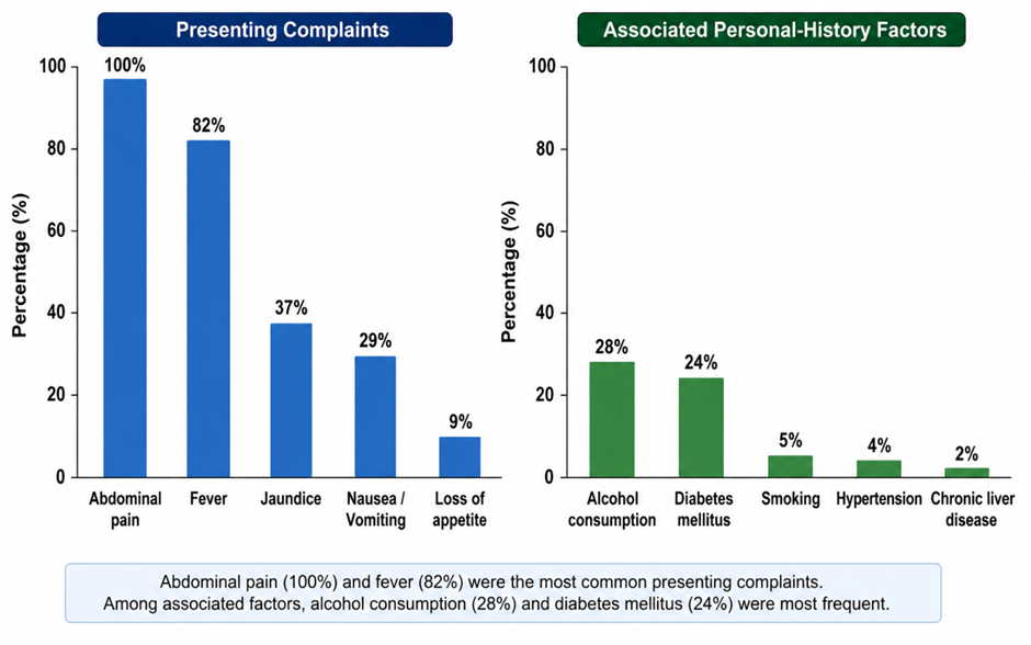 Figure 1. Frequency of presenting complaints and associated personal-history factors among patients with liver abscess
Figure 1 illustrates the frequency of presenting complaints and associated personal-history factors among the 100 patients diagnosed with liver abscess. Abdominal pain was the universal presenting complaint and was reported by all patients (100%), followed by fever in 82% and jaundice in 37%. Less frequent symptoms included vomiting (9%), breathlessness (5%), loss of appetite (4%), diarrhoea (2%), and abdominal distension (1%). Among the associated personal-history factors, alcohol consumption was the most common and was documented in 28% of patients, followed by diabetes mellitus in 25%. Other associated factors were reported in only 1% of the study population. Overall, the figure demonstrates that abdominal pain and fever constituted the dominant clinical presentation, whereas alcohol consumption and diabetes mellitus were the principal associated factors.
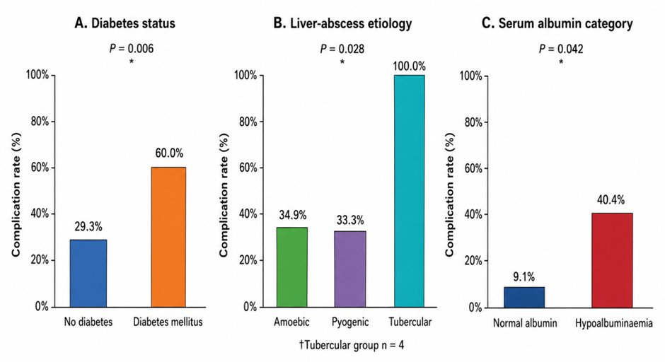
Figure 2. Complication rates according to diabetes status, liver-abscess etiology and serum albumin category
Figure 2presents the proportion of patients who developed complications according to diabetes mellitus status, liver-abscess etiology, and serum albumin category. Complications occurred in 60.0% of patients with diabetes mellitus, compared with 29.3% of patients without diabetes, demonstrating a statistically significant association between diabetes and adverse outcomes (P = 0.006). With respect to etiology, complications were recorded in 34.9% of patients with amoebic liver abscess and 33.3% of those with pyogenic liver abscess. All four patients with tubercular liver abscess developed complications, resulting in a complication rate of 100%; the association between abscess etiology and complications was statistically significant (P = 0.028). However, this finding should be interpreted cautiously because of the small number of patients in the tubercular group.
DISCUSSION:
The present study provides a comprehensive overview of the clinical spectrum, etiological distribution, laboratory abnormalities, microbiological findings, management practices, and factors associated with complications among 100 patients with liver abscess treated at a tertiary-care centre in Central India. The principal findings were a marked male predominance, concentration of cases in the economically productive middle-age groups, predominance of amoebic liver abscess, frequent right-lobe and solitary involvement, and substantial use of image-guided drainage. Diabetes mellitus, abscess etiology, and hypoalbuminaemia were significantly associated with complications in the unadjusted analysis. These findings reinforce the continuing clinical importance of liver abscess in the Indian subcontinent, where amoebic and pyogenic disease frequently coexist within the same clinical setting.
The highest proportion of patients in this study belonged to the 41–50-year age group, and 84% were male. This demographic pattern is consistent with the profile reported in a recent Northern Indian study in which liver abscess predominantly affected men between 20 and 60 years of age. That study also found amoebic liver abscess to be more common than pyogenic liver abscess [13]. Mukhopadhyay et al. similarly observed that 91.7% of patients with amoebic liver abscess were male, with a mean age of approximately 44 years [14]. The marked male predominance may be influenced by greater occupational exposure to contaminated food and water, differences in healthcare-seeking behaviour, alcohol consumption, and possible biological differences in susceptibility to invasive amoebiasis. Nevertheless, these explanations were not directly examined in the present study and should therefore be treated as possible rather than proven mechanisms.
Abdominal pain was present in all patients, while fever occurred in 82% and jaundice in 37%. These results show that abdominal pain and fever remain the most useful clinical indicators for suspecting liver abscess, although neither finding can reliably distinguish amoebic from pyogenic disease. Sarawat et al. also identified right upper-quadrant pain and fever as the dominant presenting manifestations of both amoebic and pyogenic liver abscesses [13]. Mukhopadhyay et al. reported abdominal pain in 83.3% and fever in 80.6% of patients with amoebic liver abscess [14]. In the present study, fever was numerically more frequent among patients with pyogenic abscess, whereas jaundice occurred in approximately half of the pyogenic group. However, neither difference reached statistical significance. The higher numerical frequency of jaundice in pyogenic disease may reflect greater systemic inflammation, biliary involvement, hepatic dysfunction, or sepsis, but the relatively small etiological subgroups reduced the statistical power to confirm these differences.
Amoebic liver abscess accounted for 63% of cases, pyogenic liver abscess for 33%, and tubercular liver abscess for 4%. The predominance of amoebic disease is consistent with its endemicity in India and with the findings of Sarawat et al., who reported amoebic and pyogenic etiologies in 61.6% and 25.3% of patients, respectively [13]. The persistence of amoebic liver abscess in India may be related to faecal contamination of food and water, inadequate sanitation, delayed treatment of intestinal amoebiasis, and variations in socioeconomic conditions. However, the finding that one-third of patients had pyogenic disease demonstrates that bacterial liver abscess also represents a substantial burden and requires prompt microbiological evaluation and empirical antimicrobial coverage.
Alcohol consumption was documented in 28% of patients. Although alcohol use has frequently been reported among Indian patients with amoebic liver abscess, it was not significantly associated with complications in this study. Therefore, alcohol consumption may be more relevant as a predisposing or associated characteristic than as a direct determinant of an adverse short-term outcome. Alcohol can contribute to malnutrition, impaired hepatic defence, altered intestinal permeability, and delayed presentation, but the present dataset did not contain sufficient information about the duration, quantity, or pattern of alcohol use to evaluate a dose-response relationship.
Leukocytosis was observed in 75% of patients, hyperbilirubinaemia in 38%, elevated INR in 41%, and hypoalbuminaemia in 89%. These findings demonstrate that liver abscess frequently produces both systemic inflammatory changes and biochemical evidence of impaired hepatic or nutritional status. Sarawat et al. reported that pyogenic liver abscess was associated with low serum albumin and elevated inflammatory markers, while amoebic disease was frequently associated with altered liver enzymes and hyperbilirubinaemia[13]. An earlier Indian laboratory study also found an altered albumin-to-globulin ratio in most patients with liver abscess, while liver-function abnormalities alone had limited etiological specificity [15]. Thus, biochemical parameters may be more valuable for evaluating disease severity and prognosis than for establishing the precise cause of the abscess.
The microbiological findings require careful interpretation. Culture was sterile in 71% of patients, whereas Klebsiella species and Escherichia coli were the most frequently reported bacterial isolates. A high sterile-culture rate is common in liver-abscess cohorts, particularly when patients receive antibiotics before aspiration, when aspirated material is not transported promptly, when anaerobic cultures are unavailable, or when the abscess is amoebic rather than bacterial. Mehta et al. reported sterile pus cultures in 151 of 219 evaluated liver-abscess cases, while bacterial growth was detected in only 68 [15]. Culture-negative pyogenic liver abscess is also a recognized clinical category, and outcomes may remain favourable when patients receive timely empirical antimicrobial therapy and adequate source control [16]. Consequently, a sterile aspirate should not be considered sufficient evidence to exclude pyogenic infection, especially when clinical and radiological findings suggest a bacterial process.
The identification of Klebsiella and E. coli as the principal bacterial pathogens is consistent with contemporary Asian data. Diabetes mellitus is particularly associated with pyogenic liver abscess and with Klebsiellapneumoniae infection. In a large population-based cohort, patients with newly diagnosed type 2 diabetes had nearly three times the risk of developing pyogenic liver abscess compared with those without diabetes, and K. pneumoniae was the most common pathogen among diabetic patients [17]. These findings support the need for early glucose assessment, glycaemic control, blood cultures, and appropriate Gram-negative antimicrobial coverage in patients presenting with suspected pyogenic abscess.
Radiologically, 61% of patients had isolated right-lobe involvement, 23% had bilobar disease, and 65% had a solitary cavity. The predominance of solitary right-lobe lesions is well documented in amoebic liver abscess. Mukhopadhyay et al. reported solitary cavities in 94.4% and right-lobe involvement in 85.5% of their cases [14]. The greater frequency of right-lobe disease is commonly attributed to its larger volume and preferential portal blood flow. The substantial proportion of bilobar and multiple abscesses in the present study may reflect the inclusion of pyogenic and tubercular cases in addition to amoebic disease. Multiple or bilobar abscesses may represent a greater burden of infection and can complicate effective drainage, although the present study did not separately test lesion number or location as predictors of complications.
The management findings demonstrate the central role of minimally invasive drainage. Percutaneous catheter drainage was the most frequently reported modality, followed by medical treatment, needle aspiration, and surgery. Randomized clinical studies have generally demonstrated that catheter drainage provides earlier clinical improvement and faster reduction of the abscess cavity than repeated needle aspiration, particularly for large, partially liquefied, or thick collections [18–20]. Kulhari and Mandia reported shorter time to clinical improvement and more rapid cavity reduction with pigtail catheter drainage than with needle aspiration [18]. Similar advantages were observed in randomized studies by Ahmed et al. and Singh et al. [19,20]. These findings support the frequent use of catheter drainage in the present cohort, although the choice between medical treatment, aspiration, catheter drainage, and surgery should remain individualized according to abscess size, liquefaction, location, number, suspected etiology, clinical stability, and response to initial therapy.
Surgical intervention was reported in 20% of patients, which is relatively high in the modern era of image-guided management. This may indicate that the study centre received patients with rupture, peritonitis, failed percutaneous drainage, multiloculated cavities, or other complicated presentations. However, the thesis dataset did not provide a detailed comparison of the indications, timing, or outcomes of surgery relative to percutaneous treatment. Furthermore, the reported management frequencies totalled 102 for a cohort of 100 patients. This may indicate that some patients received sequential or multiple treatment modalities, but the source data did not explicitly clarify this issue. Management percentages should consequently be interpreted as reported treatment records rather than necessarily mutually exclusive patient groups.
Complications occurred in 37% of patients. Recurrence was the most frequently documented adverse outcome, affecting 24%, followed by typhlitis in 11%, with pancreatitis and sinus-tract formation occurring rarely. The recurrence rate appears clinically important. Recurrence may arise from incomplete resolution, inadequate luminal treatment in amoebiasis, resistant or inadequately treated organisms, persistent biliary pathology, multiple collections, or insufficient drainage. Singh et al. reported recurrent amoebic liver abscess in a North Indian cohort and suggested that larger abscess size and reduced antimicrobial susceptibility could contribute to recurrence [21]. Long-term studies of pyogenic liver abscess have also shown that recurrence is strongly influenced by underlying disease, particularly persistent biliary pathology [22]. However, the present study did not report the timing of recurrence, microbiological confirmation, treatment adherence, biliary evaluation, or whether the recurrent lesion represented relapse or reinfection. These factors should be incorporated into future prospective studies.
Diabetes mellitus was significantly associated with complications: 60% of diabetic patients developed a complication compared with 29.3% of non-diabetic patients. This is one of the most clinically relevant findings of the study. Hyperglycaemia can impair neutrophil chemotaxis, phagocytosis, intracellular microbial killing, and tissue repair, potentially allowing more aggressive infection and delayed abscess resolution. Du et al. found that diabetes was independently associated with poorer short-term survival among patients with pyogenic liver abscess [23]. Lee et al. also identified diabetes and hypoalbuminaemia as independent factors associated with prolonged hospitalization in pyogenic liver abscess [24]. The present results therefore support early identification of diabetes, careful glucose control, and closer surveillance of diabetic patients for treatment failure, dissemination, and recurrence. Nevertheless, because only unadjusted chi-square analysis was performed, diabetes should be described as an associated factor rather than an independent predictor in the present cohort.
Hypoalbuminaemia was another significant factor associated with complications. Complications occurred in 40.4% of patients with low albumin compared with 9.1% of patients with normal albumin. Serum albumin may reflect nutritional reserve, systemic inflammation, hepatic synthetic function, capillary leakage, and severity of acute illness. Lee et al. similarly reported that hypoalbuminaemia was independently related to prolonged hospitalization in pyogenic liver abscess [24]. Long-term follow-up of amoebic liver abscess has also shown that hypoalbuminaemia may delay radiological resolution of the abscess cavity [25]. Albumin may therefore serve as a simple and inexpensive marker for identifying patients who require nutritional evaluation, more intensive monitoring, and repeated imaging. However, serum albumin is influenced by multiple acute and chronic conditions and should not be interpreted as a disease-specific marker.
Abscess etiology was significantly associated with complications, largely because all four patients with tubercular liver abscess developed complications. This observation should be interpreted cautiously because the tubercular group was extremely small. The apparent 100% complication rate may reflect delayed diagnosis, chronicity, atypical presentation, or referral bias rather than an inherently uniform prognosis. Tubercular liver abscess is a rare form of extrapulmonary tuberculosis and can be difficult to distinguish from pyogenic abscess, malignancy, or other hepatic lesions. The discrepancy between five microbiological reports of M. tuberculosis and four patients classified etiologically as tubercular abscess in the thesis also indicates the need to verify the final diagnostic classification before manuscript submission.
Age, sex, alcohol consumption, leukocyte count, bilirubin, and INR were not significantly associated with complications. Hyperbilirubinaemia nevertheless showed a clinically relevant numerical pattern, with complications in 50% of patients with elevated bilirubin compared with 29.5% of those with normal levels. Failure to reach statistical significance may have resulted from the modest sample size and categorization of continuous biochemical variables into broad groups. Future studies should retain continuous values and use multivariable logistic regression to determine whether bilirubin, albumin, leukocyte count, INR, abscess size, lesion number, and diabetes independently predict complications.
The strengths of this study include the inclusion of amoebic, pyogenic, and tubercular etiologies and the simultaneous evaluation of clinical, biochemical, microbiological, radiological, therapeutic, and outcome variables. However, several limitations should be acknowledged. The single-centre cross-sectional design limits generalizability and prevents causal inference. The sample size was modest, particularly within the tubercular subgroup. The statistical analysis was restricted to unadjusted categorical comparisons, creating a risk of confounding. Continuous laboratory measurements, abscess diameter, volume, segmental location, time to intervention, duration of antimicrobial therapy, glycaemic control, length of hospitalization, and mortality were not incorporated into the outcome analysis. The definitions and timing of recurrence and other complications were not fully described. Finally, inconsistencies in the management totals, albumin labels, and tubercular classification should be resolved against the master data before journal submission.
Despite these limitations, the findings have practical clinical implications. Patients with diabetes mellitus, hypoalbuminaemia, atypical etiology, recurrent disease, or extensive radiological involvement should be considered for closer clinical observation, early source control, nutritional assessment, and structured follow-up. The predominance of amoebic disease highlights the continuing need for sanitation and safe-water interventions, whereas the substantial pyogenic burden emphasizes culture-guided antimicrobial therapy and antimicrobial stewardship. Overall, timely diagnosis, appropriate etiological classification, early image-guided drainage when indicated, optimization of comorbidities, and surveillance for recurrence remain central to reducing complications from liver abscess in Central India.
CONCLUSION:
Liver abscess remains an important cause of morbidity among adults presenting to tertiary-care surgical services in Central India. In the present study, the disease predominantly affected middle-aged men and commonly presented with abdominal pain and fever. Amoebic liver abscess was the most frequent etiological type, followed by pyogenic and tubercular abscesses. Most lesions were solitary and involved the right hepatic lobe. Leukocytosis, hypoalbuminaemia, altered coagulation parameters, and hyperbilirubinaemia were common laboratory abnormalities, while a substantial proportion of aspirate cultures remained sterile. Among culture-positive cases, Klebsiella species and Escherichia coli were the principal bacterial isolates.
REFERENCES:
1. Haque R, Huston CD, Hughes M, Houpt E, Petri WA Jr. Amebiasis. N Engl J Med. 2003;348(16):1565-73. doi: 10.1056/NEJMra022710.
2. Roediger R, Lisker-Melman M. Pyogenic and amebic infections of the liver. GastroenterolClin North Am. 2020;49(2):361-77. doi: 10.1016/j.gtc.2020.01.013.
3. Jindal A, Pandey A, Sharma MK, Mukund A, Vijayaraghavan R, Arora V, et al. Management practices and predictors of outcome of liver abscess in adults: a series of 1630 patients from a liver unit. J ClinExpHepatol. 2021;11(3):312-20. doi: 10.1016/j.jceh.2020.10.002.
4. Chaudhary S, Noor MT, Jain S, Kumar R, Thakur BS. Amoebic liver abscess: a report from Central India. Trop Doct. 2016;46(1):12-5. doi: 10.1177/0049475515592283.
5. Ghosh S, Sharma S, Gadpayle AK, Gupta HK, Mahajan RK, Sahoo R, et al. Clinical, laboratory, and management profile in patients of liver abscess from Northern India. J Trop Med. 2014;2014:142382. doi: 10.1155/2014/142382.
6. Sahu V, Pipal DK, Singh Y, Verma V, Singaria M, Pipal VR, et al. Epidemiology, clinical features, and outcome of liver abscess: a single-center experience. Cureus. 2022;14(10):e29812. doi: 10.7759/cureus.29812.
7. Sharma MP, Dasarathy S, Verma N, Saksena S, Shukla DK. Prognostic markers in amebic liver abscess: a prospective study. Am J Gastroenterol. 1996;91(12):2584-8.
8. Serraino C, Elia C, Bracco C, Rinaldi G, Pomero F, Silvestri A, et al. Characteristics and management of pyogenic liver abscess: a European experience. Medicine (Baltimore). 2018;97(19):e0628. doi: 10.1097/MD.0000000000010628.
9. Chung YFA, Tan YM, Lui HF, Tay KH, Lo RHG, Kurup A, et al. Management of pyogenic liver abscesses: percutaneous or open drainage? Singapore Med J. 2007;48(12):1158-65.
10. Rajak CL, Gupta S, Jain S, Chawla Y, Gulati M, Suri S. Percutaneous treatment of liver abscesses: needle aspiration versus catheter drainage. AJR Am J Roentgenol. 1998;170(4):1035-9. doi: 10.2214/ajr.170.4.9530055.
11. Bammigatti C, Ramasubramanian NS, Kadhiravan T, Das AK. Percutaneous needle aspiration in uncomplicated amebic liver abscess: a randomized trial. Trop Doct. 2013;43(1):19-22. doi: 10.1177/0049475513481767.
12. Trillos-Almanza MC, Restrepo Gutierrez JC. How to manage: liver abscess. Frontline Gastroenterol. 2021;12(3):225-31.
13. Sarawat D, Varghese G, Sahu C, Tejan N, Singh S, Patel SS, et al. Profile of amoebic vs pyogenic liver abscess and comparison of demographical, clinical, and laboratory profiles of these patients from a tertiary care center in Northern India. J ClinExpHepatol. 2023;13(6):1025-31. doi: 10.1016/j.jceh.2023.07.002.
14. Mukhopadhyay M, Saha AK, Sarkar A, Mukherjee S. Amoebic liver abscess: presentation and complications. Indian J Surg. 2010;72(1):37-41. doi: 10.1007/s12262-010-0007-6.
15. Mehta RB, Parija SC, Smile SR, Vasanthakumari D, Rao RS. A study on laboratory diagnosis of liver abscess in Pondicherry. J Indian Med Assoc. 1989;87(1):4-6.
16. Shelat VG, Wang Q, Chia CLK, Wang Z, Low JK, Woon WWL. Patients with culture-negative pyogenic liver abscess have the same outcomes compared with those with Klebsiellapneumoniae pyogenic liver abscess. HepatobiliaryPancreat Dis Int. 2016;15(5):504-11. doi: 10.1016/S1499-3872(16)60127-3.
17. Wang TY, Lai HC, Chen HH, Wang ML, Hsieh MC, Chang CT, et al. Pyogenic liver abscess risk in patients with newly diagnosed type 2 diabetes mellitus: a nationwide, population-based cohort study. Front Med (Lausanne). 2021;8:675345. doi: 10.3389/fmed.2021.675345.
18. Kulhari M, Mandia R. Prospective randomized comparative study of pigtail catheter drainage versus percutaneous needle aspiration in treatment of liver abscess. ANZ J Surg. 2019;89(3). doi: 10.1111/ans.14917.
19. Ahmed M, Alam J, Hussain S, Aslam M. Prospective randomized comparative study of percutaneous catheter drainage and percutaneous needle aspiration in the treatment of liver abscess. ANZ J Surg. 2021;91(3). doi: 10.1111/ans.16461.
20. Singh S, Chaudhary P, Saxena N, Khandelwal S, Poddar DD, Biswal UC. Treatment of liver abscess: prospective randomized comparison of catheter drainage and needle aspiration. Ann Gastroenterol. 2013;26(4):332-9.
21. Singh A, Banerjee T, Shukla SK. Factors associated with high rates of recurrence of amebic liver abscess in North India. Am J Trop Med Hyg. 2021;104(4):1383-7. doi: 10.4269/ajtmh.20-0074.
22. Cheng HC, Chang WL, Chen WY, Kao AW, Chuang CH, Sheu BS. Long-term outcome of pyogenic liver abscess: factors related with abscess recurrence. J ClinGastroenterol. 2008;42(10):1110-5. doi: 10.1097/MCG.0b013e31815819ba.
23. Du Z, Zhou X, Zhao J, Bi J, Ren Y, Zhang J, et al. Effect of diabetes mellitus on short-term prognosis of 227 pyogenic liver abscess patients after hospitalization. BMC Infect Dis. 2020;20:145. doi: 10.1186/s12879-020-4855-9.
24. Lee CH, Jo HG, Cho EY, Song JS, Jung GM, Cho YK, et al. Maximal diameter of liver abscess independently predicts prolonged hospitalization and poor prognosis in patients with pyogenic liver abscess. BMC Infect Dis. 2021;21:171. doi: 10.1186/s12879-021-05873-7.
25. Sharma MP, Ahuja V, Soni P. Long-term follow-up of amebic liver abscess: clinical and ultrasound patterns of resolution. Trop Gastroenterol. 1995;16(4):24-8.