Association of Obesity and Hematological Parameters with Gallstone Disease and Histopathological Changes in the Gallbladder.
- Surabhi M. , Assistant Professor, Department of Pathology, Farookh Academy of Medical Education Mysore-Madikeri Road, Mysore, Karnataka- 571103, India.
- N. Usha , Assistant Professor, Department of Pathology, Farookh academy of medical education Hospital and research centre, Mysore 571103, Karnataka, India.
- Suman Nayak , Assistant Professor, Department of Pathology, IQ City medical college and Hospital, Durgapur, India.
Article Information:
Abstract:
Background: Gallstone disease is a common hepatobiliary disorder influenced by metabolic, demographic, hematological, and environmental factors. Obesity promotes biliary cholesterol supersaturation and impaired gallbladder motility, while haemolytic and other forms of anaemia may contribute to pigment-stone formation. Persistent mechanical and chemical irritation by gallstones may produce a broad spectrum of inflammatory, hyperplastic, metaplastic, dysplastic, and neoplastic changes in the gallbladder. Aim: To assess the association of obesity and hematological parameters with gallstone characteristics and histopathological changes in the gallbladder. Materials and Methods: This hospital-based prospective observational study included 161 cholecystectomy specimens with cholelithiasis received in the Department of Pathology, Mysore Medical College and Research Institute, Mysore, over 18 months. Demographic details, body mass index, hematological parameters, lipid profile, and clinical information were recorded. Gallstones were evaluated for number, size, shape, colour, and morphological type. Representative sections from the neck, body, fundus, and all abnormal areas were processed routinely and stained with haematoxylin and eosin. Categorical variables were compared using the chi-square or Fisher’s exact test. Cramér’s V was used to quantify the strength of association. A p value <0.05 was considered statistically significant. Results: The mean age was 42.12±14.25 years. Overweight and obesity were present in 96 patients (59.6%), while anaemia was identified in 72 (44.7%). Leucocytosis occurred in 46 (28.6%), and 137 (85.1%) had normal platelet counts. Pigment stones were the most common type, occurring in 81 patients (50.3%), followed by cholesterol stones in 51 (31.7%) and mixed stones in 29 (18.0%). Multiple stones were found in 121 cases (75.2%). BMI category (χ²=6.96, p=0.325), anaemia (χ²=3.16, p=0.206), and stone multiplicity (χ²=5.24, p=0.073) were not significantly associated with gallstone type. Stone size (χ²=25.70, p<0.001; Cramér’s V=0.283) and shape (χ²=282.77, p<0.001; Cramér’s V=0.937) were significantly associated with gallstone type. Mixed stones were predominantly multifaceted, pigment stones were irregular, and cholesterol stones were round-ovoid. Chronic non-specific cholecystitis was the predominant histopathological lesion, occurring in 146 cases (90.7%). Detailed histopathological diagnosis was not significantly associated with gallstone type (p=0.495). No metaplasia, dysplasia, or malignancy was identified. Conclusion: Overweight/obesity and anaemia were common among patients with gallstone disease but were not significantly associated with gallstone type. Stone size and shape were the principal characteristics distinguishing the morphological stone categories. Chronic non-specific cholecystitis was the predominant histopathological lesion, regardless of stone type. Routine histopathological examination of cholecystectomy specimens remains essential for detecting clinically unsuspected gallbladder lesions.
Keywords:
Article :
INTRODUCTION:
Gallstone disease is a common disorder of the hepatobiliary system caused by precipitation of cholesterol, bilirubin, calcium salts, and other constituents of bile. Gallstones are broadly classified as cholesterol, pigment, or mixed stones, and their formation is influenced by genetic, metabolic, demographic, and environmental factors. Although many gallstones remain asymptomatic, persistent obstruction or irritation may cause biliary colic, acute or chronic cholecystitis, pancreatitis, cholangitis, and other clinically significant complications.[1] The burden of gallstone disease varies considerably across geographical and ethnic populations, with age, female sex, parity, family history, dietary factors, metabolic disorders, and obesity being important determinants.[2]
Obesity is a well-established modifiable risk factor for gallstone formation. Increased body mass index (BMI) promotes hepatic cholesterol synthesis and biliary cholesterol supersaturation and may impair gallbladder motility. A large Mendelian-randomization study supported a potentially causal relationship between elevated BMI and symptomatic gallstone disease, particularly among women.[3] However, gallstones may also occur in individuals with normal BMI, and the risk appears to increase progressively with increasing BMI rather than being confined to persons classified as obese.[4]
Abnormal hematological findings may coexist with gallstone disease. Chronic haemolysis increases bilirubin production and predisposes to pigment-stone formation, while anaemia and alterations in leukocyte counts may reflect underlying haematological disease, nutritional factors, or gallbladder inflammation. Consequently, assessment of haemoglobin, red-cell indices, total and differential leukocyte counts, and platelet counts may provide useful clinical information and help identify associated abnormalities.
Long-standing gallstones produce repeated mechanical irritation and inflammation of the gallbladder mucosa. The resulting histopathological spectrum includes acute and chronic cholecystitis, cholesterolosis, Rokitansky-Aschoff sinuses, epithelial hyperplasia, adenomyomatous hyperplasia, metaplasia, dysplasia, and occasionally incidental carcinoma.[5] Because apparently benign gallbladders may contain clinically important microscopic lesions, routine histopathological examination of cholecystectomy specimens remains valuable. The present study was therefore undertaken to evaluate obesity and hematological parameters in patients with gallstone disease and to correlate these factors, along with gallstone characteristics, with histopathological changes in the gallbladder.
AIM
To assess the association of obesity and hematological parameters with gallstone characteristics and histopathological changes in the gallbladder.
OBJECTIVES
1. To determine the BMI distribution and hematological profile of patients undergoing cholecystectomy for gallstone disease.
2. To describe the number, size, shape, colour, and morphological type of gallstones and the spectrum of histopathological changes in the gallbladder.
3. To evaluate the associations of BMI and hematological parameters with gallstone characteristics and gallbladder histopathological lesions.
MATERIALS AND METHODS:
Source of Data
The study data were obtained from patients who underwent open or laparoscopic cholecystectomy for gallstone disease and whose gallbladder specimens were received in the Department of Pathology, Mysore Medical College and Research Institute, Mysore. Clinical information, anthropometric measurements, hematological findings, operative details, and relevant laboratory results were obtained from medical records and the study proforma. Gross and microscopic findings were obtained through examination of the corresponding cholecystectomy specimens.
Study Design
This was a hospital-based prospective observational study.
Study Location
The study was conducted in the Department of Pathology, Mysore Medical College and Research Institute, Mysore, Karnataka. Cholecystectomy specimens received from the affiliated surgical departments were examined.
Study Duration
The study was conducted over 18 months, from December 2019 to May 2021.
Study Population
The study population comprised patients of all age groups who underwent cholecystectomy for clinically or radiologically diagnosed gallstone disease and whose resected gallbladder specimens contained one or more gallstones.
Sample Size
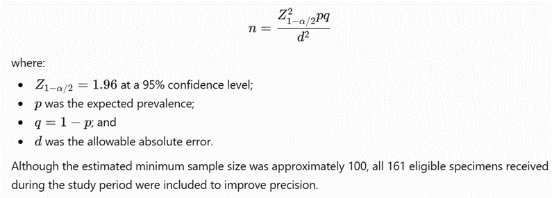
A total of 161 eligible cholecystectomy specimens with cholelithiasis were included. All eligible specimens received during the defined study period were enrolled through consecutive sampling.
Inclusion Criteria
· Patients of any age and either sex who underwent open or laparoscopic cholecystectomy.
· Cholecystectomy specimens containing one or more gallstones.
· Specimens received with adequate clinical and identification details.
· Specimens adequately preserved and suitable for routine gross and microscopic examination.
· Cases for which relevant anthropometric and hematological information was available.
Exclusion Criteria
· Gallbladder specimens without gallstones.
· Gallbladder tissues obtained during autopsy.
· Inadequately fixed, extensively autolysed, or severely damaged specimens that prevented reliable histopathological assessment.
· Specimens without adequate patient identification or essential clinical information.
· Cases with unavailable BMI or essential hematological data were excluded from analyses specifically involving those variables.
Study Variables
The principal variables included:
· Age and sex;
· Height, weight, and BMI;
· Haemoglobin, haematocrit/packed-cell volume, RBC count, total WBC count, differential leukocyte count, platelet count, MCV, MCH, MCHC, and RDW;
· Number, size, colour, shape, and morphological type of gallstones;
· Gross appearance, dimensions, wall thickness, mucosal appearance, and abnormal focal lesions of the gallbladder;
· Histopathological diagnosis and associated inflammatory, hyperplastic, metaplastic, dysplastic, or neoplastic changes.
BMI was calculated as:
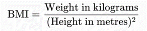
For adult participants, BMI was classified according to the prespecified study criteria into underweight, normal weight, overweight, and obesity. Age-appropriate BMI interpretation was used for participants younger than 18 years.
Procedure and Methodology
Eligible cases were identified when cholecystectomy specimens were received in the pathology department. Patient identity and specimen-label details were verified against the histopathology requisition form. Clinical history, age, sex, operative approach, anthropometric measurements, and laboratory findings were recorded using a structured study proforma.
Height and weight recorded during the preoperative assessment were used to calculate BMI. Hematological findings were obtained from the complete blood count performed during the preoperative period. Anaemia and abnormalities of leukocyte, erythrocyte, and platelet parameters were classified according to the laboratory reference ranges applicable to the patient’s age and sex.
Each gallbladder was examined grossly after adequate fixation. Its length, maximum external diameter, wall thickness, external surface, mucosal appearance, and contents were recorded. Areas of congestion, haemorrhage, necrosis, ulceration, thickening, polypoid growth, or other abnormalities were documented.
The gallstones were removed, washed when necessary, and examined for number, maximum diameter, colour, external surface, consistency, and shape. Based on gross morphological characteristics, the stones were categorized as cholesterol, pigment, or mixed stones. When multiple stones were present, the predominant morphological pattern was recorded.
Representative tissue sections were taken from the neck, body, and fundus of each gallbladder. Additional sections were obtained from all abnormal-looking areas, including thickened, ulcerated, nodular, necrotic, or polypoid regions. Histopathological findings were assessed in relation to BMI, hematological parameters, and gallstone characteristics.
Sample Processing
The received gallbladder specimens were opened and fixed overnight in 10% neutral-buffered formalin. After gross examination, at least three representative tissue sections one each from the neck, body, and fundus were obtained. Additional sections were taken from any grossly abnormal area.
The tissue samples were processed using routine histopathological procedures, including dehydration through graded alcohol, clearing in xylene, and paraffin-wax impregnation. Paraffin blocks were prepared, and sections of approximately 5 µm thickness were cut using a rotary microtome.
The sections were mounted on glass slides, placed on a slide warmer, deparaffinized in xylene, and rehydrated through descending grades of alcohol. They were stained routinely with haematoxylin and eosin. After dehydration and clearing, the slides were mounted and examined under light microscopy.
Microscopic examination included assessment of:
· Acute and chronic inflammatory infiltrates;
· Mucosal ulceration and fibrosis;
· Rokitansky-Aschoff sinuses;
· Cholesterolosis;
· Xanthogranulomatous or granulomatous inflammation;
· Epithelial or adenomyomatous hyperplasia;
· Gastric or intestinal metaplasia;
· Dysplasia; and
· Benign or malignant neoplasms.
The final histopathological diagnosis was recorded for each specimen.
Data Collection
Data were collected prospectively using a structured and pretested study proforma. The following information was recorded:
1. Demographic and clinical information;
2. Height, weight, and calculated BMI;
3. Complete blood count and red-cell indices;
4. Gross gallbladder findings;
5. Gallstone number, size, colour, shape, and type; and
6. Microscopic findings and final histopathological diagnosis.
Each patient was assigned a study identification number. Data were checked for completeness and consistency before entry into the electronic database. Patient-identifying information was kept confidential.
Statistical Methods
Data were entered into Microsoft Excel and analysed using IBM SPSS Statistics for Windows, version 22.0.
· Continuous variables were summarized using mean and standard deviation or median and interquartile range, according to their distribution.
· Categorical variables were expressed as frequencies and percentages.
· BMI and hematological parameters were compared across gallstone types and major histopathological categories.
· The chi-square test was used to assess associations between categorical variables.
· Fisher’s exact test was used when expected cell frequencies were less than five.
· Independent-samples t test or one-way analysis of variance was used for normally distributed continuous variables.
· Mann-Whitney U or Kruskal-Wallis tests were used for non-normally distributed continuous variables.
· Effect estimates were reported, wherever applicable, with 95% confidence intervals.
· A two-sided p value of less than 0.05 was considered statistically significant.
Methodological note: Because all 161 participants had gallstone disease, the study could evaluate obesity and hematological parameters in relation to gallstone type and histopathological changes, but it could not independently estimate their association with the occurrence of gallstone disease compared with people without gallstones.
RESULTS:
Table 1: Overall associations of obesity and hematological parameters with gallstone characteristics and gallbladder histopathology (N=161)
|
Association examined |
Relevant distribution, n (%) |
Effect estimate (95% CI) |
Test of significance |
P value |
|
BMI category versus gallstone type |
Underweight 7 (4.4%); normal 58 (36.0%); overweight 88 (54.7%); obese 8 (5.0%) |
Cramér’s V=0.147 |
χ²=6.96, df=6 |
0.325 |
|
Overweight/obesity versus gallstone type |
Mixed 15/29 (51.7%); pigment 51/81 (63.0%); cholesterol 30/51 (58.8%) |
Mixed: 34.4%-68.6%; pigment: 52.1%-72.7%; cholesterol: 45.2%-71.2% |
χ²=1.14, df=2 |
0.565 |
|
Anaemia versus gallstone type |
Mixed 17/29 (58.6%); pigment 32/81 (39.5%); cholesterol 23/51 (45.1%) |
Mixed: 40.7%-74.5%; pigment: 29.6%-50.4%; cholesterol: 32.3%-58.6% |
χ²=3.16, df=2 |
0.206 |
|
Gallstone type versus number of stones |
Multiple: mixed 26/29 (89.7%); pigment 61/81 (75.3%); cholesterol 34/51 (66.7%) |
Cramér’s V=0.180 |
χ²=5.24, df=2 |
0.073 |
|
Gallstone type versus stone size |
Size categories: <0.5, 0.5 and >0.5 cm |
Cramér’s V=0.283 |
χ²=25.70, df=4 |
<0.001* |
|
Gallstone type versus stone shape |
Mixed predominantly multifaceted; cholesterol predominantly round-ovoid; pigment predominantly irregular |
Cramér’s V=0.937 |
χ²=282.77, df=4 |
<0.001* |
|
Gallstone type versus detailed histopathological diagnosis |
Chronic non-specific cholecystitis: mixed 27/29 (93.1%); pigment 77/81 (95.1%); cholesterol 42/51 (82.4%) |
Cramér’s V=0.188 |
χ²=11.40, df=12† |
0.495 |
|
Gallbladder wall thickening versus chronic cholecystitis |
Thickened wall in 11/161 (6.8%) |
|
χ² test, as reported |
0.538 |
*Statistically significant.
†The original dissertation reported
Table 1 presents the overall associations of obesity and hematological parameters with gallstone characteristics and gallbladder histopathology among 161 patients. Most participants were overweight (54.7%), followed by those with normal BMI (36.0%), while 5.0% were obese and 4.4% were underweight. BMI category was not significantly associated with gallstone type (χ²=6.96, df=6, p=0.325; Cramér’s V=0.147). The combined prevalence of overweight and obesity was highest among patients with pigment stones (63.0%; 95% CI: 52.1%-72.7%), followed by cholesterol stones (58.8%; 95% CI: 45.2%-71.2%) and mixed stones (51.7%; 95% CI: 34.4%-68.6%); however, this difference was not statistically significant (χ²=1.14, df=2, p=0.565).
Anaemia was more frequent among patients with mixed stones (58.6%; 95% CI: 40.7%-74.5%) than among those with cholesterol stones (45.1%; 95% CI: 32.3%-58.6%) or pigment stones (39.5%; 95% CI: 29.6%-50.4%). Nevertheless, anaemia was not significantly associated with gallstone type (χ²=3.16, df=2, p=0.206). Multiple stones were observed in 89.7% of mixed-stone cases, 75.3% of pigment-stone cases, and 66.7% of cholesterol-stone cases. This association approached but did not reach statistical significance (χ²=5.24, df=2, p=0.073; Cramér’s V=0.180).
Stone size differed significantly according to gallstone type (χ²=25.70, df=4, p<0.001; Cramér’s V=0.283), indicating a moderate association. A very strong association was also observed between gallstone type and shape (χ²=282.77, df=4, p<0.001; Cramér’s V=0.937): mixed stones were predominantly multifaceted, cholesterol stones were predominantly round-ovoid, and pigment stones were predominantly irregular. Chronic non-specific cholecystitis was the most common histopathological diagnosis in all three groups, occurring in 93.1% of mixed, 95.1% of pigment, and 82.4% of cholesterol-stone cases. Detailed histopathological diagnosis did not differ significantly between gallstone types (χ²=11.40, df=12, p=0.495). Similarly, gallbladder wall thickening, observed in 11 patients (6.8%), was not significantly associated with chronic cholecystitis (p=0.538).
Table 2: BMI distribution and hematological profile of patients undergoing cholecystectomy for gallstone disease (N=161)
|
Parameter |
n (%) or Mean (SD) |
95% CI |
Test of significance |
P value |
|
Age, years |
42.12 (14.25) |
39.90-44.34 |
One-sample t=−2.57‡ |
0.011* |
|
Underweight, BMI <18.5 kg/m² |
7 (4.4%) |
2.1%-8.7% |
Proportion z=−11.59§ |
<0.001* |
|
Normal BMI, 18.5-24.9 kg/m² |
58 (36.0%) |
29.0%-43.7% |
Proportion z=−3.55§ |
<0.001* |
|
Overweight, BMI 25.0-29.9 kg/m² |
88 (54.7%) |
46.9%-62.2% |
Proportion z=1.18§ |
0.237 |
|
Obese, BMI ≥30 kg/m² |
8 (5.0%) |
2.5%-9.5% |
Proportion z=−11.43§ |
<0.001* |
|
Combined overweight or obesity |
96 (59.6%) |
51.9%-66.9% |
Proportion z=2.44§ |
0.015* |
|
Haemoglobin <10 g/dL |
23 (14.3%) |
9.7%-20.5% |
Proportion z=−9.06§ |
<0.001* |
|
Haemoglobin 10-<12 g/dL |
49 (30.4%) |
23.9%-37.9% |
Proportion z=−4.97§ |
<0.001* |
|
Haemoglobin ≥12 g/dL |
89 (55.3%) |
47.6%-62.7% |
Proportion z=1.34§ |
0.180 |
|
Anaemia, haemoglobin <12 g/dL |
72 (44.7%) |
37.3%-52.4% |
Proportion z=−1.34§ |
0.180 |
|
Microcytosis among anaemic patients, MCV <75 fL |
31/72 (43.1%) |
32.3%-54.5% |
Proportion z=−1.18§ |
0.239 |
|
Hypochromia among anaemic patients, MCH <27 pg |
44/72 (61.1%) |
49.6%-71.5% |
Proportion z=1.89§ |
0.059 |
|
Low total leukocyte count |
2 (1.2%) |
0.3%-4.4% |
Proportion z=−12.37§ |
<0.001* |
|
Normal total leukocyte count |
113 (70.2%) |
62.7%-76.7% |
Proportion z=5.12§ |
<0.001* |
|
Leucocytosis |
46 (28.6%) |
22.2%-36.0% |
Proportion z=−5.44§ |
<0.001* |
|
Neutrophilic leucocytosis |
32 (19.9%) |
14.4%-26.7% |
Proportion z=−7.63§ |
<0.001* |
|
Neutrophilia |
54 (33.5%) |
26.7%-41.2% |
Proportion z=−4.18§ |
<0.001* |
|
Lymphopenia |
46 (28.6%) |
22.2%-36.0% |
Proportion z=−5.44§ |
<0.001* |
|
Thrombocytopenia |
7 (4.4%) |
2.1%-8.7% |
Proportion z=−11.59§ |
<0.001* |
|
Normal platelet count |
137 (85.1%) |
78.8%-89.8% |
Proportion z=8.91§ |
<0.001* |
|
Thrombocytosis |
17 (10.6%) |
6.7%-16.3% |
Proportion z=−10.01§ |
<0.001* |
‡Age was compared with a reference value of 45 years.
§One-sample proportion test against a reference proportion of 50%;
Table 2 describes the BMI distribution and hematological profile of patients undergoing cholecystectomy for gallstone disease. The mean age was 42.12±14.25 years, with a 95% CI of 39.90-44.34 years. When compared with the specified reference age of 45 years, the mean age was significantly lower (t=−2.57, p=0.011). Regarding BMI, 88 patients (54.7%; 95% CI: 46.9%-62.2%) were overweight and eight (5.0%; 95% CI: 2.5%-9.5%) were obese. Overall, 96 patients (59.6%; 95% CI: 51.9%-66.9%) were either overweight or obese, which was significantly greater than the specified 50% reference proportion (z=2.44, p=0.015). Normal BMI was recorded in 58 patients (36.0%), whereas only seven (4.4%) were underweight.
Haemoglobin concentration was below 10 g/dL in 23 patients (14.3%), between 10 and <12 g/dL in 49 (30.4%), and ≥12 g/dL in 89 (55.3%). Thus, anaemia defined as haemoglobin <12 g/dL was present in 72 patients (44.7%; 95% CI: 37.3%-52.4%). The prevalence of anaemia did not differ significantly from the 50% reference proportion (z=−1.34, p=0.180). Among the 72 anaemic patients, 31 (43.1%; 95% CI: 32.3%-54.5%) had microcytosis and 44 (61.1%; 95% CI: 49.6%-71.5%) had hypochromia. Neither microcytosis (p=0.239) nor hypochromia (p=0.059) differed significantly from the respective 50% reference proportion, although hypochromia showed a borderline predominance.
The total leukocyte count was normal in 113 patients (70.2%; 95% CI: 62.7%-76.7%), representing a significant majority (z=5.12, p<0.001). Leucocytosis was found in 46 patients (28.6%), while only two (1.2%) had a low leukocyte count. Neutrophilic leucocytosis was observed in 32 patients (19.9%), neutrophilia in 54 (33.5%), and lymphopenia in 46 (28.6%). Platelet counts were normal in 137 patients (85.1%; 95% CI: 78.8%-89.8%), which was significantly greater than 50% (z=8.91, p<0.001). Thrombocytosis was recorded in 17 patients (10.6%), whereas seven (4.4%) had thrombocytopenia.
Table 3: Gallstone characteristics and spectrum of histopathological changes in the gallbladder (N=161)
|
Characteristic |
n (%) |
95% CI |
Test of significance |
P value |
|
Morphological type |
||||
|
Pigment stones |
81 (50.3%) |
42.7%-57.9% |
χ²=26.93, df=2¶ |
<0.001* |
|
Cholesterol stones |
51 (31.7%) |
25.0%-39.2% |
||
|
Mixed stones |
29 (18.0%) |
12.8%-24.7% |
||
|
Number of stones |
||||
|
Single |
40 (24.8%) |
18.8%-32.1% |
χ²=40.75, df=1¶ |
<0.001* |
|
Multiple |
121 (75.2%) |
67.9%-81.2% |
||
|
Maximum stone size |
||||
|
<0.5 cm |
43 (26.7%) |
20.5%-34.0% |
χ²=9.98, df=2¶ |
0.007* |
|
0.5 cm |
46 (28.6%) |
22.2%-36.0% |
||
|
>0.5 cm |
72 (44.7%) |
37.3%-52.4% |
||
|
Stone shape |
||||
|
Round-ovoid |
55 (34.2%) |
27.3%-41.8% |
χ²=21.29, df=2¶ |
<0.001* |
|
Polyhedral/multifaceted |
29 (18.0%) |
12.8%-24.7% |
||
|
Irregular |
77 (47.8%) |
40.3%-55.5% |
||
|
Histopathological diagnosis |
||||
|
Chronic non-specific cholecystitis |
146 (90.7%) |
85.2%-94.3% |
χ²=819.52, df=7¶ |
<0.001* |
|
Acute cholecystitis |
3 (1.9%) |
0.6%-5.3% |
||
|
Gangrenous cholecystitis |
2 (1.2%) |
0.3%-4.4% |
||
|
Granulomatous cholecystitis |
1 (0.6%) |
0.1%-3.4% |
||
|
Follicular cholecystitis with adenomyomatous hyperplasia |
1 (0.6%) |
0.1%-3.4% |
||
|
Chronic cholecystitis with cholesterolosis |
3 (1.9%) |
0.6%-5.3% |
||
|
Chronic cholecystitis with adenomyomatous hyperplasia |
1 (0.6%) |
0.1%-3.4% |
||
|
Chronic cholecystitis with both cholesterolosis and adenomyomatous hyperplasia |
4 (2.5%) |
1.0%-6.2% |
||
|
Any cholesterolosis |
7 (4.4%) |
2.1%-8.7% |
Proportion z=−11.59§ |
<0.001* |
|
Any adenomyomatous hyperplasia |
6 (3.7%) |
1.7%-7.9% |
Proportion z=−11.74§ |
<0.001* |
|
Metaplasia or dysplasia |
0 (0.0%) |
0.0%-2.3% |
Not applicable |
|
|
Malignant lesion |
0 (0.0%) |
0.0%-2.3% |
Not applicable |
|
Goodness-of-fit chi-square test against equal expected proportions across the categories.
Table 3 summarizes the morphological characteristics of gallstones and the histopathological spectrum of gallbladder lesions. Pigment stones were the most common morphological type, accounting for 81 cases (50.3%; 95% CI: 42.7%-57.9%), followed by cholesterol stones in 51 (31.7%; 95% CI: 25.0%-39.2%) and mixed stones in 29 (18.0%; 95% CI: 12.8%-24.7%). The distribution of gallstone types was significantly non-uniform (χ²=26.93, df=2, p<0.001).
Multiple stones were present in 121 patients (75.2%; 95% CI: 67.9%-81.2%), while 40 (24.8%; 95% CI: 18.8%-32.1%) had a single stone. This difference was statistically significant (χ²=40.75, df=1, p<0.001), demonstrating a clear predominance of multiple stones. Regarding maximum stone size, 43 stones (26.7%) measured <0.5 cm, 46 (28.6%) measured 0.5 cm, and 72 (44.7%) measured >0.5 cm. The size distribution was significantly non-uniform (χ²=9.98, df=2, p=0.007), with stones larger than 0.5 cm constituting the largest category.
Irregular stones were most common, occurring in 77 cases (47.8%; 95% CI: 40.3%-55.5%), followed by round-ovoid stones in 55 (34.2%; 95% CI: 27.3%-41.8%) and polyhedral or multifaceted stones in 29 (18.0%; 95% CI: 12.8%-24.7%). Stone-shape distribution differed significantly across these categories (χ²=21.29, df=2, p<0.001).
Chronic non-specific cholecystitis was the predominant histopathological diagnosis, identified in 146 specimens (90.7%; 95% CI: 85.2%-94.3%). Acute cholecystitis and chronic cholecystitis with cholesterolosis were each observed in three cases (1.9%), while gangrenous cholecystitis was found in two (1.2%). Granulomatous cholecystitis, follicular cholecystitis with adenomyomatous hyperplasia, and chronic cholecystitis with adenomyomatous hyperplasia were each detected in one case (0.6%). Four patients (2.5%) had chronic cholecystitis accompanied by both cholesterolosis and adenomyomatous hyperplasia. The overall distribution of histopathological diagnoses was significantly non-uniform (χ²=819.52, df=7, p<0.001), owing to the marked predominance of chronic non-specific cholecystitis.
Considering overlapping lesions, cholesterolosis was identified in seven patients (4.4%; 95% CI: 2.1%-8.7%), and adenomyomatous hyperplasia in six (3.7%; 95% CI: 1.7%-7.9%). No metaplasia, dysplasia, or malignant lesion was identified. The upper limit of the 95% CI for each absent finding was 2.3%, indicating that although no such lesion was observed in this sample, a very low underlying prevalence could not be completely excluded.
Table 4: Associations of BMI and hematological abnormalities with gallstone type and histopathological lesions (N=161)
|
Factor |
Mixed stones (n=29), n (%) |
Pigment stones (n=81), n (%) |
Cholesterol stones (n=51), n (%) |
Association estimate |
Test of significance |
P value |
|
Underweight |
0 (0.0%) |
5 (6.2%) |
2 (3.9%) |
Cramér’s V=0.147 |
χ²=6.96, df=6 |
0.325 |
|
Normal BMI |
14 (48.3%) |
25 (30.9%) |
19 (37.3%) |
|||
|
Overweight |
13 (44.8%) |
49 (60.5%) |
26 (51.0%) |
|||
|
Obese |
2 (6.9%) |
2 (2.5%) |
4 (7.8%) |
|||
|
Anaemia, Hb <12 g/dL |
17 (58.6%) |
32 (39.5%) |
23 (45.1%) |
Cramér’s V=0.140 |
χ²=3.16, df=2 |
0.206 |
|
Normal Hb, ≥12 g/dL |
12 (41.4%) |
49 (60.5%) |
28 (54.9%) |
|||
|
Elevated triglycerides |
15 (51.7%) |
33 (40.7%) |
25 (49.0%) |
Cramér’s V=0.095 |
χ²=1.45, df=2 |
0.485† |
|
Elevated total cholesterol |
13 (44.8%) |
18 (22.2%) |
16 (31.4%) |
Cramér’s V=0.184 |
χ²=5.45, df=2 |
0.066 |
|
Elevated LDL cholesterol |
9 (31.0%) |
13 (16.0%) |
10 (19.6%) |
Cramér’s V=0.137 |
χ²=3.01, df=2 |
0.222 |
|
Low HDL cholesterol |
13 (44.8%) |
24 (29.6%) |
19 (37.3%) |
Cramér’s V=0.121 |
χ²=2.38, df=2 |
0.305 |
|
Chronic non-specific cholecystitis |
27 (93.1%) |
77 (95.1%) |
42 (82.4%) |
Cramér’s V=0.188 |
χ²=11.40, df=12 |
0.495‡ |
|
Any other inflammatory/hyperplastic lesion |
2 (6.9%) |
4 (4.9%) |
9 (17.6%) |
|||
|
Multiple stones |
26 (89.7%) |
61 (75.3%) |
34 (66.7%) |
Cramér’s V=0.180 |
χ²=5.24, df=2 |
0.073 |
|
Predominant characteristic shape |
Multifaceted: 28 (96.6%) |
Irregular: 76 (93.8%) |
Round-ovoid: 50 (98.0%) |
Cramér’s V=0.937 |
χ²=282.77, df=4 |
<0.001* |
|
Predominant size category |
>0.5 cm: 16 (55.2%) |
<0.5 cm: 35 (43.2%) |
>0.5 cm: 27 (52.9%) |
Cramér’s V=0.283 |
χ²=25.70, df=4 |
<0.001* |
*Statistically significant at .
†The dissertation reported for triglycerides, whereas recalculation from the displayed counts gave χ²=1.45 and . The original electronic dataset should be checked before publication.
‡The dissertation reported.
Table 4 evaluates the relationships of BMI and hematological abnormalities with gallstone type and histopathological lesions. Among patients with mixed stones, 48.3% had normal BMI, 44.8% were overweight, and 6.9% were obese. Among pigment-stone cases, 60.5% were overweight, 30.9% had normal BMI, 6.2% were underweight, and 2.5% were obese. Among patients with cholesterol stones, 51.0% were overweight, 37.3% had normal BMI, 7.8% were obese, and 3.9% were underweight. Despite the predominance of overweight patients, BMI distribution did not differ significantly by gallstone type (χ²=6.96, df=6, p=0.325; Cramér’s V=0.147).
Anaemia was most frequent in mixed-stone cases (58.6%), followed by cholesterol-stone (45.1%) and pigment-stone cases (39.5%). However, the difference was not statistically significant (χ²=3.16, df=2, p=0.206; Cramér’s V=0.140). Elevated triglycerides were detected in 51.7% of patients with mixed stones, 40.7% with pigment stones, and 49.0% with cholesterol stones, with no significant association (χ²=1.45, df=2, p=0.485). Elevated total cholesterol was more common with mixed stones (44.8%) than with cholesterol (31.4%) or pigment stones (22.2%); however, the association was borderline and did not reach statistical significance (χ²=5.45, df=2, p=0.066). Neither elevated LDL cholesterol (χ²=3.01, p=0.222) nor low HDL cholesterol (χ²=2.38, p=0.305) was significantly associated with gallstone type. The corresponding Cramér’s V values were small, indicating weak relationships.
Chronic non-specific cholecystitis was identified in 93.1% of mixed-stone, 95.1% of pigment-stone, and 82.4% of cholesterol-stone cases. Other inflammatory or hyperplastic lesions occurred in 6.9%, 4.9%, and 17.6%, respectively. Nevertheless, the detailed histopathological distribution did not differ significantly by stone type (χ²=11.40, df=12, p=0.495; Cramér’s V=0.188).
Multiple stones were most frequent among mixed-stone cases (89.7%), followed by pigment-stone (75.3%) and cholesterol-stone cases (66.7%). This relationship was of small magnitude and did not reach statistical significance (χ²=5.24, df=2, p=0.073; Cramér’s V=0.180). In contrast, gallstone shape showed a very strong association with morphological type (χ²=282.77, df=4, p<0.001; Cramér’s V=0.937). Mixed stones were predominantly multifaceted (96.6%), pigment stones were predominantly irregular (93.8%), and cholesterol stones were predominantly round-ovoid (98.0%). Stone size was also significantly associated with gallstone type (χ²=25.70, df=4, p<0.001; Cramér’s V=0.283): stones >0.5 cm predominated among mixed and cholesterol stones, whereas stones <0.5 cm formed the largest size category among pigment stones. Overall, stone morphology and size, rather than BMI or hematological abnormalities, were the principal characteristics significantly associated with gallstone type.
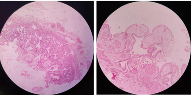
Figure 1 Figure 2
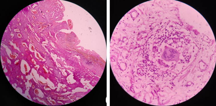
Figure 3 Figure 4
Figure 1: Microscopic picture of Adenomyomatous hyperplasia along with Rokitansky aschoff sinuses with associated cholesterolosis at low magnification
Figure 2: Microscopic picture of cholesterolosis at 10x magnification
Figure 3: Microscopic picture of Gangrenous cholecystitis
Figure 4: Microscopic picture of chronic granulomatous cholecystitis shows a giant cell surrounded by lymphocytes.
DISCUSSION:
Table 1: Overall associations of obesity and hematological parameters with gallstone characteristics and histopathology
In the present study, 59.6% of patients were overweight or obese, but BMI category was not significantly associated with gallstone type (χ²=6.96, p=0.325). Similarly, the prevalence of overweight/obesity did not differ significantly among patients with mixed, pigment, and cholesterol stones (p=0.565). These findings indicate that excess body weight was common among patients with cholelithiasis but did not determine the morphological composition of the stones. Kharga et al. (2016)[1] similarly observed that symptomatic cholelithiasis occurred across all BMI categories, although its probability increased progressively with rising BMI, age, and female sex. The absence of an association with stone type in the present study may be related to the predominance of pigment stones, whereas obesity is biologically more closely linked with cholesterol supersaturation and cholesterol-stone formation. Moreover, because the present study did not include a non-gallstone control group, it could not estimate the independent risk of developing gallstone disease associated with obesity.
Anaemia was present in 44.7% of patients and was most frequent among patients with mixed stones (58.6%), followed by cholesterol (45.1%) and pigment stones (39.5%). However, the association between anaemia and stone type was not statistically significant (χ²=3.16, p=0.206). Although chronic haemolytic anaemias are recognized causes of pigment stones, many anaemic patients in this study had microcytic or hypochromic changes, suggesting that nutritional iron deficiency rather than haemolysis may have been common. The lack of reticulocyte count, bilirubin, lactate dehydrogenase, haptoglobin, ferritin, and peripheral-smear classification limited differentiation between haemolytic and non-haemolytic anaemia. Therefore, haemoglobin level alone may not demonstrate a specific relationship with pigment-stone formation.
Multiple stones were more frequent with mixed stones (89.7%) than with pigment (75.3%) or cholesterol stones (66.7%), but the association was not statistically significant (p=0.073). Singh et al. (2019)[2] evaluated gallstone number, size, and type in relation to gallbladder mucosal responses and demonstrated that gallstones were accompanied by inflammation, hyperplasia, metaplasia, and other epithelial alterations. In the present study, stone size and shape were significantly associated with stone type (both p<0.001). Mixed stones were predominantly multifaceted, cholesterol stones round-ovoid, and pigment stones irregular, producing a very strong association for shape (Cramér’s V=0.937). Navyashree and Giriyan (2020)[3], in their four-year study of 178 calculous gallbladders, reported mixed stones as the most common type (64.04%) and found round-to-oval stones to be the predominant morphology. The difference from the present study, in which pigment and irregular stones predominated, may reflect geographical, dietary, metabolic, and infective differences.
Chronic non-specific cholecystitis was the dominant lesion in every stone group, but the detailed histopathological diagnosis was not significantly associated with gallstone type (p=0.495). This is consistent with the concept that chronic irritation can be produced by all stone types. Singh et al. (2019)[2] also concluded that the pathological response of the gallbladder mucosa was related to cholelithiasis, although particular responses varied with stone characteristics. Wall thickening was present in only 6.8% of the current cases and was not significantly associated with chronic cholecystitis (p=0.538). Histological inflammation may therefore occur without gross wall thickening, demonstrating the limited sensitivity of gross examination alone.
Table 2: BMI distribution and hematological profile
The mean age of the patients was 42.12±14.25 years, indicating that gallstone disease requiring cholecystectomy predominantly affected middle-aged individuals. Beena et al. (2017)[4] similarly reported that gallbladder diseases were most frequent among women in the fourth and fifth decades. Navyashree and Giriyan (2020)[3] found higher frequencies in the fifth and seventh decades, with women accounting for 69% of cases. Variations in peak age may be related to referral patterns, population structure, reproductive factors, dietary exposure, and timing of surgical treatment.
Overweight was the most frequent BMI category in the present study, affecting 54.7% of patients, while 5.0% were obese. Together, overweight and obesity affected 59.6% and significantly exceeded the prespecified 50% reference proportion (p=0.015). This finding supports the metabolic contribution of excess body weight to gallstone disease. Kharga et al. (2016)[1] reported an increasing risk of symptomatic cholelithiasis with increasing BMI but emphasized that normal-weight individuals were not protected. Thus, the presence of 36.0% of patients with normal BMI in the current study is compatible with the multifactorial origin of gallstones. However, the one-sample proportion tests in Table 2 only indicate whether categories differed from an arbitrary 50% benchmark and should not be interpreted as comparisons with the general population.
Anaemia was found in 44.7% of patients; 14.3% had haemoglobin below 10 g/dL and 30.4% had values between 10 and <12 g/dL. Among anaemic patients, 43.1% had microcytosis and 61.1% had hypochromia, making iron deficiency a plausible explanation in many cases. Because serum ferritin and iron studies were not available, this interpretation requires confirmation. Normal leukocyte counts were recorded in 70.2%, although leucocytosis occurred in 28.6%, neutrophilia in 33.5%, and neutrophilic leucocytosis in 19.9%. These abnormalities may reflect acute inflammation, secondary infection, tissue necrosis, physiological stress, or associated illness. The relatively low frequency of acute and gangrenous cholecystitis explains why most patients nevertheless had normal leukocyte counts.
Platelet counts were normal in 85.1% of cases, while thrombocytosis and thrombocytopenia occurred in 10.6% and 4.4%, respectively. Reactive thrombocytosis may accompany inflammation or iron deficiency, whereas thrombocytopenia may be associated with liver disease, infection, medication, or unrelated hematological disorders. Because the study was cross-sectional and lacked postoperative measurements, it could not establish whether these abnormalities were caused by gallbladder inflammation or represented pre-existing conditions.
Table 3: Gallstone morphology and histopathological spectrum
Pigment stones were the most frequent type in the present study (50.3%), followed by cholesterol (31.7%) and mixed stones (18.0%), with a significantly non-uniform distribution (p<0.001). Kumar et al. (2015)[5] also reported pigment stones as the most frequent type in their evaluation of 400 cholecystectomy specimens. In contrast, Navyashree and Giriyan (2020)[3] found mixed stones in 64.04% of 178 calculous specimens. Such differences support geographical heterogeneity in stone composition. Pigment stones may be influenced by infection, haemolysis, bilirubin metabolism, and socioeconomic factors, whereas cholesterol stones are more strongly related to metabolic and dietary factors.
Multiple stones constituted 75.2% of cases, significantly exceeding single stones (24.8%; p<0.001). Mixed stones showed the highest proportion of multiplicity. The maximum stone size was >0.5 cm in 44.7% of patients, while 26.7% had stones <0.5 cm. Stone-size distribution was statistically significant (p=0.007). Singh et al. (2019)[2] emphasized that the number and size of stones may influence the extent and pattern of mucosal injury. Multiple stones can create repeated contact at different mucosal sites, whereas larger stones may exert greater localized mechanical pressure. Nevertheless, the clinical importance of morphology depends on stone duration, mobility, biochemical composition, and associated infection.
Irregular stones were most common (47.8%), followed by round-ovoid (34.2%) and multifaceted stones (18.0%). The marked relationship between shape and stone type supports the internal consistency of gross classification: pigment stones were predominantly irregular, cholesterol stones round-ovoid, and mixed stones multifaceted. Morphological classification remains practical in routine pathology, although chemical or spectroscopic analysis would provide more accurate compositional confirmation.
Chronic non-specific cholecystitis was identified in 90.7% of specimens, substantially exceeding all other diagnoses. This agrees with Mondal et al. (2016)[6], who reported chronic cholecystitis in 79.8% of 786 gallstone cases, followed by acute-on-chronic cholecystitis (6.1%), cholesterolosis (2.9%), xanthogranulomatous cholecystitis (1.7%), metaplasia (4.7%), dysplasia (2.2%), and gallbladder carcinoma (0.6%). Kumar et al. (2015)[5] reported chronic cholecystitis in 66.75%, chronic active cholecystitis in 20.25%, acute cholecystitis in 6%, and carcinoma in 1.25% of 400 specimens. Shah et al. (2016)[7] similarly found chronic cholecystitis in 82.25%, acute cholecystitis or abscess in 10%, and adenocarcinoma in 2% of 400 specimens.
Beena et al. (2017)[4] found chronic cholecystitis to be the major lesion among 200 cholecystectomy specimens. Kumari et al. (2018)[8] also reported chronic inflammatory lesions as the predominant category. Agarwal et al. (2018)[9], in a large series of 1,693 cholecystectomy specimens, demonstrated a broad morphological spectrum, supporting careful sampling of all specimens. Ahadi et al. (2020)[10] examined 1,004 specimens and reported chronic calculous cholecystitis in 61.18%, metaplasia in 19.55%, and invasive carcinoma in 0.2%.
The present study identified cholesterolosis in 4.4% and adenomyomatous hyperplasia in 3.7%. However, no metaplasia, dysplasia, or malignancy was detected. This contrasts with several larger studies that identified small but clinically important proportions of premalignant and malignant lesions.[6,9,10] Dincel et al. (2018)[11] demonstrated that unexpected gallbladder carcinoma may be found during routine microscopic examination, reinforcing the need for histopathological evaluation even when the specimen appears grossly benign. The absence of malignancy in the present study may be related to its modest sample size, regional incidence, exclusion pattern, or chance rather than proof that the risk was absent. Awasthi (2015)[12] and Sharma and Choudhury (2015)[13] likewise reported chronic cholecystitis as the dominant finding but identified less frequent incidental lesions that could not reliably be predicted clinically.
Table 4: Associations across gallstone types
Although overweight patients predominated within each stone group, BMI distribution did not vary significantly among mixed, pigment, and cholesterol stones (p=0.325). This suggests that BMI may influence the overall probability of symptomatic gallstone disease without reliably predicting the stone’s gross type. Similarly, anaemia was not associated with stone type (p=0.206). Further studies should classify anaemia etiologically, because grouping iron-deficiency, chronic-disease, and haemolytic anaemia together may obscure a specific association between haemolysis and pigment stones.
Elevated triglycerides, total cholesterol, LDL, and low HDL were not significantly associated with stone type. Total cholesterol showed the strongest but still non-significant relationship (p=0.066). These findings indicate that circulating lipid concentrations may not directly reflect biliary cholesterol saturation at a single time point. Lipid-lowering treatment, fasting status, diet, diabetes, and duration of disease could also modify the observed associations. The triglyceride result should be interpreted using the value recalculated from the displayed cells (χ²=1.45, p=0.485), while the discrepancy with the dissertation value of p=0.188 should be resolved from the original dataset before publication.
Chronic non-specific cholecystitis predominated across all three stone types and did not show a significant association with stone composition (p=0.495). Similar observations across histopathological series suggest that chronic inflammation is a common endpoint of persistent gallstone irritation rather than a response restricted to a particular stone category.[2,5,6] In contrast, stone shape and size were strongly associated with stone type. The shape association was particularly strong (Cramér’s V=0.937), confirming that gross morphology distinguished the three stone categories effectively.
CONCLUSION:
Overweight and obesity were common among patients undergoing cholecystectomy for gallstone disease, affecting 59.6% of the study population. Anaemia was also frequent, being present in 44.7% of patients, while leucocytosis and thrombocytosis occurred in smaller proportions. However, BMI category, anaemia, and lipid abnormalities were not significantly associated with the morphological type of gallstone. Pigment stones were the most frequent type, and most patients had multiple stones. Stone size and shape were significantly associated with gallstone type: mixed stones were predominantly multifaceted, cholesterol stones were round-ovoid, and pigment stones were irregular. Chronic non-specific cholecystitis was the predominant histopathological lesion, irrespective of gallstone type. No metaplasia, dysplasia, or malignancy was identified. These findings indicate that obesity and hematological abnormalities are common accompanying features of gallstone disease, but stone morphology and size are more useful than BMI or routine hematological parameters for differentiating gallstone types. Routine histopathological examination of all cholecystectomy specimens remains important for identifying inflammatory, hyperplastic, premalignant, and incidental malignant lesions.
LIMITATIONS
1. This was a single-centre, hospital-based study; therefore, the findings may not be generalizable to the wider population or other geographical regions.
2. The study included only patients with gallstone disease who underwent cholecystectomy. The absence of a non-gallstone control group prevented estimation of whether obesity, anaemia, or other hematological abnormalities independently increased the risk of developing gallstones.
3. The observational, cross-sectional nature of the study did not permit conclusions regarding temporal sequence or causality.
4. The sample size of 161 was relatively small, particularly for uncommon histopathological lesions such as dysplasia and gallbladder carcinoma.
5. Gallstones were classified using gross morphological characteristics without chemical, crystallographic, or spectroscopic analysis. Consequently, some stones may have been misclassified.
6. Anaemia was primarily defined according to haemoglobin concentration. Serum ferritin, iron studies, reticulocyte count, peripheral-smear findings, bilirubin fractions, lactate dehydrogenase, and haptoglobin were not consistently assessed; hence, iron-deficiency, haemolytic, and anaemia of chronic disease could not be reliably differentiated.
7. Potential confounding variables such as diet, socioeconomic status, parity, oral-contraceptive use, diabetes mellitus, metabolic syndrome, rapid weight loss, haemolytic disorders, medication use, and family history were not comprehensively adjusted for.
8. BMI was used as the only measure of obesity and did not account for central adiposity, body-fat percentage, waist circumference, or waist-to-hip ratio.
9. Preoperative and postoperative hematological parameters were not compared, making it difficult to determine whether the abnormalities were related to gallbladder inflammation or pre-existing conditions.
10. The duration of gallstone disease and the duration and severity of symptoms were not correlated with mucosal alterations.
11. Some contingency-table cells contained very small numbers, which reduced statistical power and the reliability of chi-square approximations for uncommon histopathological diagnoses.
12. Minor discrepancies existed between the reported and recalculated p values for triglycerides and the association between gallstone type and detailed histopathology. These findings should be verified against the original electronic dataset before publication.
REFERENCES:
1. Kharga B, Sharma BK, Singh VK, Nishant K, Bhutia P, Tamang R, et al. Obesity not necessary, risk of symptomatic cholelithiasis increases as a function of BMI. J Clin Diagn Res. 2016;10(10):PC28-PC32.
2. Singh A, Singh G, Kaur K, Goyal G, Saini G, Sharma D. Histopathological changes in gallbladder mucosa associated with cholelithiasis: a prospective study. Niger J Surg. 2019;25(1):21-5. Full text
3. Navyashree N, Giriyan SS. Morphological characterization of gallstones in cholecystectomy specimens: a four years study. Saudi J Pathol Microbiol. 2020;5(2):43-9. Article
4. Beena D, Shetty J, Jose V. Histopathological spectrum of diseases in gallbladder. Natl J Lab Med. 2017;6(4):PO06-PO09.
5. Kumar H, Kini H, Tiwari A. Histological evaluation of 400 cholecystectomy specimens. J Pathol Nepal. 2015;5(10):834-40. Article
6. Mondal B, Maulik D, Biswas BK, Sarkar GN, Ghosh D. Histopathological spectrum of gallstone disease from cholecystectomy specimens in rural areas of West Bengal, India: an approach to the association between gallstone disease and gallbladder carcinoma. Int J Community Med Public Health. 2016;3(11):3229-35. Article
7. Shah H, Khan MA, Shah W. Histopathological pattern of 400 cholecystectomy specimens. J Postgrad Med Inst. 2016;30(3):250-3.
8. Kumari G, Deshpande KA, Roy S. Clinicopathological study of gallbladder lesions: two years study. Ann Pathol Lab Med. 2018;5(5):A456-62.
9. Agarwal S, Pandey P, Ralli M, Agarwal R, Saxena P. Morphologic characterisation of 1693 cholecystectomy specimens: a study from a tertiary care center in Northern India. J Clin Diagn Res. 2018;12(1):EC05-EC09.
10. Ahadi M, Ghaffarzadegan K, Aghajanzadeh P, Taheri D. Histopathologic features of 1000 cholecystectomy specimens. Iran J Cancer Prev. 2020;13(1):e103522. Article
11. Dincel O, Goksu M, Hatipoglu HS. Importance of routine histopathological examination of a gallbladder surgical specimen: unexpected gallbladder cancer. J Cancer Res Ther. 2018;14(6):1325-9.
12. Awasthi N. A retrospective histopathological study of cholecystectomies. Int J Health Allied Sci. 2015;4(3):203-6.
13. Sharma I, Choudhury D. Histopathological patterns of gallbladder diseases with special reference to incidental cases: a hospital-based study. Int J Res Med Sci. 2015;3(12):3553-7.