Clinical Profile and Antimicrobial Resistance Patterns of Ventilator-Associated Pneumonia in a Tertiary Care Center: A Prospective Observational Study.
- Shubhangi H , Professor and HOD, Dr PDMMC Amravati.
- Ajay Daphale , Professor, Dr PDMMC AMRAVATI.
- Sharwari Bodkhey , Junior Resident 3rd year Dr PDMMC, Amravati.
- Vikram Kokate , Associate Professor, Dr PDMMC Amravati.
- Ganesh Bansod , Assisstant Professor, Dr PDMMC Amravati.
- Shrikant Awasarmol ,
Article Information:
Abstract:
Background: Ventilator-associated pneumonia (VAP) is a major healthcare-associated infection among critically ill patients receiving mechanical ventilation and is associated with considerable morbidity, mortality, and antimicrobial resistance. Knowledge of the clinical profile, causative microorganisms, antimicrobial susceptibility, and resistance patterns is essential for appropriate empirical therapy and antimicrobial stewardship. Objective: To evaluate the clinical profile, microbiological spectrum, antimicrobial resistance patterns, and clinical outcomes of adult patients with ventilator-associated pneumonia admitted to the intensive care units of a tertiary care hospital, and to assess the association of antimicrobial resistance with clinical outcomes. Materials and Methods: A prospective longitudinal observational study was conducted over 18 months in the Medical and Surgical Intensive Care Units of a tertiary care teaching hospital. A total of 174 adult patients who developed VAP after at least 48 hours of invasive mechanical ventilation were included. Demographic and clinical characteristics, timing of VAP onset, microbiological findings, antimicrobial susceptibility patterns, resistance phenotypes, and clinical outcomes were recorded. Endotracheal aspirates were processed using standard microbiological methods, and antimicrobial susceptibility testing was performed according to standard laboratory procedures. Clinical outcomes were compared according to causative organisms and antimicrobial resistance status. Results: Among the 174 patients, 106 (60.9%) were male and 68 (39.1%) were female, with a mean age of 51.0 ± 15.0 years. Medical ICU patients accounted for 93 (53.4%) cases, while 81 (46.6%) were admitted to the Surgical ICU. Early-onset VAP occurred in 50 (28.7%) patients and late-onset VAP in 124 (71.3%). Gram-negative organisms predominated (63.8%), with Acinetobacterbaumannii and Pseudomonas aeruginosa being the most frequently isolated pathogens, accounting for 30 (17.2%) cases each. Among 142 culture-positive cases, 74 (52.1%) demonstrated one or more clinically significant antimicrobial resistance phenotypes. Mortality occurred in 56 (32.2%) patients. Organism-wise mortality was highest among Escherichia coli (42.9%) and Acinetobacterbaumannii (40.0%) among the major pathogens. Mortality was significantly higher among patients with MDR/XDR/CRE/ESBL-associated isolates than among those with non-MDR isolates (40.5% vs. 22.1%; p = 0.029), and MODS was also significantly more frequent among patients with resistant organisms (24.3% vs. 10.3%; p = 0.049). The mean SOFA score at 72 hours was significantly higher among non-survivors than survivors (9.8 ± 2.5 vs. 6.5 ± 2.1; p < 0.001). Mortality did not differ significantly between early- and late-onset VAP (30.0% vs. 33.1%; p = 0.832). Conclusion: Ventilator-associated pneumonia in this cohort was characterized by a predominance of Gram-negative pathogens and a substantial burden of antimicrobial resistance. Antimicrobial resistance was associated with higher mortality and MODS in unadjusted analyses, while worsening SOFA scores at 72 hours were strongly associated with mortality. However, MDR status was not an independent predictor of mortality after multivariable adjustment. These findings emphasize the importance of continuous microbiological surveillance, local antimicrobial susceptibility data, early recognition of clinical deterioration, and targeted antimicrobial stewardship in the management of VAP.
Keywords:
Article :
INTRODUCTION:
Among patients receiving invasive mechanical ventilation in critical care units (ICUs), ventilator-associated pneumonia (VAP) is one of the most prevalent healthcare-associated infections. It is characterized as pneumonia that develops 48 hours or more after endotracheal intubation and initiation of mechanical ventilation and remains an important contributor to morbidity, prolonged hospital stay, increased healthcare expenditure, and mortality among critically ill patients. Despite advances in critical care medicine, VAP continues to pose substantial diagnostic and therapeutic challenges, particularly with the increasing prevalence of antimicrobial-resistant organisms.¹⁻³
The development of VAP is multifactorial and involves microaspiration of contaminated oropharyngeal secretions, bacterial biofilm formation on endotracheal tubes, impaired mucociliary clearance, and compromised host immune responses. Prolonged mechanical ventilation, previous exposure to broad-spectrum antibiotics, extended ICU stay, invasive procedures, underlying comorbidities, and microorganism transmission within the ICU further increase the risk of VAP. These factors not only predispose critically ill patients to infection but may also facilitate colonization and infection with multidrug-resistant (MDR) organisms, thereby complicating antimicrobial treatment.⁴⁻⁶
The microbiological profile of VAP varies considerably across geographical regions, hospitals, and individual ICUs because of differences in antimicrobial prescribing practices, infection prevention measures, local epidemiology, and patient characteristics. Gram-negative bacilli, particularly Acinetobacterbaumannii, Klebsiellapneumoniae, Pseudomonas aeruginosa, and Escherichia coli, are frequently reported among the major pathogens associated with VAP. Gram-positive organisms, including Staphylococcus aureus and methicillin-resistant S. aureus (MRSA), also contribute to the disease burden in healthcare settings. The increasing prevalence of carbapenem-resistant organisms, extended-spectrum β-lactamase (ESBL)-producing Enterobacterales, and other MDR bacteria has further complicated empirical antimicrobial therapy and represents a major public health concern.⁷⁻¹⁰
The selection of appropriate empirical antimicrobial therapy is an important determinant of clinical management in patients with VAP. Delayed initiation of effective therapy or use of antimicrobials that are inactive against the causative organism has been associated with adverse clinical consequences, whereas inappropriate or prolonged use of broad-spectrum antibiotics contributes to the emergence and dissemination of antimicrobial resistance. Therefore, assessment of the clinical characteristics of affected patients together with knowledge of the local microbiological spectrum and antimicrobial susceptibility patterns is essential for selecting appropriate empirical therapy, optimizing antimicrobial use, and strengthening antimicrobial stewardship.¹¹⁻¹⁴
In resource-limited settings, where antimicrobial resistance is increasing and local epidemiological data remain limited, continued surveillance of VAP is particularly important. Routine evaluation of ICU-specific microbiological profiles and antibiograms can assist clinicians in selecting appropriate empirical antimicrobial therapy while minimizing unnecessary exposure to broad-spectrum agents. Such surveillance also helps healthcare institutions monitor changing resistance patterns and supports infection prevention and antimicrobial stewardship strategies.¹⁵⁻¹⁷
The present study was therefore undertaken to evaluate the clinical and microbiological profile of ventilator-associated pneumonia among adult intensive care unit patients in a tertiary care setting, with particular emphasis on antimicrobial susceptibility and resistance patterns. The study also assessed clinical outcomes, including mortality, sepsis, septic shock, and multiple organ dysfunction syndrome, and evaluated the relationship of these outcomes with causative organisms and antimicrobial resistance. In addition, the study examined SOFA score trajectory and independent predictors of mortality to provide a more comprehensive assessment of factors associated with adverse outcomes in patients with VAP.
MATERIALS AND METHODS:
Study Design and Setting
This prospective observational study was conducted in the intensive care units of a tertiary care hospital over a period of 18 months. The study included adult patients who developed ventilator-associated pneumonia after at least 48 hours of mechanical ventilation. Patients were followed prospectively for their clinical course, microbiological findings, antimicrobial susceptibility patterns, and clinical outcomes.
Study Population
A total of 174 adult patients diagnosed with ventilator-associated pneumonia were included in the study. VAP was defined as pneumonia developing after at least 48 hours of mechanical ventilation in patients who were not diagnosed with pneumonia at the time of admission. Patients admitted with pneumonia and those with sepsis at admission were excluded according to the predefined study criteria.
Sample Size
The study included 174 adult patients diagnosed with ventilator-associated pneumonia during the study period. Patients fulfilling the predefined eligibility criteria were enrolled using convenience sampling.
Data Collection
Demographic and clinical data were recorded prospectively for each patient, including age, sex, ICU type, comorbidities, indication for mechanical ventilation, timing of VAP onset, duration of mechanical ventilation, clinical findings, laboratory parameters, and SOFA score. Microbiological samples were collected from patients with suspected VAP and processed according to standard laboratory procedures. The isolated organisms and their antimicrobial susceptibility patterns were recorded. Patients were followed for clinical outcomes, including survival or mortality, sepsis, septic shock, MODS, duration of mechanical ventilation, ICU stay, and hospital stay.
Inclusion Criteria
• Adult patients (≥18 years) admitted to the Medical or Surgical ICU.
• Patients receiving invasive mechanical ventilation for at least 48 hours.
• Patients diagnosed with ventilator-associated pneumonia according to the predefined diagnostic criteria.
Molecular Detection of Respiratory Pathogens
Exclusion Criteria
• Patients with pneumonia before initiation of mechanical ventilation.
• Patients with evidence of active infection or sepsis at the time of ICU admission
Data Collection
Patients with a new or progressive pulmonary infiltrate on chest radiography following at least 48 hours of mechanical ventilation and at least two of the following clinical features—fever (>38°C) or hypothermia (<36°C), leukocytosis (>10,000 cells/mm³) or leukopenia (<5,000 cells/mm³), purulent endotracheal secretions, or deteriorating oxygenation—were diagnosed with ventilator-associated pneumonia.
VAP was further categorized as early-onset VAP, occurring within the first four days of mechanical ventilation, and late-onset VAP, developing on or after the fifth day of mechanical ventilation.
Clinical Profile Assessment
The clinical profile of patients with VAP was assessed using demographic and clinical characteristics recorded in the study proforma. These included age, sex, type of ICU (medical or surgical), duration of mechanical ventilation before development of VAP, and timing of VAP onset. These variables were summarized descriptively to characterize the study population.
Microbiological Processing
Respiratory specimens obtained from patients with suspected ventilator-associated pneumonia were processed in the microbiology laboratory according to standard microbiological procedures. Samples were examined for bacterial and fungal growth, and the isolated organisms were identified using routine laboratory methods. The causative microorganisms were recorded for each patient, including cases with polymicrobial growth and samples showing no microbial growth.
Antimicrobial Susceptibility Testing
Antimicrobial susceptibility testing was performed for the bacterial isolates using the laboratory's standard susceptibility-testing procedures. The susceptibility results were recorded for the antimicrobial agents tested, and the proportion of susceptible isolates was calculated for the major Gram-negative pathogens. The antimicrobial resistance profile was subsequently categorized according to the resistance definitions used in the study, including multidrug-resistant (MDR), extensively drug-resistant (XDR), extended-spectrum β-lactamase (ESBL)-producing, carbapenem-resistant Enterobacterales (CRE), and pandrug-resistant (PDR) phenotypes where applicable.
Data Collection
A prevalidated semi-structured case record form was used to document demographic characteristics, ICU category, duration of mechanical ventilation, timing of VAP onset, microbiological findings, isolated pathogens, antimicrobial susceptibility results, and resistance profiles.
Outcome Measures
The primary objective of the study was to evaluate the clinical and microbiological profile of ventilator-associated pneumonia in adult intensive care unit patients. Secondary objectives included assessment of antimicrobial susceptibility patterns, frequency of clinically significant antimicrobial resistance phenotypes, and clinical outcomes including survival, mortality, sepsis, septic shock, and multiple organ dysfunction syndrome (MODS). The association of clinical outcomes with causative organisms and antimicrobial resistance status was also evaluated. In addition, the relationship between VAP onset, SOFA score trajectory, and clinical outcome was assessed, and multivariate logistic regression was performed to identify independent predictors of mortality.
Ethical Considerations
The study protocol was approved by the Institutional Ethics Committee before initiation of the study. Patient confidentiality and anonymity were maintained throughout the study, and the collected data were analyzed without personal identifiers.
Statistical Analysis
Data were analyzed using SPSS version 27.0. Continuous variables were expressed as mean ± standard deviation or median with interquartile range, as appropriate, while categorical variables were presented as frequencies and percentages. Comparisons between categorical variables were performed using the chi-square test or Fisher's exact test, as appropriate. Continuous variables were compared using the independent-samples t-test or an appropriate non-parametric test based on data distribution.
Clinical and microbiological characteristics were compared between early-onset and late-onset VAP. Associations between antimicrobial resistance status and clinical outcomes, including mortality and MODS, were also assessed. SOFA scores at VAP diagnosis and at 72 hours were compared between survivors and non-survivors.
Multivariate logistic regression analysis was performed to identify independent predictors of mortality. Adjusted odds ratios (ORs) with 95% confidence intervals (CIs) were reported. A two-sided p-value < 0.05 was considered statistically significant.
RESULTS:
Clinical Profile of Patients with Ventilator-Associated Pneumonia
A total of 174 adult patients with ventilator-associated pneumonia were included in the study. The mean age of the study population was 51.0 ± 15.0 years, with 106 (60.9%) males and 68 (39.1%) females. Of the total patients, 93 (53.4%) were admitted to the Medical ICU and 81 (46.6%) to the Surgical ICU. Late-onset VAP was more frequent than early-onset VAP, occurring in 124 (71.3%) and 50 (28.7%) patients, respectively. The mean duration of mechanical ventilation before development of VAP was significantly longer among patients with late-onset VAP than among those with early-onset VAP (7.7 ± 2.6 vs. 3.5 ± 0.5 days; p < 0.001). The total duration of mechanical ventilation was also significantly longer in patients with late-onset VAP (17.3 ± 4.7 vs. 13.6 ± 4.1 days; p < 0.001). The clinical profile included assessment of underlying comorbidities, indication for mechanical ventilation, clinical presentation, and severity of illness. Common presenting clinical features included fever, tachypnea, hypoxemia, purulent respiratory secretions, and radiological evidence of new or progressive pulmonary infiltrates. Severity of illness was assessed using the Sequential Organ Failure Assessment (SOFA) score.
Table 1. Clinical characteristics of patients with ventilator-associated pneumonia (N = 174)
|
Variable |
n (%) / Mean ± SD |
|
Age (years) |
51.0 ± 15.0 |
|
18–30 years |
16 (9.2) |
|
31–45 years |
45 (25.9) |
|
46–60 years |
61 (35.1) |
|
>60 years |
52 (29.9) |
|
Median age (IQR) |
50.0 (40.2–62.0) |
|
Sex |
|
|
Male |
106 (60.9) |
|
Female |
68 (39.1) |
|
ICU type |
|
|
Medical ICU |
93 (53.4) |
|
Surgical ICU |
81 (46.6) |
|
Comorbidities / risk factors |
|
|
Hypertension |
56 (32.2) |
|
Diabetes mellitus |
52 (29.9) |
|
Smoking history |
49 (28.2) |
|
COPD |
25 (14.4) |
|
CKD |
17 (9.8) |
|
Prior antibiotic exposure |
85 (48.9) |
|
Prior surgery |
65 (37.4) |
|
Trauma |
34 (19.5) |
|
VAP onset |
|
|
Early-onset VAP |
50 (28.7) |
|
Late-onset VAP |
124 (71.3) |
|
Mechanical ventilation duration before VAP |
|
|
Early-onset VAP |
3.5 ± 0.5 days |
|
Late-onset VAP |
7.7 ± 2.6 days |
|
Total mechanical ventilation duration |
|
|
Early-onset VAP |
13.6 ± 4.1 days |
|
Late-onset VAP |
17.3 ± 4.7 days |
|
SOFA score at VAP diagnosis |
7.4 ± 1.9 |
|
Low SOFA (0–5) |
29 (16.7) |
|
Moderate SOFA (6–9) |
118 (67.8) |
|
High SOFA (10–14) |
27 (15.5) |
|
Very high SOFA (≥15) |
0 (0) |
Microbiological Profile of Ventilator-Associated Pneumonia
Among the 174 patients with VAP, Gram-negative organisms predominated, accounting for 111 (63.8%) cases, followed by Gram-positive organisms in 28 (16.1%) cases. Candida species were isolated in 3 (1.7%) patients, while 32 (18.4%) samples showed no microbial growth. Polymicrobial infection was identified in 50 (28.7%) patients.
Among the individual pathogens, Acinetobacterbaumannii and Pseudomonas aeruginosa were the most frequently isolated organisms, with 30 (17.2%) cases each, followed by Klebsiellapneumoniae in 24 (13.8%) and Escherichia coli in 14 (8.0%) cases. Other bacterial pathogens included Haemophilusinfluenzae, Streptococcus pneumoniae, methicillin-resistant Staphylococcus aureus (MRSA), methicillin-sensitive Staphylococcus aureus (MSSA), Klebsiellaoxytoca, Enterobacter spp., Citrobacter spp., Proteus mirabilis, Enterococcus spp., and Serratiamarcescens.
The predominance of Gram-negative pathogens, particularly A. baumannii and P. aeruginosa, represented the major microbiological pattern observed in the study.
Table 2. Distribution of Microorganisms Isolated from Patients with VAP (N = 174)
|
Microorganism |
n (%) |
|
Acinetobacterbaumannii |
30 (17.2) |
|
Pseudomonas aeruginosa |
30 (17.2) |
|
Klebsiellapneumoniae |
24 (13.8) |
|
Escherichia coli |
14 (8.0) |
|
Haemophilusinfluenzae |
7 (4.0) |
|
Streptococcus pneumoniae |
7 (4.0) |
|
MRSA |
6 (3.4) |
|
MSSA |
6 (3.4) |
|
Klebsiellaoxytoca |
4 (2.3) |
|
Enterobacter spp. |
4 (2.3) |
|
Candida spp. |
3 (1.7) |
|
Citrobacter spp. |
2 (1.1) |
|
Proteus mirabilis |
2 (1.1) |
|
Enterococcus spp. |
2 (1.1) |
|
Serratiamarcescens |
1 (0.6) |
|
No growth |
32 (18.4) |
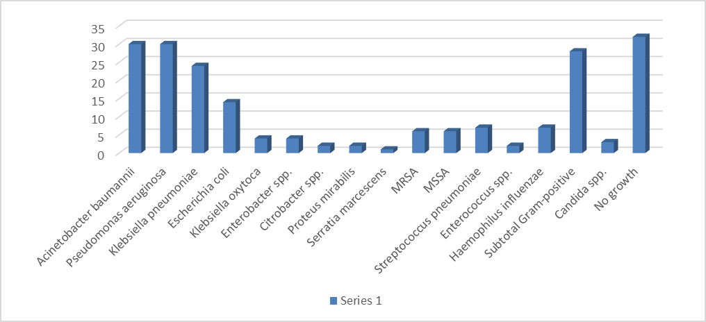
Figure 1. Distribution of microorganisms isolated from patients with VAP
Antibiotic Susceptibility Pattern
Antimicrobial susceptibility testing demonstrated variable susceptibility among the major Gram-negative pathogens. Overall, imipenem showed the highest susceptibility (52.3%), followed by meropenem (51.4%), gentamicin (45.9%), amikacin (43.2%), piperacillin–tazobactam (41.4%), and ciprofloxacin (39.6%). Susceptibility to several third- and fourth-generation cephalosporins was comparatively lower.
Among the major pathogens, Acinetobacterbaumannii demonstrated marked resistance across most tested antimicrobial agents. Susceptibility to imipenem and meropenem was 13.3% and 20.0%, respectively, while susceptibility to ciprofloxacin and levofloxacin was 6.7% and 16.7%, respectively. Pseudomonas aeruginosa showed comparatively higher susceptibility to meropenem and imipenem (60.0% each), while susceptibility to levofloxacin was 63.3%.
Klebsiellapneumoniae demonstrated susceptibility of 58.3% to both imipenem and meropenem, whereas Escherichia coli showed the highest susceptibility to these carbapenems, at 85.7% each. These findings demonstrate considerable variation in antimicrobial susceptibility among individual pathogens and support the importance of local antibiogram data in guiding empirical antimicrobial therapy for VAP.
Table 3. Antibiotic susceptibility pattern of major Gram-negative isolates
|
Antibiotic |
A. baumannii (n=30) |
P. aeruginosa (n=30) |
K. pneumoniae (n=24) |
E. coli (n=14) |
Overall sensitivity (%) |
|
Amikacin |
30.0 |
50.0 |
50.0 |
50.0 |
43.2 |
|
Gentamicin |
13.3 |
66.7 |
50.0 |
57.1 |
45.9 |
|
Ciprofloxacin |
6.7 |
53.3 |
41.7 |
57.1 |
39.6 |
|
Levofloxacin |
16.7 |
63.3 |
20.8 |
35.7 |
36.0 |
|
Meropenem |
20.0 |
60.0 |
58.3 |
85.7 |
51.4 |
|
Imipenem |
13.3 |
60.0 |
58.3 |
85.7 |
52.3 |
|
Piperacillin–Tazobactam |
16.7 |
56.7 |
41.7 |
35.7 |
41.4 |
|
Cefepime |
6.7 |
53.3 |
25.0 |
28.6 |
28.8 |
|
Ceftazidime |
16.7 |
56.7 |
29.2 |
28.6 |
34.2 |
|
Cefotaxime |
13.3 |
30.0 |
29.2 |
21.4 |
26.1 |
|
Cefoxitin |
16.7 |
36.7 |
33.3 |
57.1 |
35.1 |
|
Aztreonam |
10.0 |
43.3 |
29.2 |
28.6 |
30.0 |
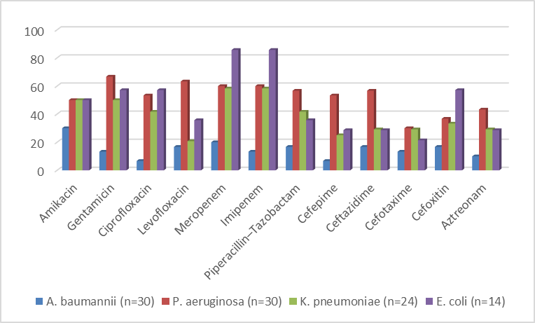
Figure 2: Antibiotic susceptibility of major Gram-negative isolates.
Antimicrobial Resistance Patterns
Among the 142 culture-positive cases, 74 (52.1%) demonstrated one or more clinically significant antimicrobial resistance phenotypes. Multidrug resistance (MDR) was identified in 26 (18.3%) isolates, extensively drug-resistant (XDR) isolates in 16 (11.3%), extended-spectrum β-lactamase (ESBL) production in 16 (11.3%), carbapenem-resistant Enterobacterales (CRE) in 10 (7.0%), and pandrug resistance (PDR) in 6 (4.2%).
The highest proportion of resistant isolates was observed among Acinetobacterbaumannii (76.7%), followed by Escherichia coli (64.3%), Klebsiellapneumoniae (62.5%), and Pseudomonas aeruginosa (53.3%). The frequency of resistant organisms was significantly higher among patients with late-onset VAP than among those with early-onset VAP (61.2% vs. 31.8%; p = 0.002).
Among the resistant isolates, A. baumannii demonstrated the greatest overall resistance burden, with MDR/XDR and carbapenem-resistant phenotypes being particularly prominent. These findings indicate a substantial burden of antimicrobial resistance among VAP-associated pathogens in the study setting.
Table 4. Antimicrobial resistance patterns among culture-positive isolates
|
Resistance category |
Number |
Percentage (%) |
|
Non-MDR |
68 |
47.9 |
|
MDR |
26 |
18.3 |
|
XDR |
16 |
11.3 |
|
ESBL |
16 |
11.3 |
|
CRE |
10 |
7.0 |
|
PDR |
6 |
4.2 |
Table 5. Antimicrobial Resistance According to VAP Onset
|
Resistance status |
Early-onset VAP (n=50) |
Late-onset VAP (n=124) |
p-value |
|
Resistant isolates |
16 (31.8%) |
58 (61.2%) |
0.002 |
|
Non-resistant isolates |
34 (68.2%) |
66 (38.8%) |
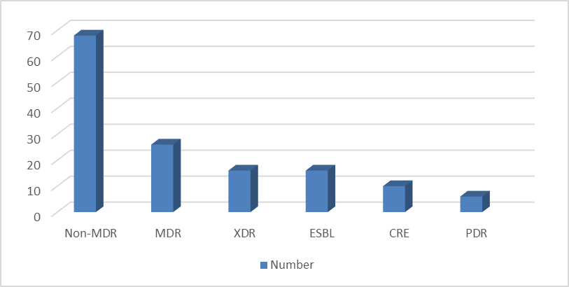
Figure 3: Distribution of antimicrobial resistance phenotypes among culture-positive isolates.
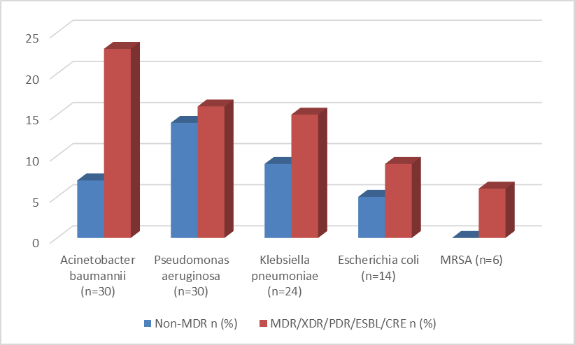
Figure 4: Comparison of antimicrobial resistance patterns between early- and late-onset ventilator-associated pneumonia.
Clinical Outcomes of Patients with Ventilator-Associated Pneumonia.
Among the 174 patients included in the study, 118 (67.8%) survived and 56 (32.2%) died. Sepsis was documented in 52 (29.9%) patients, while septic shock and multiple organ dysfunction syndrome (MODS) occurred in 30 (17.2%) and 31 (17.8%) patients, respectively.
The mean ICU stay was 19.3 ± 5.9 days, the mean hospital stay was 25.6 ± 8.6 days, and the mean total duration of mechanical ventilation was 16.2 ± 4.6 days. Late-onset VAP was associated with significantly longer ICU stay (20.5 ± 5.7 vs. 17.0 ± 5.5 days; p < 0.001), hospital stay (26.7 ± 8.6 vs. 23.4 ± 9.1 days; p = 0.024), and total duration of mechanical ventilation (17.3 ± 4.7 vs. 13.6 ± 4.1 days; p < 0.001) compared with early-onset VAP.
Mortality did not differ significantly between early-onset and late-onset VAP (30.0% vs. 33.1%; p = 0.832). Similarly, the frequencies of sepsis, septic shock, and MODS did not differ significantly according to VAP onset.
Among the 56 patients who died, septic shock was the most frequent documented cause of death, accounting for 18 (32.1%) deaths, followed by refractory hypoxemia in 12 (21.4%), MODS in 11 (19.6%), cardiac arrest in 6 (10.7%), refractory ARDS in 5 (8.9%), and disseminated intravascular coagulation in 4 (7.1%).
Table 6. Clinical Outcomes of Patients with VAP
|
Clinical outcome |
n (%) / Mean ± SD |
|
Survived |
118 (67.8) |
|
Died |
56 (32.2) |
|
Sepsis |
52 (29.9) |
|
Septic shock |
30 (17.2) |
|
MODS |
31 (17.8) |
|
ICU stay (days) |
19.3 ± 5.9 |
|
Hospital stay (days) |
25.6 ± 8.6 |
|
Total mechanical ventilation (days) |
16.2 ± 4.6 |
Table 7. Causes of Death Among Patients with VAP
|
Cause of death |
n (%) |
|
Septic shock |
18 (32.1) |
|
Refractory hypoxemia |
12 (21.4) |
|
MODS |
11 (19.6) |
|
Cardiac arrest |
6 (10.7) |
|
Refractory ARDS |
5 (8.9) |
|
Disseminated intravascular coagulation |
4 (7.1) |
|
Total |
56 (100) |
Clinical Outcomes According to Causative Organism
Clinical outcomes varied across the different microorganisms isolated from patients with VAP. Among the major pathogens, mortality was highest among patients with Escherichia coli (42.9%) and Acinetobacterbaumannii (40.0%), followed by Klebsiellapneumoniae (29.2%) and Pseudomonas aeruginosa (26.7%). Among less frequently isolated organisms, mortality was observed in 75.0% of patients with Klebsiellaoxytoca, 50.0% with Enterobacter spp., and 50.0% with Citrobacter spp. Mortality was 33.3% among patients with MRSA and Candida spp., while no deaths were recorded among patients with Streptococcus pneumoniae, Proteus mirabilis, or Enterococcus spp.
Among patients with no microbial growth, 11 of 32 patients (34.4%) died. Because several individual organisms were isolated from small numbers of patients, the organism-specific mortality percentages should be interpreted cautiously. These findings describe the observed outcome pattern according to the causative organism and do not establish a statistically significant association between individual organisms and mortality.
Table 8. Clinical Outcomes According to Causative Organism
|
Organism |
Total n |
Deaths n |
Mortality (%) |
|
Acinetobacterbaumannii |
30 |
12 |
40.0 |
|
Pseudomonas aeruginosa |
30 |
8 |
26.7 |
|
Klebsiellapneumoniae |
24 |
7 |
29.2 |
|
Escherichia coli |
14 |
6 |
42.9 |
|
Haemophilusinfluenzae |
7 |
2 |
28.6 |
|
Streptococcus pneumoniae |
7 |
0 |
0 |
|
MSSA |
6 |
1 |
16.7 |
|
MRSA |
6 |
2 |
33.3 |
|
Klebsiellaoxytoca |
4 |
3 |
75.0 |
|
Enterobacter spp. |
4 |
2 |
50.0 |
|
Candida spp. |
3 |
1 |
33.3 |
|
Citrobacter spp. |
2 |
1 |
50.0 |
|
Proteus mirabilis |
2 |
0 |
0 |
|
Enterococcus spp. |
2 |
0 |
0 |
|
Serratiamarcescens |
1 |
0 |
0 |
|
No growth |
32 |
11 |
34.4 |
Association of Antimicrobial Resistance with Clinical Outcomes
Clinical outcomes were compared between patients with resistant isolates and those with non-MDR isolates. Mortality was significantly higher among patients with MDR/XDR/CRE/ESBL-associated isolates than among those with non-MDR isolates (40.5% vs. 22.1%; p = 0.029). Similarly, multiple organ dysfunction syndrome (MODS) occurred significantly more frequently among patients with resistant organisms than among those with non-MDR isolates (24.3% vs. 10.3%; p = 0.049).
The mean ICU stay was 19.1 ± 5.0 days among patients with resistant isolates compared with 19.8 ± 6.3 days among those with non-MDR isolates (p = 0.470). Similarly, the mean hospital stay was 24.6 ± 8.2 and 26.8 ± 8.8 days, respectively (p = 0.126). Sepsis was observed in 31.1% of patients with resistant isolates compared with 23.5% of those with non-MDR isolates (p = 0.413).
These findings indicate that antimicrobial resistance was significantly associated with increased mortality and MODS, whereas differences in sepsis, ICU stay, and hospital stay were not statistically significant.
Table 9. Clinical Outcomes According to Antimicrobial Resistance Status
|
Clinical outcome |
Resistant isolates* |
Non-MDR isolates |
p-value |
|
Mortality |
30 (40.5%) |
15 (22.1%) |
0.029 |
|
Sepsis |
23 (31.1%) |
16 (23.5%) |
0.413 |
|
MODS |
18 (24.3%) |
7 (10.3%) |
0.049 |
|
ICU stay (days) |
19.1 ± 5.0 |
19.8 ± 6.3 |
0.470 |
|
Hospital stay (days) |
24.6 ± 8.2 |
26.8 ± 8.8 |
0.126 |
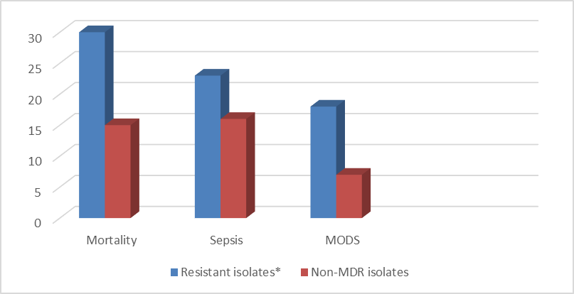
Figure 5. Clinical outcomes according to antimicrobial resistance status among patients with ventilator-associated pneumonia
SOFA Score and Clinical Outcome
The severity of illness was assessed using the Sequential Organ Failure Assessment (SOFA) score at the time of VAP diagnosis and at 72 hours. The mean SOFA score at the time of VAP diagnosis did not differ significantly between survivors and non-survivors (7.4 ± 1.9 vs. 7.6 ± 2.0; p = 0.513). In contrast, the mean SOFA score at 72 hours was significantly higher among non-survivors than survivors (9.8 ± 2.5 vs. 6.5 ± 2.1; p < 0.001).
Worsening of the SOFA score at 72 hours was observed in all non-survivors compared with only 5.1% of survivors (p < 0.001). These findings indicate that deterioration in organ function during the early course of VAP was strongly associated with mortality, whereas the initial SOFA score at the time of VAP diagnosis was not significantly different between survivors and non-survivors.
Table 10. SOFA Score According to Clinical Outcome
|
SOFA parameter |
Survivors (n=118) |
Non-survivors (n=56) |
p-value |
|
SOFA at VAP diagnosis |
7.4 ± 1.9 |
7.6 ± 2.0 |
0.513 |
|
SOFA at 72 hours |
6.5 ± 2.1 |
9.8 ± 2.5 |
<0.001 |
|
Worsening SOFA at 72 h |
6 (5.1%) |
56 (100%) |
<0.001 |
Multivariate Predictors of Mortality
Multivariate logistic regression analysis was performed to identify independent predictors of mortality among patients with ventilator-associated pneumonia. Multiple organ dysfunction syndrome (MODS) was independently associated with mortality and represented the strongest predictor in the model (adjusted OR 4.07, 95% CI 2.84–8.32; p = 0.001). A lower PaO₂/FiO₂ ratio was also independently associated with mortality (adjusted OR 0.69, 95% CI 0.39–0.95; p = 0.024).
In contrast, age, SOFA score at the time of VAP diagnosis, serum creatinine, serum albumin, MDR status, and late-onset VAP were not independently associated with mortality after adjustment. MDR status, although significantly associated with mortality in the unadjusted analysis, did not retain statistical significance in the multivariate model (adjusted OR 1.76, 95% CI 0.83–3.77; p = 0.162). Similarly, late-onset VAP was not independently associated with mortality (adjusted OR 1.01, 95% CI 0.50–2.05; p = 0.984).
Table 11. Multivariate Logistic Regression Analysis of Predictors of Mortality
|
Predictor |
Adjusted OR |
95% CI |
p-value |
|
Age |
1.02 |
0.99–1.05 |
0.210 |
|
SOFA score at VAP diagnosis |
1.08 |
0.97–1.20 |
0.158 |
|
Serum creatinine |
1.21 |
0.91–1.62 |
0.190 |
|
Serum albumin |
0.78 |
0.52–1.16 |
0.218 |
|
MODS |
4.07 |
2.84–8.32 |
0.001 |
|
PaO₂/FiO₂ ratio |
0.69 |
0.39–0.95 |
0.024 |
|
MDR status |
1.76 |
0.83–3.77 |
0.162 |
|
Late-onset VAP |
1.01 |
0.50–2.05 |
0.984 |
DISCUSSION:
Ventilator-associated pneumonia (VAP) remains an important healthcare-associated infection among critically ill patients receiving mechanical ventilation and is associated with considerable morbidity, mortality, prolonged hospitalization, and antimicrobial use. In the present study, 174 adult patients with VAP were evaluated with respect to their clinical profile, microbiological characteristics, antimicrobial susceptibility, resistance patterns, and clinical outcomes. The study population had a mean age of 51.0 ± 15.0 years, with males constituting 60.9% of the cohort. Medical ICU patients accounted for 53.4% of cases, while 46.6% were admitted to the Surgical ICU. Late-onset VAP was more common than early-onset VAP (71.3% vs. 28.7%). These findings provide an important clinical context for understanding the microbiological and resistance patterns observed in the study.18
Gram-negative organisms predominated in the present study, accounting for 63.8% of the isolates, whereas Gram-positive organisms accounted for 16.1%. Acinetobacterbaumannii and Pseudomonas aeruginosa were the most frequently isolated organisms, with 30 (17.2%) isolates each, followed by Klebsiellapneumoniae (13.8%) and Escherichia coli (8.0%). The predominance of Gram-negative pathogens, particularly non-fermenting Gram-negative bacilli, is an important finding because these organisms are frequently associated with difficult-to-treat infections in the ICU setting. The high representation of A. baumannii and P. aeruginosa also emphasizes the importance of continuous microbiological surveillance in individual ICUs.19
The antimicrobial susceptibility profile demonstrated substantial resistance among the Gram-negative isolates. Overall susceptibility was highest for imipenem (52.3%) and meropenem (51.4%), followed by gentamicin (45.9%), amikacin (43.2%), and piperacillin–tazobactam (41.4%). Susceptibility to several cephalosporins and fluoroquinolones was comparatively low. A. baumannii showed particularly poor susceptibility to most tested antimicrobial agents, whereas E. coli demonstrated comparatively higher susceptibility to carbapenems. These findings indicate that empirical antimicrobial treatment of VAP should be guided by local susceptibility data rather than relying solely on generalized antibiotic recommendations.20
A substantial burden of antimicrobial resistance was observed in the present study. Among the 142 culture-positive cases, 74 (52.1%) demonstrated one or more clinically significant antimicrobial resistance phenotypes. MDR was identified in 26 (18.3%) isolates, XDR in 16 (11.3%), ESBL production in 16 (11.3%), CRE in 10 (7.0%), and PDR in 6 (4.2%). The highest proportion of resistant isolates was observed among A. baumannii (76.7%), followed by E. coli (64.3%), K. pneumoniae (62.5%), and P. aeruginosa (53.3%). This substantial resistance burden represents an important therapeutic challenge and highlights the need for regular ICU-specific antibiograms and antimicrobial stewardship interventions.21
Clinical Outcomes and Relationship with Antimicrobial Resistance.
The present study demonstrated a substantial clinical burden associated with VAP, with an overall mortality of 32.2%. Sepsis was observed in 29.9% of patients, while septic shock and MODS occurred in 17.2% and 17.8%, respectively. Among the patients who died, septic shock was the most frequent documented cause of death, followed by refractory hypoxemia and MODS. These findings highlight the importance of early recognition of systemic deterioration and organ dysfunction in patients with VAP.22
Mortality varied across the microorganisms isolated in the study. Among the major pathogens, the highest mortality was observed among patients with Escherichia coli (42.9%) and Acinetobacterbaumannii (40.0%), followed by Klebsiellapneumoniae (29.2%) and Pseudomonas aeruginosa (26.7%). However, the interpretation of organism-specific mortality should be cautious because several less frequently isolated organisms were represented by small numbers of patients. Therefore, these findings describe the observed outcome pattern rather than establishing an independent association between individual organisms and mortality.23
An important finding of the present study was the significant association between antimicrobial resistance and adverse clinical outcomes. Mortality was significantly higher among patients with MDR/XDR/CRE/ESBL-associated isolates than among those with non-MDR isolates (40.5% vs. 22.1%; p = 0.029). MODS was also significantly more frequent among patients with resistant organisms (24.3% vs. 10.3%; p = 0.049). These findings suggest that antimicrobial resistance may contribute to poorer outcomes by limiting effective antimicrobial treatment options. Nevertheless, the association should be interpreted as an observational relationship rather than evidence of causation.24
The timing of VAP onset also demonstrated an important relationship with antimicrobial resistance. Resistant isolates were significantly more frequent among patients with late-onset VAP than among those with early-onset VAP (61.2% vs. 31.8%; p = 0.002). However, mortality did not differ significantly between early- and late-onset VAP (30.0% vs. 33.1%; p = 0.832). This suggests that although late-onset VAP was associated with a greater resistance burden, the timing of VAP onset alone was not sufficient to predict mortality in this cohort.25
The trajectory of organ dysfunction appeared to be more closely related to outcome. Although the SOFA score at VAP diagnosis was similar between survivors and non-survivors, the SOFA score at 72 hours was significantly higher among non-survivors (9.8 ± 2.5 vs. 6.5 ± 2.1; p < 0.001). Worsening of the SOFA score at 72 hours was observed in all non-survivors compared with only 5.1% of survivors (p < 0.001). These findings indicate that early deterioration in organ function may provide greater prognostic information than the initial severity assessment alone.25
The multivariate analysis further clarified the factors independently associated with mortality. MODS was the strongest independent predictor of mortality (adjusted OR 4.07, 95% CI 2.84–8.32; p = 0.001), while a lower PaO₂/FiO₂ ratio was also independently associated with mortality (adjusted OR 0.69, 95% CI 0.39–0.95; p = 0.024). In contrast, MDR status was not independently associated with mortality after adjustment (adjusted OR 1.76, 95% CI 0.83–3.77; p = 0.162). Thus, antimicrobial resistance was associated with mortality in the unadjusted analysis but did not remain an independent predictor after accounting for other clinical factors. This distinction is important because it suggests that the adverse effect of resistant infections may be mediated or confounded by the severity of critical illness and organ dysfunction.12
Strengths and Limitations
The major strength of this study is the integrated assessment of the clinical profile, microbiological spectrum, antimicrobial susceptibility, resistance patterns, and clinical outcomes of adult patients with VAP. The inclusion of organism-specific mortality and comparison of outcomes according to antimicrobial resistance provides clinically relevant information for ICU practice and antimicrobial stewardship.
However, the study was conducted at a single tertiary-care centre, which may limit the generalizability of the findings to other institutions. Some organism-specific mortality estimates were based on small numbers of isolates and should therefore be interpreted cautiously. Molecular characterization of resistance mechanisms was not performed, and the study relied primarily on conventional microbiological and phenotypic susceptibility methods. Furthermore, antimicrobial resistance patterns may change over time, emphasizing the need for continued institutional surveillance.
CONCLUSION:
The present study demonstrated that ventilator-associated pneumonia among adult intensive care unit patients was characterized by a predominance of Gram-negative organisms, particularly Acinetobacterbaumannii and Pseudomonas aeruginosa, together with a substantial burden of antimicrobial resistance. More than half of the culture-positive cases demonstrated one or more clinically significant antimicrobial resistance phenotypes, with particularly high resistance observed among A. baumannii.
The study also demonstrated an important clinical impact of antimicrobial resistance. Overall mortality was 32.2%, and mortality was significantly higher among patients with MDR/XDR/CRE/ESBL-associated isolates than among those with non-MDR isolates (40.5% vs. 22.1%; p = 0.029). MODS was also significantly more frequent among patients with resistant organisms (24.3% vs. 10.3%; p = 0.049). Organism-wise mortality was highest among E. coli and A. baumannii among the major pathogens, although mortality estimates for less frequently isolated organisms should be interpreted cautiously because of their small numbers.
Among the major pathogens, mortality was highest among patients with Escherichia coli and Acinetobacterbaumannii. Although higher mortality percentages were observed for some less frequently isolated organisms, these estimates should be interpreted cautiously because of their small sample sizes. These findings highlight the importance of integrating the patient's clinical profile, microbiological findings, antimicrobial susceptibility results, and clinical severity in the management of VAP.
REFERENCES:
1. Torres A, Niederman MS, Chastre J, Ewig S, Fernandez-Vandellos P, Hanberger H, et al. International ERS/ESICM/ESCMID/ALAT guidelines for the management of hospital-acquired pneumonia and ventilator-associated pneumonia. EurRespir J. 2017;50(3):1700582.
2. Kalil AC, Metersky ML, Klompas M, Muscedere J, Sweeney DA, Palmer LB, et al. Management of adults with hospital-acquired and ventilator-associated pneumonia: 2016 clinical practice guidelines by the IDSA and ATS. Clin Infect Dis. 2016;63(5):e61-e111.
3. Papazian L, Klompas M, Luyt CE. Ventilator-associated pneumonia in adults: a narrative review. Intensive Care Med. 2020;46(5):888-906.
4. Fernando SM, Tran A, Cheng W, Klompas M, Kyeremanteng K, Mehta S, et al. Diagnosis of ventilator-associated pneumonia in critically ill adult patients: a systematic review and meta-analysis. Intensive Care Med. 2020;46(6):1170-1179.
5. Klompas M. Ventilator-associated pneumonia: diagnosis, treatment, and prevention. BMJ. 2023;381:e075158.
6. Semet C, Boissier F, de Jong A, Razazi K, Mahjoub Y, et al. The ongoing challenge of ventilator-associated pneumonia: epidemiology, prevention, and risk factors for mortality. Infect PrevPract. 2023;5:100320.
7. Bassetti M, Righi E, Vena A, Giacobbe DR. Epidemiology and treatment of hospital-acquired and ventilator-associated pneumonia. CurrOpin Infect Dis. 2023;36(2):154-162.
8. Wałaszek M, Różańska A, Wałaszek MZ, Wójkowska-Mach J, Domańska J, Dubiel G, et al. Epidemiology of ventilator-associated pneumonia, microbiological diagnostics and duration of antimicrobial treatment in Polish ICUs. BMC Infect Dis. 2018;18:308.
9. Rit K, Chakraborty B, Saha R, Majumder U. Ventilator-associated pneumonia in a tertiary care hospital in India: incidence, etiology, risk factors and role of multidrug-resistant pathogens. Int J Med Public Health. 2014;4:51-56.
10. Patil HV, Patil VC. Incidence, bacteriology and clinical outcome of ventilator-associated pneumonia at a tertiary care hospital. J Nat SciBiol Med. 2017;8(1):46-55.
11. Melsen WG, Rovers MM, Groenwold RH, Bergmans DC, Camus C, Bauer TT, et al. Attributable mortality of ventilator-associated pneumonia: a meta-analysis. Lancet Infect Dis. 2013;13(8):665-671.
12. Timsit JF, Esaied W, Neuville M, Bouadma L, Mourvillier B. Update on ventilator-associated pneumonia. F1000Res. 2017;6:2061.
13. Barbier F, Andremont A, Wolff M, Bouadma L. Hospital-acquired pneumonia and ventilator-associated pneumonia: recent advances in epidemiology and management. CurrOpinPulm Med. 2013;19(3):216-228.
14. Chastre J, Fagon JY. Ventilator-associated pneumonia. Am J RespirCrit Care Med. 2002;165(7):867-903.
15. Kollef MH. Ventilator-associated pneumonia: a multivariable analysis. Chest. 1993;103(2):547-552.
16. World Health Organization. Global antimicrobial resistance and use surveillance system (GLASS) report 2024. Geneva: WHO; 2024.
17. European Centre for Disease Prevention and Control. Healthcare-associated infections acquired in intensive care units. HAI-Net Annual Epidemiological Report 2023. Stockholm: ECDC; 2024.
18. Magill SS, O'Leary E, Janelle SJ, Thompson DL, Dumyati G, Nadle J, et al. Changes in prevalence of healthcare-associated infections in U.S. hospitals. N Engl J Med. 2018;379(18):1732-1744.
19. Magiorakos AP, Srinivasan A, Carey RB, Carmeli Y, Falagas ME, Giske CG, et al. Multidrug-resistant, extensively drug-resistant and pandrug-resistant bacteria: international expert proposal for interim standard definitions. ClinMicrobiol Infect. 2012;18(3):268-281.
20. Clinical and Laboratory Standards Institute. Performance Standards for Antimicrobial Susceptibility Testing. 35th ed. CLSI supplement M100. Wayne (PA): CLSI; 2025.
21. Cilloniz C, Dominedò C, Torres A. Multidrug-resistant Gram-negative bacteria in hospital-acquired and ventilator-associated pneumonia. SeminRespirCrit Care Med. 2021;42(2):269-283.
22. Kalanuria AA, Ziai W, Mirski M. Ventilator-associated pneumonia in the ICU. Crit Care. 2014;18(2):208.
23. Wu D, Wu C, Zhang S, Zhong Y. Risk factors of ventilator-associated pneumonia in critically ill patients. Front Pharmacol. 2019;10:482.
24. Martin-Loeches I, Torres A. What is new in ventilator-associated pneumonia? EurRespir Rev. 2024;33:230187.
25. Metersky ML, Kalil AC. Hospital-acquired and ventilator-associated pneumonia: advances in diagnosis and antimicrobial therapy. Clin Chest Med. 2023;44(3):453-468.