Histopathological Spectrum of Lung Lesions in Medico-Legal Autopsies: A Three-Year Retrospective Study.
- Bikash Ranjan Panda , Associate Professor, Department of Pathology, Veer Surendra Sai Institute of Medical Sciences and Research, Burla, Odisha
- Jyotirmayee Mishra , Professor, Department of Pathology Veer Surendra Sai Institute of Medical Sciences and Research, Burla, Odisha
- Yespal Sharma , Assistant Professor, Department of Pathology Veer Surendra Sai Institute of Medical Sciences and Research, Burla, Odisha.
Article Information:
Abstract:
Background: Preventable respiratory disorders contribute significantly to global morbidity and mortality. Because clinical manifestations and radiological findings in pulmonary conditions often overlap, achieving a precise antemortem diagnosis can be challenging. Postmortem histopathological evaluation remains vital for uncovering pulmonary pathology, clarifying internal organ changes, and determining the underlying cause of death. Objectives: This study aimed to analyze the spectrum and prevalence of histopathological changes observed in lung tissues retrieved during medico-legal autopsies. Materials and Methods: A retrospective study of 150 autopsy lung specimens was conducted in the Department of Pathology, VIMSAR, Burla, over a three-year period (January 2020 to December 2022). Preserved tissue samples were fixed in 10% neutral buffered formalin, routinely processed, paraffin-embedded, and stained with Hematoxylin and Eosin (H&E). Special stains (Ziehl-Neelsen and Periodic Acid-Schiff) were utilized where indicated. Microscopic findings were documented and systematically analyzed. Results: Of the 150 cases evaluated, microscopic changes were identified in 88.6% (n=133). Unremarkable tissue was observed in 7.3% (n=11), while 4.0% (n=6) showed autolysis. Males constituted 63.3% (n=95) of cases and females accounted for 36.7% (n=55). The predominant histopathological finding was pulmonary congestion/edema/interstitial inflammation (64.0%), followed by pneumonia (12.7%), emphysema (7.3%), tuberculosis (2.7%), acute respiratory distress syndrome (ARDS; 1.3%), and other granulomatous lesions (0.7%). Conclusion: Histopathological examination of autopsy lung specimens provides critical diagnostic clarity regarding incidental pulmonary conditions and primary causes of death. Despite advances in diagnostic modalities, autopsy examination remains an indispensable tool for refining clinical diagnosis and quality assurance.
Keywords:
Article :
INTRODUCTION:
Respiratory pathologies encompass a wide array of inflammatory, vascular, infectious, and neoplastic conditions. Furthermore, due to the close physiological coupling of the cardiovascular and pulmonary systems, the lungs frequently exhibit secondary alterations during terminal events. Pathological processes affecting the pulmonary parenchyma typically manifest as inflammatory responses, progressive interstitial fibrosis, or granulomatous reactions. While clinical history, laboratory parameters, and radiological imaging provide helpful diagnostic orientation, definitive evaluation and prognostic assessment rely heavily on histopathological examination.
Certain early or subtle tissue-level alterations are difficult to capture through routine clinical biopsies, leaving gaps in the understanding of early disease progression. In this context, postmortem histopathological analysis serves as an informative tool to elucidate underlying disease mechanisms, clarify cause-of-death determinations in forensic practice, and expand pulmonary pathology knowledge.
Gross examination of autopsy lungs offers initial insights into tissue aeration (hyperinflation versus atelectasis), vascular congestion, parenchymal consolidation, nodular lesions, bullous changes, cavitation, and pleural or bronchial involvement. Integrating gross observations with microscopic analysis allows for precise diagnostic categorization. The primary aim of this study was to establish the prevalence and structural patterns of pathological changes in lung specimens submitted for medico-legal postmortem examination.
MATERIALS AND METHODS:
Study Design and Setting
This retrospective study was conducted in the Department of Pathology at VIMSAR, Burla, encompassing a three-year timeframe from January 2020 to December 2022.
Case Selection
All consecutive autopsy lung specimens received during the study period were included, without restriction on age or sex. A total of 150 lung samples were evaluated. Medico-legal autopsies were conducted by qualified forensic specialists, and representative pulmonary tissue samples were preserved in 10% neutral buffered formalin prior to transfer to the pathology department.
Processing and Microscopic Examination
For each case, a minimum of two tissue blocks per lung were sampled, regardless of whether macroscopic lesions were visibly discernible. Tissues underwent standard automated processing and paraffin embedding. Microtome sections were cut at a thickness of 4 μ"m" , stained with standard Hematoxylin and Eosin (H&E), and mounted. Auxiliary histochemical stains, including Ziehl-Neelsen (ZN) for acid-fast bacilli and Periodic Acid-Schiff (PAS) for fungal elements, were performed when clinically or morphologically indicated. Light microscopy was performed to categorize histopathological changes.
RESULTS:
A total of 150 autopsy lung specimens were analyzed. Overall, 133 cases (88.6%) demonstrated significant histopathological abnormalities. Eleven cases (7.3%) revealed normal pulmonary histology, and six cases (4.0%) were autolyzed, preventing reliable microscopic evaluation. Male subjects predominated the study population ( ; 63.3%) compared to female subjects ( ; 36.7%).
Table 1. Gender-Wise Distribution of Histopathological Lung Lesions
|
Lesion Type |
Male (n, %) |
Female (n, %) |
Total (n, %) |
|
Congestion / Edema / Interstitial Inflammation |
60 (40.0%) |
36 (24.0%) |
96 (64.0%) |
|
Pneumonia |
13 (8.7%) |
6 (4.0%) |
19 (12.7%) |
|
Emphysema |
6 (4.0%) |
5 (3.3%) |
11 (7.3%) |
|
Tuberculosis |
3 (2.0%) |
1 (0.7%) |
4 (2.7%) |
|
Acute Respiratory Distress Syndrome (ARDS) |
2 (1.3%) |
0 (0.0%) |
2 (1.3%) |
|
Other Inflammatory Lesions (Fungal / Granulomatous) |
1 (0.7%) |
0 (0.0%) |
1 (0.7%) |
|
Autolyzed |
4 (2.7%) |
2 (1.3%) |
6 (4.0%) |
|
Normal Histology |
6 (4.0%) |
5 (3.3%) |
11 (7.3%) |
|
Total |
95 (63.3%) |
55 (36.7%) |
150 (100.0%) |
Table 2. Age-Wise Distribution of Histopathological Lung Lesions
|
Lesion Type |
0–15 yrs |
16–30 yrs |
31–45 yrs |
46–60 yrs |
> 60 yrs |
Total |
|
Congestion / Edema / Interstitial Inflammation |
4 |
20 |
18 |
26 |
28 |
96 |
|
Pneumonia |
3 |
7 |
2 |
4 |
3 |
19 |
|
Emphysema |
0 |
0 |
7 |
4 |
0 |
11 |
|
Tuberculosis |
0 |
1 |
0 |
2 |
1 |
4 |
|
ARDS |
0 |
0 |
0 |
0 |
2 |
2 |
|
Other Inflammatory Lesions |
0 |
0 |
0 |
1 |
0 |
1 |
|
Autolyzed |
0 |
3 |
2 |
1 |
0 |
6 |
|
Normal Histology |
4 |
0 |
4 |
3 |
0 |
11 |
|
Total |
9 |
31 |
31 |
45 |
34 |
150 |
Histopathological Findings
- Congestion, Edema, and Terminal Changes: Present in 64.0% ( ) of cases, representing the most common finding across both sexes. These changes were concentrated in individuals aged 16 to 60+ years.
- Pneumonia: Identified in 12.7% ( ) of cases, making it the second most frequent lesion. Peak incidence was noted in the 16–30 age group ( ). Microscopic evaluation revealed acute alveolar exudation, diffuse alveolar damage, and interstitial inflammatory infiltrates.
- Emphysema: Observed in 11 cases (7.3%), with 6 males and 5 females. Histology demonstrated enlarged airspaces with alveolar septal disruption and loss of pulmonary parenchyma. Cases were identified in the 31–45 and 46–60 age brackets.
- Tuberculosis: Detected in 4 cases (2.7%; 3 males, 1 female), predominantly within the 46–60 age group. Microscopic sections showed characteristic caseating granulomatous inflammation.
- ARDS: Confirmed in 2 male patients (> 60 years), showing prominent intra-alveolar hyaline membranes and lymphocytic interstitial infiltrates.
- Neoplastic Lesions: No primary or metastatic malignant neoplasms were identified in this series.
Microscopic Observations
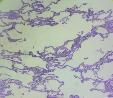
- Figure 1 (Emphysema): Histology shows marked enlargement of airspaces with fragmentation and loss of alveolar septa, characteristic of emphysema.
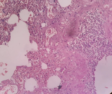
- Figure 2 (Interstitial Pneumonia): Photomicrograph demonstrates diffuse alveolar damage, interstitial fibrosis, and infiltration by mononuclear inflammatory cells.
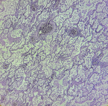
- Figure 3 (ARDS): Microscopic field highlights dense hyaline membrane formation lining the alveolar ducts alongside intra-alveolar inflammation, indicative of subacute ARDS.
DISCUSSION:
Postmortem histopathological examination offers valuable insights into disease prevalence, terminal pathophysiological events, and diagnostic correlation. In the present study, male subjects accounted for the majority of autopsy cases (63.3%), a finding consistent with studies by Kurawar et al., Selvambigai et al., Garg et al., and Chauhan et al. Higher male representation in medico-legal autopsies often relates to greater occupational exposure, environmental hazards, and trauma risk factors.
Terminal pulmonary changes—comprising vascular congestion, pulmonary edema, interstitial inflammation, and focal hemorrhage—were the predominant findings (64.0%). These non-specific terminal features align with reports by Kurawar et al., Garg et al., and Chauhan et al.. Environmental pollution, tobacco usage, underlying cardiovascular disease, and terminal hemodynamic failure contribute to these changes. Pneumonia constituted the second most common entity (12.7%), which compares well with findings from Chauhan et al. and Kurawar et al.. Pulmonary tuberculosis was noted in 2.7% of cases, with a higher prevalence in males, reflecting findings described by Hjorth et al.
ARDS was identified in 1.3% of cases, comparable to low rates reported by Sachdev et al. and Khare et al. Emphysema was detected in 7.3% of cases, similar to observations by Khare et al. The majority of individuals presenting with pulmonary pathology fell into the working-age demographic of 16 to 60 years, consistent with observations by Selvambigai et al. and Kurawar et al.
Limitations
Several limitations were encountered during this study:
1. Partial Tissue Sampling: In certain instances, only targeted or partial tissue specimens were submitted rather than whole organs, restricting comprehensive architectural assessment.
2. Autolysis: Postmortem tissue degradation rendered 4.0% of specimens non-diagnostic.
3. Clinical History Constraints: Incomplete antemortem clinical records occasionally limited clinicopathological correlation.
CONCLUSION:
This study underscores the role of postmortem histopathology in identifying both clinically silent and fatal pulmonary conditions. Non-specific terminal changes (congestion and edema), pneumonia, and emphysema represented the most frequent pathological features. Because a significant proportion of lesions occurred in the active working population (16–60 years), early screening and preventive healthcare strategies remain critical. Postmortem tissue examination continues to serve as an essential tool for clinical audit, diagnostic refinement, and legal cause-of-death determination.
REFERENCES:
1. Bal MS, Sethi PS, Suri AK, Bodal VK, Kaur G. Histopathological pattern in lung autopsies. JPAFMAT. 2008;8(2):29-31.
2. Kumar V, Abbas AK, Aster JC. Robbins and Cotran Pathologic Basis of Disease. 9th ed. Elsevier; 2014.
3. Kasper DL, Fauci AS, Hauser SL, Longo DL, Jameson JL, Loscalzo J. Harrison's Principles of Internal Medicine. 19th ed. McGraw-Hill; 2015.
4. Ghosal R, Kloer P, Lewis KE. A review of novel biological tools used in screening for the early detection of lung cancer. Postgrad Med J. 2009;85(1004):358-363.
5. Reddy KSN, Murty OP. The Essentials of Forensic Medicine and Toxicology. 33rd ed. Jaypee Brothers Medical Publishers; 2014.
6. Kurawar RR, Vasaikar MS. Spectrum of histomorphological changes in lungs at autopsy: A 5 year study. J Med Sci Clin Res. 2017;5(12):31304-31308.
7. Selvambigai G, Amudhavalli S, Deepak Chakravarthi CD, Ravi S. Histopathological study of lung in autopsy cases: a prospective study. Int J Res Med Sci. 2016;4(11):4816-4819.
8. Garg P, Sharma A, Kundal RK. Spectrum of pulmonary histopathological lesions: A study of 100 autopsy cases. J Med Sci Clin Res. 2017;5(12):31400-31405.
9. Chauhan G, Agrawal M, Thakkar N, Parghi B. Spectrum of histopathological lesions in lung autopsy. KRMDS. 2015;3(2):109-112.
10. Hjorth L, Jensen HS, Noer H. Acute pulmonary infections at autopsy. A study of clinical and microscopic diagnoses at autopsy compared with microscopic autopsy findings. Ugeskr Laeger. 1995;157(49):6873-6876.
11. Sachdev S, Pandit SP. Acute respiratory distress syndrome: an autopsy study. J Postgrad Med Edu Res. 2014;48(1):8-13.
12. Khare P, Gupta R, Ahuja M, Khare N, Agarwal S, Bansal D. Prevalence of lung lesions at autopsy: A histopathological study. J Clin Diagn Res. 2017;11(5):EC13-EC16.