Diagnostic and Prognostic Role of Fetal Venous Doppler in Fetal Growth Restriction: A Prospective Observational Study
- Rakesh Vijayvargiya , Professor, Department of Radiodiagnosis, MGMMC and MYH Indore (M.P)
- Alka Agrawal , HOD and professor, Department of Radiodiagnosis, MGMMC and MYH Indore (M.P)
- Neelesh Shrivastava , Resident, Department of Radiodiagnosis, MGMMC and MYH Indore (M.P)
- Yashasvi Sharma , Resident, Department of Radiodiagnosis, MGMMC and MYH Indore (M.P)
- Jatin Rawat , Resident, Department of Radiodiagnosis, MGMMC and MYH Indore (M.P)
Article Information:
Abstract:
Background: Fetal growth restriction is associated with placental insufficiency, fetal hypoxia, and adverse perinatal outcomes. Venous Doppler may identify cardiovascular deterioration in affected fetuses. This study evaluated umbilical vein and ductus venosus Doppler findings and their ability to predict adverse outcomes. Methods: This prospective observational study included 170 pregnant women between 27 and 37 weeks of gestation with a singleton fetus having an estimated fetal weight below the 10th percentile. Fetal biometry and Doppler assessment of the umbilical artery, umbilical vein, and ductus venosus were performed at approximately 32 and 36 weeks. Participants were followed until delivery. Associations were assessed using the chi-square test, with p<0.05 considered significant. Results: Normal umbilical vein flow decreased from 97.1% at 32 weeks to 80.0% at 36 weeks, while normal ductus venosus pulsatility decreased from 87.1% to 70.0%. At 36 weeks, triphasic umbilical vein pulsations were present in 17.6%, and absent or reversed ductus venosus a-wave was observed in 21.2%. Abnormal umbilical vein and ductus venosus findings were significantly associated with lower estimated fetal weight (p<0.001 and p=0.003, respectively). Both parameters showed 80% sensitivity for adverse outcomes. Umbilical vein Doppler showed 94% specificity, compared with 84% for ductus venosus Doppler. Conclusion: Serial fetal venous Doppler provides useful information about worsening fetal compromise and may improve risk assessment and delivery planning in fetal growth restriction.
Keywords:
Article :
INTRODUCTION:
|
|
This article is an open access article distributed under the terms and conditions of the Creative Commons Attribution (CC-BY) license (http://creativecommons.org/licenses/by/4.0/). |
INTRODUCTION
Fetal growth restriction (FGR) is an important complication of pregnancy and a major cause of perinatal illness and death. It occurs when a fetus fails to achieve its expected growth potential and is commonly defined as an estimated fetal weight below the 10th percentile for gestational age. However, not every small fetus is growth restricted. Distinguishing a constitutionally small fetus from pathological FGR is important because true FGR increases the risk of fetal distress, stillbirth, neonatal acidosis, intensive care admission, and long-term neurodevelopmental problems.1,2
FGR may result from maternal, fetal, or placental factors, although placental insufficiency is the most common cause. Reduced placental perfusion leads to chronic fetal hypoxia and inadequate nutrient supply. The fetus responds by redistributing blood toward vital organs, particularly the brain and heart. When these compensatory mechanisms fail, progressive cardiovascular deterioration may occur.3,4
Doppler ultrasonography provides a non-invasive assessment of fetal and placental circulation. Umbilical artery Doppler is widely used to identify increased placental resistance, but abnormalities may become apparent only after placental disease is advanced. Fetal venous Doppler, particularly examination of the ductus venosus and umbilical vein, provides information about cardiac function and fetal decompensation. Increased ductus venosus pulsatility or absent or reversed flow during atrial contraction is associated with fetal acidemia and adverse outcomes. Venous abnormalities may also appear before changes in cardiotocography or biophysical profile.5,6
Therefore, this study evaluated fetal venous Doppler findings in FGR and examined their relationship with fetal compromise and perinatal outcomes.
MATERIALS AND METHODS:
This hospital-based, time-bound, prospective observational study was conducted in the Department of Radiodiagnosis, Mahatma Gandhi Memorial Medical College and Maharaja Yeshwantrao Hospital, Indore, Madhya Pradesh, India. The study was carried out over one year after obtaining approval from the Institutional Scientific Review Board and Institutional Ethics Committee. Written informed consent was obtained from every participant before enrolment.
The study included 170 pregnant women referred for antenatal ultrasonography. Women between 27 and 37 weeks of gestation with a singleton viable fetus and fetal growth restriction were eligible. FGR was defined as an estimated fetal weight below the 10th percentile for gestational age. Pregnant women with maternal risk factors, including hypertension, diabetes mellitus, hypothyroidism, anaemia, and heart disease, were also included. Women without evidence of FGR and those who did not provide consent were excluded.
The sample size was estimated using Cochran’s formula: N = Z²p(1 − p)/d², where Z was 1.96 at a 95% confidence level, the expected prevalence of FGR was 12.5% (p=0.125), and the absolute precision was 5% (d=0.05). The minimum calculated sample size was 168, which was rounded to 170 participants. The calculation was performed with assistance from a statistician at MGM Medical College, Indore.
Participants meeting the eligibility criteria received information about the study in English or Hindi. After informed consent and completion of the required Form F under the Pre-Conception and Pre-Natal Diagnostic Techniques Act, obstetric ultrasonography was performed. Each participant was examined in the supine position in a quiet setting using a high-end ultrasound system equipped with a low-frequency curvilinear transducer. An adequate quantity of ultrasound gel was applied, and the transducer was placed gently over the maternal abdomen.
Initially, standard fetal biometric parameters were recorded and the estimated fetal weight was calculated. Participants were subsequently followed at approximately 32 and 36 weeks of gestation. Doppler examination included assessment of the umbilical artery, umbilical vein, and ductus venosus. All participants were followed until delivery to record the perinatal outcome. The final fetal biometry, estimated fetal weight, and Doppler findings obtained before delivery were used for analysis (Figure 1).
Data were entered into Microsoft Excel and analysed using SPSS version 25.0 (IBM Corp., Armonk, NY, USA). Continuous variables were presented as mean and standard deviation, while categorical variables were expressed as frequencies and percentages. The association of estimated fetal weight categories with umbilical vein and ductus venosus findings was assessed using Pearson’s chi-square test or Fisher’s exact test, as appropriate. The diagnostic performance of venous Doppler for predicting adverse perinatal outcomes was evaluated by calculating sensitivity, specificity, positive predictive value, and negative predictive value. A two-sided p-value <0.05 was considered statistically significant.
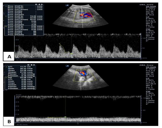
Figure 1. Doppler findings in a 27-year-old multigravida at 36 weeks of gestation with fetal growth restriction (estimated fetal weight: 1900 g) and no identifiable risk factors. (A) Ductus venosus Doppler showing a mildly increased pulsatility index. (B) Umbilical vein Doppler demonstrating normal monophasic flow. Cesarean delivery was performed because of a non-reassuring fetal heart rate, and the neonate required a brief NICU admission.
RESULTS:
The study included 170 pregnant women with fetal growth restriction. The mean maternal age was 27.8 ± 5.4 years, and the mean gestational age at presentation was 33.8 ± 2.7 weeks. Most women were nulliparous (56.5%), and 68.8% had at least one maternal risk factor. Hypertensive disorders were the most common, affecting 53.5%. Most fetuses had an estimated weight between 1500 and 1999 g (38.2%). Caesarean delivery was performed in 54.1% of cases. (Table 1)
At 32 weeks, normal umbilical artery, umbilical vein, and ductus venosus findings were observed in 75.9%, 97.1%, and 87.1% of fetuses, respectively. At 36 weeks, normal findings decreased to 56.5%, 80.0%, and 70.0%, respectively. Absent or reversed umbilical artery flow, triphasic umbilical vein pulsations, and absent or reversed ductus venosus a-wave became more frequent at 36 weeks. (Table 2)
Abnormal venous Doppler findings were more frequent among fetuses with lower estimated fetal weight. Umbilical vein abnormalities were present in 37.8% of fetuses weighing below 1500 g, compared with 1.7% in the 2000–2499 g group (p<0.001). Ductus venosus abnormalities also increased as fetal weight decreased (p=0.003). (Table 3)
Both venous Doppler parameters showed 80% sensitivity. Umbilical vein Doppler had higher specificity and positive
predictive value than ductus venosus Doppler. Both tests had high negative predictive values, indicating that normal venous Doppler findings were associated with a lower likelihood of adverse perinatal outcome. (Table 4)
Table 1. Clinical characteristics of the study population
|
Characteristic |
Value |
|
Maternal age, years |
27.8 ± 5.4 |
|
Gestational age at presentation, weeks |
33.8 ± 2.7 |
|
Nulliparous |
96 (56.5) |
|
Multiparous |
74 (43.5) |
|
Maternal risk factor present |
117 (68.8) |
|
Hypertensive disorder |
91 (53.5) |
|
No identified maternal risk factor |
53 (31.2) |
|
EFW <1500 g |
45 (26.5) |
|
EFW 1500–1999 g |
65 (38.2) |
|
EFW 2000–2499 g |
60 (35.3) |
|
Caesarean delivery |
92 (54.1) |
|
Vaginal delivery |
78 (45.9) |
Table 2. Serial fetal Doppler findings at 32 and 36 weeks
|
Vessel and Doppler finding |
32 weeks n (%) |
36 weeks n (%) |
|
|
Umbilical artery |
Normal flow |
129 (75.9) |
96 (56.5) |
|
Increased resistance |
36 (21.2) |
36 (21.2) |
|
|
Absent end-diastolic flow |
5 (2.9) |
22 (12.9) |
|
|
Reversed end-diastolic flow |
0 |
16 (9.4) |
|
|
Umbilical vein |
Normal/monophasic flow |
165 (97.1) |
136 (80.0) |
|
Biphasic pulsations |
2 (1.2) |
4 (2.4) |
|
|
Triphasic pulsations |
3 (1.8) |
30 (17.6) |
|
|
Ductus venosus |
Normal pulsatility index |
148 (87.1) |
119 (70.0) |
|
Increased pulsatility index |
15 (8.8) |
15 (8.8) |
|
|
Absent a-wave |
7 (4.1) |
17 (10.0) |
|
|
Reversed a-wave |
0 |
19 (11.2) |
|
Table 3. Association of fetal venous Doppler findings with estimated fetal weight at 36 weeks
|
Doppler finding |
<1500 g (n=45) |
1500–1999 g (n=65) |
2000–2499 g (n=60) |
p-value |
|
|
Umbilical vein |
Normal/monophasic flow |
28 (62.2) |
49 (75.4) |
59 (98.3) |
<0.001 |
|
Biphasic pulsations |
3 (6.7) |
1 (1.5) |
0 |
||
|
Triphasic pulsations |
14 (31.1) |
15 (23.1) |
1 (1.7) |
||
|
Ductus venosus |
Normal pulsatility index |
25 (55.6) |
43 (66.2) |
51 (85.0) |
0.003 |
|
Increased pulsatility index |
7 (15.6) |
5 (7.7) |
3 (5.0) |
||
|
Absent a-wave |
8 (17.8) |
6 (9.2) |
3 (5.0) |
||
|
Reversed a-wave |
5 (11.1) |
11 (16.9) |
3 (5.0) |
||
Table 4. Diagnostic performance of fetal venous Doppler for predicting adverse perinatal outcome
|
Doppler parameter |
Sensitivity (%) |
Specificity (%) |
PPV (%) |
NPV (%) |
|
Ductus venosus |
80.0 |
84.0 |
59.3 |
93.7 |
|
Umbilical vein |
80.0 |
94.0 |
80.0 |
94.0 |
DISCUSSION:
Fetal growth restriction is commonly related to placental insufficiency and may lead to progressive fetal hypoxia. Although fetal biometry identifies reduced growth, it does not directly show how well the fetus is adapting to circulatory stress.
Doppler assessment is therefore useful because arterial changes indicate increased placental resistance, while venous abnormalities reflect impaired cardiac function and more advanced fetal compromise.
In this study, the mean gestational age was 33.8 ± 2.7 weeks, and hypertensive disorders were the most frequent maternal risk factor. This agrees with hypertension and pre-eclampsia as important causes of placenta-mediated growth restriction.7 More than half of the women were nulliparous, a pattern also described by Brosens et al., and Alfirevic et al.6,8
Serial Doppler assessment showed clear deterioration between 32 and 36 weeks. Normal umbilical artery flow decreased from 75.9% to 56.5%, while absent and reversed end-diastolic flow became more frequent. Similar progressive changes have been reported by Baschat et al., and Turan et al. These findings support the concept that increasing placental resistance is followed by worsening fetal circulatory adaptation.9,10
Venous changes were uncommon at 32 weeks but increased considerably by 36 weeks. Triphasic umbilical vein pulsations increased from 1.8% to 17.6%. Similarly, absent and reversed ductus venosus a-waves increased from 4.1% and 0% to 10.0% and 11.2%, respectively. Bilardo et al., Ferrazzi et al., and Hofstaetter et al. also observed that venous abnormalities generally appear during advanced fetal compromise and are associated with cardiac dysfunction and poor outcome.11–13
Abnormal venous Doppler findings were significantly associated with lower estimated fetal weight. Umbilical vein abnormalities were more common in the lower-weight groups (p<0.001), and ductus venosus abnormalities showed a similar association (p=0.003). These findings agree with Bilardo et al., who reported that severe growth restriction is more frequently accompanied by abnormal venous flow.11
Both ductus venosus and umbilical vein Doppler showed 80% sensitivity for predicting adverse perinatal outcome. Umbilical vein Doppler had higher specificity than ductus venosus Doppler (94% versus 84%) and a higher positive predictive value (80% versus 59.3%). Both demonstrated high negative predictive values, suggesting that normal venous Doppler findings may help identify fetuses at lower immediate risk. However, Doppler findings should be interpreted together with gestational age, fetal growth, arterial Doppler, and other surveillance findings.
The study was limited by its single-centre design and absence of long-term neonatal follow-up. Nevertheless, the findings support serial venous Doppler as a useful addition to routine surveillance in fetal growth restriction.
CONCLUSION:
Fetal venous Doppler is a useful addition to the surveillance of pregnancies complicated by fetal growth restriction. Abnormal umbilical vein and ductus venosus waveforms were more frequent in fetuses with lower estimated weight and increased with advancing gestation. Both parameters showed good sensitivity and high negative predictive values for adverse perinatal outcomes, while umbilical vein Doppler demonstrated greater specificity. Serial venous Doppler assessment may help identify fetal deterioration, guide the timing of delivery, and support safer individualized obstetric management.
REFERENCES:
1. Shrivastava D, Master A. Fetal Growth Restriction. J Obstet Gynaecol India. 2020;70(2):103-110.
2. Caetano ACR, Zamarian ACP, Nardozza LMM, Miyadahira S, Tedesco GD, Rossi LD, Callado GY, Araujo Júnior E, Marcolin AC. Fetal Growth Restriction: Contemporary Evidence to Guide Delivery Timing and Intrapartum Management. Diagnostics. 2026; 16(5):806.
3. Malhotra A, Allison BJ, Castillo-Melendez M, Jenkin G, Polglase GR, Miller SL. Neonatal Morbidities of Fetal Growth Restriction: Pathophysiology and Impact. Front Endocrinol (Lausanne). 2019;10:55.
4. Miller SL, Huppi PS, Mallard C. The consequences of fetal growth restriction on brain structure and neurodevelopmental outcome. J Physiol. 2016;594(4):807-823.
5. Alfirevic Z, Stampalija T, Medley N. Fetal and umbilical Doppler ultrasound in normal pregnancy. Cochrane Database Syst Rev. 2015;2015(4):CD001450.
6. Alfirevic Z, Stampalija T, Dowswell T. Fetal and umbilical Doppler ultrasound in high-risk pregnancies. Cochrane Database Syst Rev. 2017;6(6):CD007529.
7. Cohen Y, Gutvirtz G, Avnon T, Sheiner E. Chronic Hypertension in Pregnancy and Placenta-Mediated Complications Regardless of Preeclampsia. J Clin Med. 2024;13(4):1111.
8. Brosens I, Pijnenborg R, Vercruysse L, Romero R. The “Great Obstetrical Syndromes” are associated with disorders of deep placentation. Am J Obstet Gynecol. 2011;204(3):193-201.
9. Turan OM, Turan S, Gungor S, Berg C, Moyano D, Gembruch U, et al. Progression of Doppler abnormalities in intrauterine growth restriction. Ultrasound Obstet Gynecol. 2008;32(2):160-167.
10. Baschat AA, Gembruch U, Harman CR. The sequence of changes in Doppler and biophysical parameters as severe fetal growth restriction worsens. Ultrasound Obstet Gynecol. 2001;18(6):571-577.
11. Bilardo CM, Wolf H, Stigter RH, Ville Y, Baez E, Visser GHA, et al. Relationship between monitoring parameters and perinatal outcome in severe, early intrauterine growth restriction. Ultrasound Obstet Gynecol. 2004;23(2):119-125.
12. Ferrazzi E, Bozzo M, Rigano S, Bellotti M, Morabito A, Pardi G, et al. Temporal sequence of abnormal Doppler changes in the peripheral and central circulatory systems of the severely growth-restricted fetus. Ultrasound Obstet Gynecol. 2002;19(2):140-146.
13. Hofstaetter C, Hansmann M, Eik-Nes SH, Huhta JC, Luther SL. A cardiovascular profile score in the surveillance of fetal hydrops. J Matern Fetal Neonatal Med. 2006;19(7):407-413.