Role of Ultrasonographic Umbilical Cord Diameter in the Estimation of Gestational Age: A Cross-Sectional Study
- Rakesh Vijayvargiya , Professor, Department of Radiodiagnosis, MGMMC and MYH Indore (M.P)
- Alka Agrawal , Professor and Head, Department of Radiodiagnosis, MGMMC and MYH Indore (M.P)
- Jatin Rawat , Junior resident (PG RMO), Department of Radiodiagnosis, MGMMC and MYH Indore (M.P)
- Soham Roy Chowdhury , Junior resident (PG RMO), Department of Radiodiagnosis, MGMMC and MYH Indore (M.P)
- Neelesh Shrivastava , Junior resident (PG RMO), Department of Radiodiagnosis, MGMMC and MYH Indore (M.P)
- Utkarsh Choudhary , Junior resident (PG RMO), Department of Radiodiagnosis, MGMMC and MYH Indore (M.P)
Article Information:
Abstract:
Background: Umbilical cord diameter (UCD) changes with fetal growth and may provide an additional sonographic parameter for assessing gestational age. This study evaluated the relationship between UCD and gestational age and developed gestational-age-specific reference values for a Central Indian population. Methods: This prospective cross-sectional study included 1,000 pregnant women with singleton live fetuses between 16 and 40 weeks of gestation. UCD was measured in the longitudinal plane near the fetal abdominal insertion using outer-to-outer calliper placement. Standard fetal biometric parameters and estimated fetal weight were recorded. Gestational-age-specific percentiles were calculated. Relationships between UCD, gestational age, and fetal biometric parameters were assessed using Pearson’s correlation and simple linear regression.Results: Mean UCD increased progressively from 7.02 ± 0.30 mm at 16 weeks to 19.30 ± 0.50 mm at 40 weeks. UCD showed a very strong positive correlation with gestational age (r=0.989, R²=0.978; p<0.001). Significant positive correlations were also observed with biparietal diameter (r=0.979), estimated fetal weight (r=0.939), abdominal circumference (r=0.928), femur length (r=0.834), and head circumference (r=0.583); all p<0.001. Conclusion: UCD increases consistently with advancing gestation and is closely associated with fetal biometric parameters. The derived normogram provides gestational-age-specific reference values for the Central Indian population. UCD may complement conventional fetal biometry but should not be used as an isolated measure of gestational age.
Keywords:
Article :
INTRODUCTION:
The umbilical cord is the main connection between the fetus and placenta. It normally contains two arteries and one vein surrounded by Wharton’s jelly, which protects the vessels from compression and supports the exchange of oxygen, nutrients, and metabolic waste products.1,2 Its structure and function are therefore important for normal fetal growth and well-being.
Ultrasonography allows visualization of the umbilical cord from early pregnancy and provides a safe method for assessing its structure and dimensions.3 Umbilical cord diameter (UCD) is mainly determined by the size of its vessels and the amount of Wharton’s jelly.4 Umbilical cord diameter generally increases with advancing gestational age, although its rate of growth may become slower near term. Abnormal cord diameter may be associated with altered fetal growth and adverse pregnancy outcomes.5,6
Accurate estimation of gestational age is essential for monitoring fetal growth, identifying abnormalities, and planning delivery. Conventional ultrasound parameters include biparietal diameter, head circumference, abdominal circumference, and femur length. However, their accuracy may decrease later in pregnancy because of variations in fetal growth. UCD may therefore serve as a useful additional sonographic parameter.7,8 Published information on normal UCD across gestation remains limited in the Indian population. Therefore, this study evaluated the relationship between ultrasonographically measured UCD and gestational age and aimed to develop a gestational-age-specific normogram for Indian pregnant women.
MATERIALS AND METHODS:
This prospective cross-sectional study was conducted in the Department of Radiodiagnosis, M.G.M. Medical College and M.Y. Hospital, Indore, Madhya Pradesh, India, over 12 months following ethics approval. The study protocol was approved by the Institutional Scientific Review Board and Institutional Ethics Committee. Written informed consent was obtained from all participants before enrolment. A total of 1,000 pregnant women who met the eligibility criteria were included. Pregnant women referred for routine antenatal ultrasonography were considered eligible if they had a single live intrauterine fetus between 16 and 40 completed weeks of gestation. Only Indian women with Indian partners who provided written informed consent were enrolled. Women were excluded if they had multiple pregnancy, fetal structural anomaly, intrauterine fetal death, fetal growth restriction, polyhydramnios, or oligohydramnios. Women with hypertension, gestational diabetes, other systemic diseases, or a history of drug or substance use were also excluded.
After completion of the required Pre-Conception and Pre-Natal Diagnostic Techniques Act documentation (Form F), ultrasonography was performed with the participant in the supine position. A high-end ultrasound system equipped with a low-frequency curvilinear transducer (1–6 MHz) was used. Each participant underwent a single ultrasound examination. The fetus was initially assessed for viability, position, and gross congenital anomalies (Figure 1). Standard fetal biometric measurements, including biparietal diameter, head circumference, abdominal circumference, and femur length, were obtained according to the International Society of Ultrasound in Obstetrics and Gynecology guidelines. Gestational age and estimated fetal weight were calculated using the Hadlock formula. The umbilical cord diameter was measured by a single observer to minimise interobserver variation. The cord was visualised in a longitudinal plane near its insertion into the fetal abdomen. Electronic callipers were placed perpendicular to the long axis of the cord, from its outer border to the opposite outer border. Two measurements were obtained, and their mean was recorded as the final umbilical cord diameter.
Gestational age was determined from the first day of the last menstrual period in women with regular cycles and reliable dates. When available, dating based on a first-trimester or early second-trimester ultrasound examination was considered. If the difference between menstrual and ultrasound-based gestational age exceeded seven days, sonographic dating was used. Maternal age in completed years, gestational age, fetal biometric measurements, estimated fetal weight, and umbilical cord diameter were recorded using a structured data-collection form.
Data were entered into Microsoft Excel and analysed using SPSS version 25.0 (IBM Corp., Armonk, NY, USA). Continuous variables were expressed as mean and standard deviation or median and range, while categorical variables were presented as frequencies and percentages. Gestational-age-specific means and 5th, 50th, and 95th percentiles of UCD were calculated. Pearson’s correlation coefficient was used to assess the relationship of UCD with gestational age, BPD, HC, AC, FL, and EFW. Simple linear regression was performed to derive regression equations and coefficients of determination (R²). A two-sided p-value <0.05 was considered statistically significant.
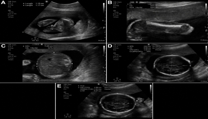
Figure 1. Ultrasonographic assessment of umbilical cord diameter and routine fetal biometric parameters in a 30-year-old pregnant woman at 22 weeks 3 days of gestation according to the dating scan. (A) Mean umbilical cord diameter (UCD), 11.55 mm. (B) Femur length (FL), corresponding to 22 weeks 0 days. (C) Abdominal circumference (AC), corresponding to 21 weeks 3 days. (D) Biparietal diameter (BPD), corresponding to 22 weeks 2 days. (E) Head circumference (HC), corresponding to 22 weeks 2 days. The mean gestational age estimated from routine fetal biometric parameters using the Hadlock formula was 22 weeks 1 day.
RESULTS:
A total of 1,000 pregnant women were included. Maternal age ranged from 18 to 41 years, with a mean of 25.80 ± 4.67 years and a median of 25 years. Most participants were aged 21–25 years (45.9%), followed by 26–30 years (30.7%). Of the participants, 420 (42.0%) were primigravida and 580 (58.0%) were multigravida. The mean gestational age was 28.01 ± 6.75 weeks. Overall, 444 (44.4%) examinations were performed during the second trimester and 556 (55.6%) during the third trimester (Table 1).
The mean umbilical cord diameter increased progressively with advancing gestational age. It rose from 7.02 ± 0.30 mm at 16 weeks to 14.19 ± 0.70 mm at 27 weeks. During the third trimester, it increased further from 14.80 ± 0.71 mm at 28 weeks to 19.30 ± 0.50 mm at 40 weeks. The median UCD showed a similar increase, from 7.0 mm at 16 weeks to 19.3 mm at 40 weeks. The 5th and 95th percentile values also increased consistently across gestation, providing gestational-age-specific reference limits for UCD (Table 2; Figure 1).
UCD showed a very strong positive correlation with gestational age (r=0.989, R²=0.978; p<0.001). Significant positive correlations were also observed with BPD (r=0.979), EFW (r=0.939), AC (r=0.928), FL (r=0.834), and HC (r=0.583); all associations were statistically significant (p<0.001). Among the fetal biometric parameters, BPD had the strongest association with UCD, whereas HC showed the weakest. Linear regression demonstrated a steady increase in UCD with advancing gestational age (Table 3; Figure 2).
Table 1. Demographic and obstetric characteristics of the study population (N=1,000)
|
Demographic and obstetric characteristics |
n (%) |
|
|
Maternal age, years |
Mean ± SD |
25.80 ± 4.67 |
|
Median |
25 |
|
|
Range |
18–41 |
|
|
18–20 |
64 (6.4) |
|
|
21–25 |
459 (45.9) |
|
|
26–30 |
307 (30.7) |
|
|
31–35 |
139 (13.9) |
|
|
≥36 |
31 (3.1) |
|
|
Gravida |
Primigravida |
420 (42.0) |
|
Multigravida |
580 (58.0) |
|
|
Gestational age, weeks |
Mean ± SD |
28.01 ± 6.75 |
|
Range |
16–40 |
|
|
Second trimester, 16–27 weeks |
444 (44.4) |
|
|
Third trimester, 28–40 weeks |
556 (55.6) |
|
Values are presented as mean ± standard deviation, median, range, or n (%).
Table 2. Gestational-age-specific reference values for ultrasonographic umbilical cord diameter
|
Trimester |
Gestational age (weeks) |
n |
Mean ± SD (mm) |
5th percentile (mm) |
Median (mm) |
95th percentile (mm) |
|
Second trimester |
16 |
36 |
7.02 ± 0.30 |
6.5 |
7.0 |
7.6 |
|
17 |
12 |
7.60 ± 0.31 |
7.1 |
7.6 |
8.2 |
|
|
18 |
36 |
8.30 ± 0.36 |
7.7 |
8.3 |
8.9 |
|
|
19 |
49 |
9.00 ± 0.40 |
8.3 |
9.0 |
9.6 |
|
|
20 |
43 |
9.80 ± 0.45 |
9.1 |
9.8 |
10.6 |
|
|
21 |
78 |
10.50 ± 0.50 |
9.6 |
10.5 |
11.3 |
|
|
22 |
60 |
11.20 ± 0.56 |
10.2 |
11.2 |
12.1 |
|
|
23 |
34 |
11.79 ± 0.59 |
10.7 |
11.8 |
12.7 |
|
|
24 |
31 |
12.39 ± 0.60 |
11.3 |
12.4 |
13.4 |
|
|
25 |
26 |
13.00 ± 0.66 |
11.9 |
13.0 |
14.1 |
|
|
26 |
16 |
13.59 ± 0.71 |
12.3 |
13.6 |
14.8 |
|
|
27 |
23 |
14.19 ± 0.70 |
12.9 |
14.2 |
15.4 |
|
|
Third trimester |
28 |
14 |
14.80 ± 0.71 |
13.5 |
14.8 |
15.9 |
|
29 |
36 |
15.19 ± 0.68 |
14.0 |
15.2 |
16.3 |
|
|
30 |
40 |
15.81 ± 0.70 |
14.5 |
15.8 |
17.0 |
|
|
31 |
41 |
16.21 ± 0.65 |
15.1 |
16.2 |
17.3 |
|
|
32 |
66 |
16.61 ± 0.71 |
15.3 |
16.6 |
17.8 |
|
|
33 |
76 |
17.01 ± 0.70 |
15.7 |
17.0 |
18.2 |
|
|
34 |
85 |
17.40 ± 0.71 |
16.1 |
17.4 |
18.7 |
|
|
35 |
77 |
17.80 ± 0.65 |
16.6 |
17.8 |
18.9 |
|
|
36 |
57 |
18.10 ± 0.60 |
17.0 |
18.1 |
19.1 |
|
|
37 |
23 |
18.40 ± 0.61 |
17.4 |
18.4 |
19.4 |
|
|
38 |
12 |
18.68 ± 0.60 |
17.6 |
18.7 |
19.7 |
|
|
39 |
16 |
18.99 ± 0.55 |
18.0 |
19.0 |
20.0 |
|
|
40 |
13 |
19.30 ± 0.50 |
18.4 |
19.3 |
20.2 |
Table 3. Association of UCD with gestational age and fetal biometric parameters
|
Parameter |
Pearson’s r |
R² |
Regression equation |
p-value |
|
Gestational age (weeks) |
0.989 |
0.978 |
UCD = 0.534 × GA − 0.800 |
<0.001 |
|
BPD (mm) |
0.979 |
0.958 |
UCD = 0.200 × BPD + 0.387 |
<0.001 |
|
HC (mm) |
0.583 |
0.339 |
UCD = 0.021 × HC + 8.755 |
<0.001 |
|
AC (mm) |
0.928 |
0.862 |
UCD = 0.044 × AC + 3.777 |
<0.001 |
|
FL (mm) |
0.834 |
0.695 |
UCD = 0.161 × FL + 5.747 |
<0.001 |
|
EFW (g) |
0.939 |
0.882 |
UCD = 0.003 × EFW + 9.352 |
<0.001 |
|
AC, abdominal circumference; BPD, biparietal diameter; EFW, estimated fetal weight; FL, femur length; GA, gestational age; HC, head circumference; UCD, umbilical cord diameter. |
||||
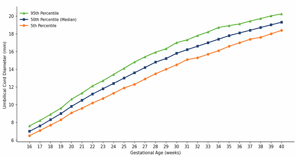
Figure 2: Reference Curves for UCD (mm) – 5th, 50th, and 95th Percentiles (16–40 Weeks)
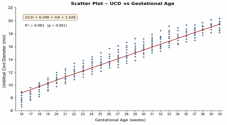
Figure 3. scatter plot - UCD (mm) vs gestational age (weeks)
DISCUSSION:
The present study evaluated ultrasonographic umbilical cord diameter (UCD) across 16–40 weeks of gestation and developed gestational-age-specific reference values for a Central Indian population. The mean maternal age was 25.80 years, and slightly more than half of the examinations were performed during the third trimester. This distribution reflects the common pattern of antenatal ultrasound referrals at tertiary hospitals, where many women undergo growth assessment during later pregnancy.9
A consistent increase in UCD was observed with advancing gestation. Mean UCD increased from 7.02 mm at 16 weeks to 19.30 mm at 40 weeks. This growth probably reflects enlargement of the umbilical vessels and increasing Wharton’s jelly needed to support fetal circulation. Cromi et al. similarly demonstrated progressive enlargement of the umbilical cord and Wharton’s jelly with advancing pregnancy.10 The increase was greater during the second trimester and became more gradual near term, suggesting that cord growth may slow during late pregnancy.
The gestational-age-specific normogram provides 5th, 50th, and 95th percentile values from 16 to 40 weeks. These reference limits may help identify cords that are unusually thin or thick for gestational age. However, values outside these limits should not be interpreted as diagnostic on their own. They should be considered together with fetal biometry, amniotic fluid volume, Doppler findings, and maternal clinical factors. Begum K et al. also reported a steady increase in UCD during the second and third trimesters and supported its use as an additional ultrasound measurement.11
UCD showed a very strong correlation with gestational age (r=0.989, p<0.001), with gestational age explaining 97.8% of its variation. Significant correlations were also found with all fetal biometric measurements. The strongest association was with BPD, followed by EFW, AC, FL, and HC. These findings indicate that umbilical cord growth occurs alongside fetal cranial, abdominal, skeletal, and overall somatic growth. Similar relationships between UCD and conventional fetal biometric parameters have been reported by Predanic et al., Udoh et al., and Raio et al.4,12,13 The comparatively weaker correlation with HC may reflect biological differences in fetal head shape and variation in its measurement.
The UCD values in this study were broadly comparable with those reported in Indian studies, although the present values were slightly higher near term.14,15 Minor differences may arise from variations in study populations, measurement techniques, gestational-age distribution, and ultrasound protocols. This supports the value of locally derived reference charts.
The study was strengthened by its large sample and measurement by a single observer. Nevertheless, it was cross-sectional and conducted at one centre. Smaller samples at some gestational weeks may have affected percentile precision, and pregnancy and neonatal outcomes were not evaluated. Therefore, multicentre longitudinal studies are required to validate this normogram and determine whether abnormal UCD independently predicts adverse outcomes. Overall, UCD appears to be a useful supplementary marker of gestational age and fetal growth, but it should complement rather than replace established fetal biometric measurements.
CONCLUSION:
Umbilical cord diameter increased progressively from 16 to 40 weeks and showed a strong positive correlation with gestational age and fetal biometric parameters. The gestational-age-specific normogram developed in this study provides reference values for the Central Indian population. Ultrasonographic measurement of UCD is a simple, non-invasive parameter that may complement conventional fetal biometry during antenatal assessment. However, it should not be used as an isolated diagnostic measure. Further multicentre longitudinal studies are required to validate these reference values and assess their clinical utility.
REFERENCES:
1. Butureanu TA. The Pathophysiology of Wharton’s Jelly and Its Impact on Fetal and Neonatal Outcomes: A Comprehensive Literature Review. Med Sci (Basel). 2025;13(4):215.
2. Debebe SK, Cahill LS, Kingdom JC, et al. Wharton’s jelly area and its association with placental morphometry and pathology. Placenta. 2020;94:34-38.
3. Weissman A Weissman A, Jakobi P, Bronshtein M, Goldstein I, Drugan A. Sonographic measurements of the umbilical cord and vessels during normal pregnancies. J Ultrasound Med. 1994;13(1):11-14.
4. Raio L Raio L, Ghezzi F, Di Naro E, Franchi M, Balestreri D, D’Addario V. Sonographic measurement of the umbilical cord and vessels during normal pregnancies. Obstet Gynecol. 1999;94(5 Pt 1):832-836.
5. Ghezzi F Ghezzi F, Raio L, Di Naro E, Franchi M, Balestreri D, D’Addario V. Nomogram of umbilical cord biometry at low-risk pregnancies. Prenat Diagn. 2001;21(10):849-852.
6. Di Naro E Di Naro E, Ghezzi F, Raio L, Franchi M, D’Addario V. Umbilical cord morphology and pregnancy outcome. Eur J Obstet Gynecol Reprod Biol. 2001;96(2):150-157.
7. Sepulveda W Sepulveda W. The umbilical cord: normal anatomy and prenatal sonographic evaluation. Ultrasound Obstet Gynecol. 1999;13(3):157-160.
8. Salomon LJ, Alfirevic Z, Berghella V, et al. ISUOG Practice Guidelines (updated): performance of the routine mid-trimester fetal ultrasound scan. Ultrasound Obstet Gynecol. 2022;59(6):840-856.
9. Vashisht BM, Bhardwaj H, Chauhan M, Anvesha, Jaiprakash. Socio-demographic determinants of full antenatal care services utilization among pregnant women delivering at a tertiary care hospital in Haryana. Public Health Rev Int J Public Health Res. 2019;6(4):139-146.
10. Cromi A, Ghezzi F, Di Naro E, Siesto G, Bergamini V, Raio L. Large cross-sectional area of the umbilical cord as a predictor of fetal macrosomia. Ultrasound Obstet Gynecol. 2007;30(6):861-866.
11. Begum K, Ahmed MU, Rahman MM, et al. Correlation between Umbilical Cord Diameter and Cross Sectional Area with Gestational Age and Foetal Anthropometric Parameters. Mymensingh Med J. 2016;25(2):190-197.
12. Predanic M, Perni SC, Chasen ST, Baergen RN, Chervenak FA. Ultrasound evaluation of abnormal umbilical cord coiling in second trimester of gestation in association with adverse pregnancy outcome. Am J Obstet Gynecol. 2005;193(2):387-394.
13. Udoh BE, Erim A, Anthony E. Sonographic assessment of umbilical cord diameter as an indicator of fetal growth and perinatal outcome. J Diagn Med Sonogr. 2021;37(1):41-45.
14. Alaspurkar S, Shelke S, Shivkumar P, Jain S, Golhar AV. Sonographic association of umbilical cord thickness in the third trimester of pregnancy with fetal outcome in a rural Indian population. Cureus. 2025;17(12):e100310.
15. Mukhiya R, Yadav S, et al. Estimation of fetal age by sonographic evaluation of umbilical cord diameter in relation to gestational age. Int J Acad Med Pharm. 2023;5(2):1195-1197.