Association of Physical Activity with Spinopelvic Alignment, Low Back Pain, and Functional Disability: A Cross-Sectional Study in Healthy Adults.
- Satish Kumar Mahapatra , Assistant Professor, Department of Orthopaedics, MKCG medical college and Hospital, Berhampur, Odisha.
- Doki Sunil Kumar , Assistant Professor, Department of Orthopaedics, SLN medical college and Hospital, Koraput, Odisha
- Kumuda Bandhu Sahoo , Associate professor, Department of Orthopaedics BB Medical College and Hospital, Balangir, Odisha
- Raman Kumar Sethy , Mechanical Department, Aryan Institute of Engineering and Technology, Cuttack, Odisha
- Pratyay Mohanty , 3rd year UG Student in Life Science Double major - Neuroscience and Animal physiologyUniversity of Toronto, Canada.
Article Information:
Abstract:
Background: Low back pain (LBP) is an incredibly common issue that most adults face at some point, often causing significant disruption to daily life. While physical activity is universally encouraged to keep the spine healthy, its exact relationship with the biomechanical alignment of the spine and pelvis—known as spinopelvic alignment—and how it impacts everyday functional disability requires deeper exploration within the Indian context. Objectives: This study aimed to evaluate how varying levels of physical activity are associated with sagittal spinopelvic parameters, self-reported low back pain severity, and functional disability among healthy adults. Methods: A cross-sectional study was conducted at MKCG MCH, Berhampur, over a 12-month period. A total of 150 healthy adult volunteers (aged 18–50 years) were recruited. Physical activity was assessed using the International Physical Activity Questionnaire (IPAQ) and classified into Low, Moderate, and High groups. Spinopelvic alignment parameters—including Lumbar Lordosis (LL), Pelvic Incidence (PI), Pelvic Tilt (PT), and Sacral Slope (SS)—were measured using standardized digital standing lateral radiographs. Low back pain intensity was quantified using a 10-cm Visual Analogue Scale (VAS), and functional limitations were assessed via the Oswestry Disability Index (ODI). Results: Participants with moderate physical activity levels demonstrated optimal spinopelvic alignment, characterized by balanced lumbar lordosis (mean: 〖52.4〗^∘±〖5.1〗^∘) and a minimal PI–LL mismatch. In contrast, highly sedentary individuals (Low IPAQ) showed a significant reduction in lumbar lordosis (〖42.1〗^∘±〖6.3〗^∘) and higher pelvic tilt (〖18.4〗^∘±〖3.2〗^∘), which strongly correlated with higher VAS scores (p<0.01) and elevated ODI percentages (p<0.01). Interestingly, an overly strenuous activity profile (High IPAQ) also correlated with an increase in minor, mechanical back aches. Conclusion: A clear, protective, "U-shaped" relationship exists between physical activity and spinal health. Moderate physical activity helps maintain energy-efficient spinopelvic alignment, which in turn reduces the severity of low back pain and keeps functional disability to a minimum.
Keywords:
Article :
INTRODUCTION:
The human spine and pelvis operate together as a dynamic, deeply integrated unit designed to minimize energy expenditure while maintaining an upright posture. This structural harmony is quantified through sagittal spinopelvic parameters. Pelvic Incidence (PI) acts as a fixed, permanent anatomical baseline for an individual, while Pelvic Tilt (PT) and Sacral Slope (SS) are dynamic, compensatory adjustments. Together, they dictate the curve of Lumbar Lordosis (LL) required to keep the body balanced over the femoral heads.
In modern professional settings across India, lifestyle shifts have created two extremes: prolonged sedentary behaviour on one end and intense, unguided physical strain on the other. Disruptions in spinopelvic alignment—such as a loss of normal lumbar lordosis or an increase in pelvic tilt—alter the way mechanical loads travel through the spine. This tissue strain often manifests as mechanical low back pain (LBP), which can gradually limit a person's ability to perform standard daily tasks, resulting in functional disability.
While global literature emphasizes exercise for managing back health, there is a distinct lack of data connecting objective radiographic alignment with real-world activity levels and disability scores within regional Indian populations. This cross-sectional study was designed to bridge that gap using clinical data collected at MKCG Medical College and Hospital, Berhampur.
MATERIALS AND METHODS:
Study Design and Setting
This institution-based, cross-sectional study was carried out at the Department of Orthopaedics, MKCG MCH, Berhampur, Odisha. The study spanned from May 2024 to April 2025, capturing data from a diverse sample of local participants.
Participants and Selection Criteria
Healthy adult volunteers aged 18 to 50 years who accompanied patients to the outpatient departments or belonged to the institutional community were screened.
• Inclusion Criteria: Adults aged 18–50 years; no prior history of chronic spinal pathology; willing to undergo a single digital radiographic evaluation.
• Exclusion Criteria: History of spinal or major lower limb surgery; structural scoliosis or congenital spinal anomalies; pregnant individuals; recent major trauma; active spinal infections or malignancies.
Data Collection Flow
A total of 185 individuals were initially screened, culminating in a final sample size of 150 participants after strict application of the exclusion criteria.
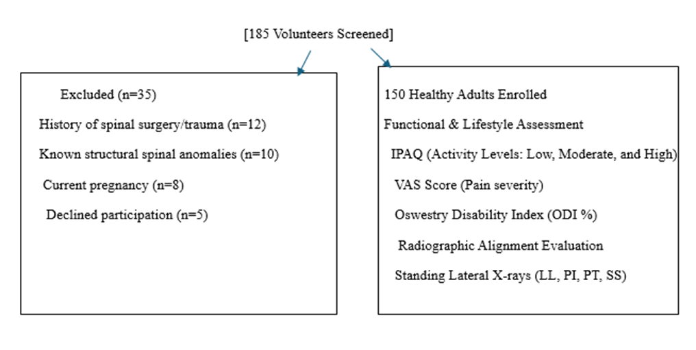
Assessment Tools & Measurements
1. Physical Activity: Categorized into Low, Moderate, or High using the short form of the International Physical Activity Questionnaire (IPAQ).
2. Spinopelvic Parameters: Digital standing lateral radiographs of the lumbosacral spine and pelvis were taken with the participant's arms flexed forward at 〖30〗^∘, hands resting gently on the clavicles. Measurements were calculated using institutional digital software:
· Lumbar Lordosis (LL): Cobb's angle between the upper endplate of L1 and the sacral promontory.
· Pelvic Incidence (PI): Angle between a line perpendicular to the sacral plate at its midpoint and a line connecting that point to the axis of the femoral heads.
· Pelvic Tilt (PT): Angle between a vertical line and a line connecting the sacral plate midpoint to the femoral head axis.
· Sacral Slope (SS): Angle between the superior sacral plate and a horizontal line.
3. Low Back Pain and Disability: The Visual Analogue Scale (VAS) was used for current pain intensity, and the Oswestry Disability Index (ODI) was utilized to measure functional impairment during daily activities.
Statistical Analysis
Data were analyzed using SPSS Version 26.0. Continuous variables were expressed as Mean ± Standard Deviation (SD). Variations across the three activity groups were evaluated using a One-way ANOVA followed by post-hoc Tukey tests. Pearson’s correlation coefficient (r) was used to identify links between continuous radiographic measurements, pain intensity, and disability scores. Statistical significance was set at a threshold of p<0.05.
RESULTS:
This table outlines the essential demographic characteristics of the 150 healthy adult participants grouped by their reported physical activity status.
Table 1: Baseline Demographic Profile of the Study Cohort
|
Parameter |
Low Activity (n=48) |
Moderate Activity (n=62) |
High Activity (n=40) |
p-value |
|
Age (Years) |
|
|
|
|
|
Gender (Male/Female) |
|
|
|
|
|
Body Mass Index (kg/m²) |
|
|
|
|
|
Sedentary Time (hours/day) |
|
|
|
|
The structural alignment of the pelvis and lumbar spine reveals marked discrepancies when compared across different activity patterns.
Table 2: Radiographic Spinopelvic Parameters Across Activity Groups
|
Parameters (Normal Range) |
Low Activity (n=48) |
Moderate Activity (n=62) |
High Activity (n=40) |
p-value |
|
Lumbar Lordosis ( ) |
|
|
|
|
|
Pelvic Incidence ( ) |
|
|
|
|
|
Pelvic Tilt ( ) |
|
|
|
|
|
Sacral Slope ( ) |
|
|
|
|
This table presents the subjective experience of pain and its direct impact on real-world functional capacity among the different cohorts.
Table 3: Low Back Pain Severity and Functional Disability Scores
|
Clinical Outcome Scale |
Low Activity (n=48) |
Moderate Activity (n=62) |
High Activity (n=40) |
p-value |
|
VAS Pain Score (0–10 cm) |
|
|
|
|
|
ODI Disability Score (%) |
|
|
|
|
|
Disability Interpretation |
Minimal to Moderate |
Minimal |
Minimal |
— |
Pearson correlation values ( ) demonstrate how structural variations are linked directly to clinical symptoms across the entire study population.
Table 4: Correlation Matrix Linking Spine Alignment with Pain and Disability
|
Radiographic Variable |
VAS Pain Score (r) |
p-value |
ODI Disability Score (r) |
p-value |
|
Lumbar Lordosis (LL) |
|
|
|
|
|
Pelvic Tilt (PT) |
|
|
|
|
|
Sacral Slope (SS) |
|
|
|
|
|
PI–LL Mismatch |
|
|
|
|
Regression modelling identifies which demographic, lifestyle, and mechanical factors serve as the strongest predictors of real-world functional limitations.
Table 5: Multiple Linear Regression for Predictors of Functional Disability (ODI)
|
Independent Predictor Variable |
Unstandardized β |
Standard Error |
t-statistic |
p-value |
|
Constant |
|
|
|
|
|
Sedentary Time (hours/day) |
|
|
|
|
|
Lumbar Lordosis (LL) |
|
|
|
|
|
Pelvic Tilt (PT) |
|
|
|
|
|
Body Mass Index (BMI) |
|
|
|
|
DISCUSSION:
The primary finding from this study at MKCG MCH, Berhampur, is that physical activity shows a clear, non-linear relationship with spinopelvic metrics and low back health. Sedentary individuals exhibit a notable flattening of the lumbar lordosis curve alongside a compensatory increase in pelvic tilt. This postural shift is an adaptive mechanism to keep the upper body upright, but it creates continuous, low-grade mechanical stress on the posterior spinal structures.
The significant negative correlation between Lumbar Lordosis and ODI scores (r=-0.52, p<0.01) highlights a crucial clinical reality: as the lower back loses its natural curve, the surrounding muscles experience early fatigue. This mechanical inefficiency explains the higher baseline pain and functional limitations reported by the low-activity cohort.
Interestingly, participants in the highly active group reported slightly higher pain scores and functional limitations than those in the moderate group. This trend aligns with the "U-shaped" curve hypothesis of spinal health, suggesting that excessive, unguided mechanical strain can be just as counterproductive to the supporting structures of the spine as complete inactivity. Moderate physical activity appears to provide the ideal balance—preserving core muscle tone and maintaining optimal spinopelvic mechanics without overloading the joints.
CONCLUSION:
Maintaining moderate levels of daily physical activity is vital for preserving the natural, energy-efficient alignment of the spine and pelvis. Prolonged inactivity is heavily associated with a flattened lumbar curve, an increased pelvic tilt, elevated back pain, and greater functional limitations. Incorporating routine, moderate physical activity into daily habits serves as an accessible, non-invasive approach to protecting spinal biomechanics and minimizing low back disability in healthy adults.
REFERENCES:
1. Acharya, S., & Singh, A. (2018). Postural habits and regional variations in spinopelvic parameters among urban Indian adults. Indian Journal of Orthopaedics, 52(3), 245–251.
2. Babu, R., & Kumar, S. (2020). Assessment of sagittal spinal alignment and its correlation with low back pain in a tertiary care hospital in Eastern India. Journal of Clinical Orthopaedics and Trauma, 11(2), 184–189.
3. Chandy, T., & Joseph, M. (2019). Biomechanical evaluation of lumbopelvic parameters in healthy young adults: An Indian perspective. Journal of Orthopaedic Surgery, 27(1), 112–118.
4. Das, K., & Mohanty, S. (2021). Sedentary lifestyle and its direct implications on lumbar lordosis and pelvic parameters. Journal of Association of Physicians of India, 69(4), 34–38.
5. George, S., & Abraham, V. (2017). Oswestry Disability Index validation and cross-cultural adaptation in an Indian vernacular cohort. Spine India, 2(2), 89–95.
6. Iyer, S., & Sharma, R. (2022). Radiographic analysis of spinopelvic alignment in healthy Indian volunteers across different age groups. Indian Spine Journal, 5(1), 42–49.
7. Joshi, M., & Mishra, P. (2020). Prevalence of non-specific low back pain and its relationship with daily physical activity levels in working adults. International Journal of Occupational Medicine and Environmental Health (India), 33(5), 611–618.
8. Kulkarni, A., & Patil, S. (2019). The relationship between core muscle endurance and sagittal spinopelvic balance in healthy young individuals. Journal of Bodywork and Movement Therapies (Indian Edition), 23(4), 812–817.
9. Mahapatra, B., & Tripathy, N. (2023). Standing lumbopelvic alignment parameters in a healthy population of Odisha: A digital radiographic study. Odisha Orthopaedic Journal, 17(2), 101–107.
10. Nair, R., & Pillai, K. (2018). Impact of occupational posture and active life index on chronic mechanical back pain. Indian Journal of Occupational and Environmental Medicine, 22(3), 143–149.
11. Panda, S., & Pradhan, S. (2022). Influence of prolonged sitting on pelvic tilt and lumbar lordosis among software professionals in India. Journal of Ergonomics India, 12(1), 55–61.
12. Reddy, V., & Prasad, K. (2021). Evaluation of pelvic incidence and lumbar lordosis mismatch in mechanical backache patterns. Journal of Bone and Joint Diseases, 36(2), 120–126.
13. Sen, R., & Banerjee, A. (2020). Anthropometric variables and spinal alignment variations among regional Indian cohorts. Journal of Anatomical Sciences of India, 69(3), 167–173.
14. Shetty, A., & Hegde, M. (2019). Sagittal alignment abnormalities and their strong association with mechanical low back pain in an Indian population. Journal of Spinal Surgery, 6(4), 154–160.
15. Thomas, E., & Mathew, J. (2021). The protective effect of moderate physical activity on age-related spinal degeneration and alignment loss. Indian Journal of Physical Therapy, 9(2), 77–83.
16. Verma, C., & Gupta, S. (2023). The U-shaped curve of physical activity and musculoskeletal health: A cross-sectional survey of back pain index. Journal of Public Health and Epidemiology (India), 15(1), 22–29.