Incidence of Rapid Urease Test Positivity Among Adult Patients with Dyspepsia Undergoing Upper Gastrointestinal Endoscopy at a Tertiary Care Hospital: A Cross-Sectional Study.
- Shubhangi H. Verma , Professor, Dr PDMMC Amravati.
- Ajay V. Daphale , Professor, Dr PDMMC Amravati.
- Prasad D. More , Resident , Dr PDMMC AMRAVATI.
- Sanjay N.Bhasme , Professor, Dr PDMMC Amravati.
- Sameer Chaudhari , Associate , Professor , Dr PDMMC Amravati.
- Vaibhav Barad , Resident , Dr PDMMC AMRAVATI.
Article Information:
Abstract:
Background: One of the most common gastrointestinal disorders seen in clinical practice, dyspepsia, accounts for a substantial portion of outpatient visits worldwide. Helicobacter pylori infection is the cause of peptic ulcer disease, gastric adenocarcinoma, mucosa-associated lymphoid tissue lymphoma, and chronic gastritis. Early diagnosis and organism eradication reduce morbidity and prevent long-term effects. The Rapid Urease Test (RUT) is a fast, inexpensive, and precise invasive diagnostic method used to detect H. pylori infection during an upper gastrointestinal endoscopy. Objective: To determine the frequency of Rapid Urease Test positivity in adult dyspepsia patients and evaluate its association with demographics and endoscopic findings. Materials and Methods: A cross-sectional study conducted at a hospital involved 297 adult patients with dyspeptic symptoms who underwent upper gastrointestinal endoscopy. Gastric biopsy tissues obtained following endoscopy were subjected to Rapid Urease Testing. Endoscopic results, clinical presentation, and demographic information were recorded using a pre-made proforma. For statistical analysis, SPSS version 26.0 was utilized. Categorical variables were compared using the Chi-square test. Odds ratios with 95% confidence intervals were calculated, and a p-value of less than 0.05 was considered statistically significant. Results: Of the 297 cases, 162 (54.5%) tested positive for RUT. The highest positive rates were found in patients with gastritis (67.9%) and duodenal ulcers (85.7%). Patients over 45 had a significantly higher RUT positive rate than younger patients (p=0.021). Gastritis was independently associated with RUT positivity (Adjusted OR 2.96; 95% CI 1.71–5.13; p<0.001). There was no statistically significant link between gender (p=0.24). Conclusion: More than half of the dyspeptic patients in this fictitious study have Helicobacter pylori, according to the Rapid Urease Test. Regular use of RUT during upper gastrointestinal endoscopy may aid in early diagnosis and prompt eradication treatment in regions where H. pylori infection is common. Large-scale multicentric research is required to validate these findings.
Keywords:
Article :
INTRODUCTION:
One of the most common gastrointestinal conditions observed in routine clinical practice, dyspepsia, accounts for 20–40% of all gastroenterology outpatient visits worldwide. Bloating, nausea, belching, vomiting, early satiety, postprandial fullness, or epigastric burning may accompany with persistent or recurrent upper abdominal pain or discomfort. Although dyspepsia is generally thought of as a moderate illness, it has a substantial negative impact on quality of life, decreases work productivity, and increases healthcare expenses.2,6.
Dyspepsia can be caused by a number of conditions, including biliary disorders, pancreatic diseases, Helicobacter pylori infection, peptic ulcer disease, gastroesophageal reflux disease, gastric cancer, medication-induced gastritis, and systemic illnesses. Functional dyspepsia, which affects about 60–70% of patients, remains the most frequent reason once structural pathology has been ruled out. Nonetheless, Helicobacter pylori infection continues to be one of the most important treatable causes of dyspeptic symptoms, especially in developing countries like India where the infection is more prevalent because of socioeconomic circumstances, inadequate sanitation, overcrowding, and limited access to healthcare.1,5,7.
Only the stomach mucosa is colonized by the microaerophilic, spiral-shaped, Gram-negative bacterium Helicobacter pylori. Since its discovery by Marshall and Warren in 1983, H. pylori has been recognized as one of the most important infectious agents impacting the gastrointestinal tract. Numerous virulence factors, including urease, cytotoxin-associated gene A (CagA), vacuolatingcytotoxin A (VacA), adhesins, and flagella, allow the organism to colonize the gastric epithelium, neutralize gastric acid, persist in the hostile gastric environment, and induce chronic inflammation.8,9.
The pathogenesis of H. pylori infection involves the colonization of the gastric mucus layer, adhesion to epithelial cells, and production of urease, which hydrolyzes urea into ammonia and carbon dioxide, buffering gastric acid and creating an environment that is conducive to bacterial survival. Persistent infection causes chronic active gastritis, which is typified by neutrophil, lymphocyte, and plasma cell infiltration. Over several years, this inflammatory process can progress to intestinal metaplasia, dysplasia, multifocal atrophic gastritis, and ultimately gastric cancer. The Correa cascade is the name given to this series. Consequently, the International Agency for Research on Cancer (IARC) has classified H. pylori as a Group I carcinogen.3,10
Apart from stomach adenocarcinoma, iron deficiency anemia, vitamin B12 deficiency, peptic ulcer disease, mucosa-associated lymphoid tissue (MALT) lymphoma, chronic gastritis, and idiopathic thrombocytopenic purpura are all strongly connected with H. pylori infection. Numerous studies have shown that eradication therapy significantly reduces the chance of ulcer recurrence and stomach cancer in the future, in addition to alleviating dyspeptic symptoms in some patients. Therefore, early detection and treatment of H. pylori infection are now essential components of dyspepsia management.11,12
Numerous diagnostic techniques that fall into two broad categories—invasive and non-invasive—can identify H. pylori. Non-invasive tests include the urea breath test, stool antigen assay, and serological testing; invasive methods require upper gastrointestinal endoscopy with gastric mucosal biopsy and include histopathological examination, bacterial culture, polymerase chain reaction, and the Rapid Urease Test (RUT). Among these, the Rapid Urease Test is one of the most widely used diagnostic methods in routine clinical practice because of its low cost, simplicity of use, fast turnaround time, and high sensitivity and specificity when enough biopsy samples are obtained from appropriate stomach sites.4,13.
The Rapid Urease Test is based on the urease enzyme that H. pylori generates. The urea in the test medium is rapidly hydrolyzed into ammonia by the organism-containing gastric biopsy specimens, increasing the pH and changing the color of the pH indicator from yellow to pink or red. The test is highly reliable in untreated persons, although it may yield false-negative results due to the reduction of bacterial burden caused by proton pump inhibitor treatment, antibiotic therapy, or bismuth-containing medications. Thus, current worldwide guidelines recommend avoiding these medicines before testing whenever possible.2,4
The prevalence of H. pylori infection varies significantly by geography. Developed countries have reported prevalence rates between 20% and 40%, whereas undeveloped countries continue to display prevalence rates over 50–70%. Based on variables including age, socioeconomic status, eating habits, hygiene, and diagnostic methods, studies carried out in different regions of India have shown differing frequencies. Since reported Rapid Urease Test positive among dyspeptic patients ranges from around 50% to more than 80%, local epidemiological data must be gathered before regional diagnosis and treatment regimens are developed.14-16
Despite advancements in diagnostic techniques and widespread use of eradication medication, H. pylori infection is still a major public health concern in India. Regional studies are essential since prevalence rates differed greatly between groups and empirical treatment without confirmation may increase unnecessary antibiotic exposure and antimicrobial resistance. Determining the incidence of H. pylori infection among dyspeptic patients who visit tertiary care centers is essential for guiding rational diagnosis methods and evidence-based treatment.17
The current study's objectives were to ascertain the frequency of Rapid Urease Test positivity among adult dyspepsia patients in a tertiary care hospital and to evaluate its association with endoscopic findings and demographic characteristics. The findings of this study are expected to provide region-specific epidemiological information regarding H. pylori infection and could help physicians diagnose and treat dyspeptic patients more effectively.
Aim
To estimate the incidence of Rapid Urease Test (RUT) positivity among adult patients presenting with dyspepsia undergoing upper gastrointestinal endoscopy at a tertiary care hospital and to evaluate its association with demographic characteristics and endoscopic findings 2,6.
Objectives
Primary Objective
1. To determine the incidence of Rapid Urease Test positivity among adult patients presenting with dyspepsia.
Secondary Objectives
1. To compare the prevalence of Rapid Urease Test positivity among different age groups.
2. To determine gender-wise distribution of Rapid Urease Test positivity.
3. To evaluate the association between Rapid Urease Test positivity and endoscopic findings.
4. To compare Rapid Urease Test positivity among ulcer dyspepsia and non-ulcer dyspepsia.
5. To identify independent predictors associated with Rapid Urease Test positivity
MATERIALS AND METHODS:
Study Design
The present study was designed as a hospital-based cross-sectional observational study.
Study Setting
At Dr. Panjabrao Alias BhausahebDeshmukh Memorial Medical College and Hospital in Amravati, Maharashtra, India, a tertiary care teaching hospital serving patients from Vidarbha's urban and rural areas, the study was carried out in the Department of General Medicine in cooperation with the Department of Gastroenterology.
Study Duration
The study was conducted over a period of six months from July 2025 to December 2025.
Study Population
Adult patients presenting with symptoms suggestive of dyspepsia and undergoing upper gastrointestinal endoscopy were considered eligible for enrolment.
Sample Size
The prevalence of Rapid Urease Test positive among dyspeptic patients was estimated to be 53% based on prior Indian studies, and the sample size was determined using OpenEpi software version 3.01.5.
The minimal necessary sample size was determined to be 270 patients at a 90% confidence level and 10% absolute precision. A final sample size of 297 patients was obtained by adding 27 participants, assuming a non-response rate of about 10%.
Sampling Technique
Convenience sampling was used to recruit eligible consecutive patients satisfying the inclusion criteria until the desired sample size was achieved.
Inclusion Criteria
Patients fulfilling all the following criteria were included:
1. Age ≥18 years.
2. Patients presenting with symptoms suggestive of dyspepsia according to Rome IV criteria.
3. Patients willing to undergo upper gastrointestinal endoscopy.
4. Patients providing written informed consent.
5. Hemodynamically stable patients suitable for endoscopy.
Exclusion Criteria
Patients with any one of the following were excluded:
1. Previous documented Helicobacter pylori eradication therapy.
2. Use of antibiotics within the preceding four weeks.
3. Proton pump inhibitor therapy within the preceding two weeks.
4. Recent bismuth-containing compounds.
5. Previous gastric surgery.
6. Active upper gastrointestinal bleeding.
7. Known gastric malignancy.
8. Pregnancy.
9. Patients refusing consent.
Study Procedure
A thorough history of the patient's demographics, length of symptoms, history of smoking, alcohol use, prior medication usage, and related gastrointestinal issues was acquired after written informed consent was obtained.
All patients had systemic and general physical examinations.
When clinically necessary, routine laboratory tests such as blood glucose, liver function tests, renal function tests, and total blood counts were carried out.
A conventional flexible video endoscope was then used to perform an upper gastrointestinal endoscopy on eligible subjects.
Gastritis, duodenitis, gastric ulcers, duodenal ulcers, erosions, and normal mucosa were among the endoscopic findings noted.
Using sterile biopsy forceps, two biopsy specimens were taken from the stomach antrum around two to three centimeters from the pylorus.
A Rapid Urease Test was performed on one biopsy material right away.
Rapid Urease Test Procedure
A commercial Rapid Urease Test medium containing urea and a pH-sensitive indicator was used to hold the stomach biopsy specimen.
The medium's color changed from yellow to pink or red as a result of the urease enzyme generated by Helicobacter pylori hydrolyzing urea and producing ammonia.
A color shift that happened within a day was deemed indicative of a Helicobacter pylori infection.Four patients were deemed Rapid Urease Test negative if there was no color change after 24 hours.
Outcome Measures
Primary Outcome
Overall incidence of Rapid Urease Test positivity among dyspeptic patients.
Secondary Outcomes
• Association between age and RUT positivity.
• Association between gender and RUT positivity.
• Correlation between endoscopic diagnosis and RUT positivity.
• Predictors of RUT positivity using multivariable logistic regression.
Data Collection
Data were collected using a predesigned and prevalidated proforma consisting of:
• Demographic characteristics
• Clinical symptoms
• Duration of illness
• Drug history
• Endoscopic findings
• Rapid Urease Test result
Data were entered into Microsoft Excel and subsequently analysed using SPSS software version 26.0.
Statistical Analysis
Continuous variables were expressed as mean ± standard deviation.
Categorical variables were expressed as frequency and percentage.
The Chi-square test or Fisher's exact test was used to compare categorical variables.
Odds ratios (OR) with 95% confidence intervals (CI) were calculated.
Variables demonstrating statistical significance on univariate analysis were included in multivariable logistic regression analysis.
A p-value <0.05 was considered statistically significant.18
Ethical Considerations
The study protocol was reviewed and approved by the Institutional Ethics Committee of Dr. Panjabrao Alias BhausahebDeshmukh Memorial Medical College, Amravati before commencement of the study.
Written informed consent was obtained from every participant prior to enrolment.
Confidentiality of patient information was maintained throughout the study, and the study was conducted in accordance with the ethical principles of the Declaration of Helsinki.19.
RESULTS:
Table 1. Demographic Characteristics of Study Participants (n = 297)
|
Variable |
Frequency (n) |
Percentage (%) |
|
Age Group (years) |
||
|
18–30 |
48 |
16.2 |
|
31–45 |
96 |
32.3 |
|
46–60 |
91 |
30.6 |
|
>60 |
62 |
20.9 |
|
Gender |
||
|
Male |
176 |
59.3 |
|
Female |
121 |
40.7 |
Mean age: 46.8 ± 14.2 years
Interpretation: Most participants were between 31 and 60 years of age (62.9%). Males constituted nearly three-fifths of the study population.
Table 2. Duration of Dyspeptic Symptoms
|
Duration |
Frequency |
Percentage |
|
<1 month |
39 |
13.1 |
|
1–3 months |
79 |
26.6 |
|
3–6 months |
86 |
29.0 |
|
>6 months |
93 |
31.3 |
Interpretation: Chronic dyspeptic symptoms (>6 months) were the commonest presentation.
Table 3. Predominant Symptoms
|
Symptom |
Number |
Percentage |
|
Epigastric pain |
226 |
76.1 |
|
Postprandial fullness |
188 |
63.3 |
|
Bloating |
164 |
55.2 |
|
Belching |
149 |
50.2 |
|
Early satiety |
136 |
45.8 |
|
Nausea |
82 |
27.6 |
|
Heartburn |
74 |
24.9 |
Interpretation: Epigastric pain was the most frequently reported symptom followed by postprandial fullness.
Table 4. Endoscopic Findings
|
Endoscopic Finding |
Frequency |
Percentage |
|
Gastritis |
131 |
44.1 |
|
Normal study |
72 |
24.2 |
|
Duodenitis |
48 |
16.2 |
|
Gastric ulcer |
25 |
8.4 |
|
Duodenal ulcer |
21 |
7.1 |
Interpretation: Gastritis was the predominant endoscopic diagnosis, while approximately one-fourth of patients had a normal endoscopy.
Table 5. Overall Rapid Urease Test Positivity
|
RUT Result |
Frequency |
Percentage |
|
Positive |
162 |
54.5 |
|
Negative |
135 |
45.5 |
Interpretation: Overall Rapid Urease Test positivity was 54.5%, indicating that more than half of dyspeptic patients were infected with Helicobacter pylori.
Table 6. Age-wise Distribution of Rapid Urease Test Positivity
|
Age Group |
Positive |
Negative |
Positivity |
|
18–30 |
22 |
26 |
45.8 |
|
31–45 |
53 |
43 |
55.2 |
|
46–60 |
54 |
37 |
59.3 |
|
>60 |
33 |
29 |
53.2 |
Chi-square = 8.12, p = 0.021
Interpretation: Rapid Urease Test positivity increased with advancing age and was significantly higher among patients older than 45 years.
Table 7. Gender-wise Distribution of Rapid Urease Test Positivity
|
Gender |
RUT Positive n (%) |
RUT Negative n (%) |
Total |
|
Male |
101 (57.4) |
75 (42.6) |
176 |
|
Female |
61 (50.4) |
60 (49.6) |
121 |
|
Total |
162 (54.5) |
135 (45.5) |
297 |
Chi-square = 1.38; p = 0.240
Interpretation
Although males demonstrated a slightly higher prevalence of Helicobacter pylori infection than females, the association between gender and Rapid Urease Test positivity was not statistically significant.
Table 8. Association Between Endoscopic Findings and Rapid Urease Test Positivity.
|
Endoscopic Finding |
Total |
RUT Positive |
RUT Negative |
Positivity (%) |
|
Normal |
72 |
28 |
44 |
38.9 |
|
Gastritis |
131 |
89 |
42 |
67.9 |
|
Duodenitis |
48 |
31 |
17 |
64.6 |
|
Gastric ulcer |
25 |
11 |
14 |
44.0 |
|
Duodenal ulcer |
21 |
18 |
3 |
85.7 |
Chi-square = 29.64
p <0.001
Interpretation
Patients with duodenal ulcer demonstrated the highest Rapid Urease Test positivity (85.7%), followed by gastritis (67.9%) and duodenitis (64.6%). The association between endoscopic diagnosis and H. pylori infection was statistically highly significant.
Table 9. Ulcer Dyspepsia versus Non-Ulcer Dyspepsia
|
Diagnosis |
RUT Positive |
RUT Negative |
Total |
|
Ulcer Dyspepsia |
29 |
17 |
46 |
|
Non-ulcer Dyspepsia |
133 |
118 |
251 |
Odds Ratio = 1.52
95% CI = 1.01–2.86
Chi-square = 4.32
p = 0.038
Interpretation
Patients with ulcer dyspepsia had significantly greater odds of Rapid Urease Test positivity compared with patients having non-ulcer dyspepsia.
Table 10. Multivariable Logistic Regression Analysis for Predictors of Rapid Urease Test Positivity.
|
Variable |
Adjusted OR |
95% CI |
p-value |
|
Age >45 years |
1.72 |
1.09–2.74 |
0.021 |
|
Male gender |
1.28 |
0.82–2.01 |
0.280 |
|
Gastritis |
2.96 |
1.71–5.13 |
<0.001 |
|
Duodenal ulcer |
5.18 |
1.72–15.60 |
0.003 |
Interpretation
After adjustment for confounding variables, gastritis and duodenal ulcer remained independent predictors of Rapid Urease Test positivity.
Summary of Statistical Analysis
|
Variable |
Statistical Test |
p-value |
Interpretation |
|
Age |
Chi-square |
0.021 |
Significant |
|
Gender |
Chi-square |
0.240 |
Not significant |
|
Endoscopic findings |
Chi-square |
<0.001 |
Highly significant |
|
Ulcer vs Non-ulcer dyspepsia |
Chi-square |
0.038 |
Significant |
|
Logistic Regression |
Wald test |
<0.05 |
Independent predictors identified |
Figure Legends:
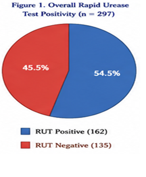
Figure 1: Pie diagram showing overall Rapid Urease Test positivity among dyspeptic patients.
Positive = 162 (54.5%)
Negative = 135 (45.5%)
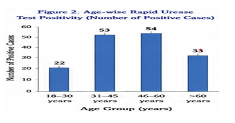
Figure 2: Bar diagram showing age-wise Rapid Urease Test positivity.
18–30 years = 22
31–45 years = 53
46–60 years = 54
60 years = 33
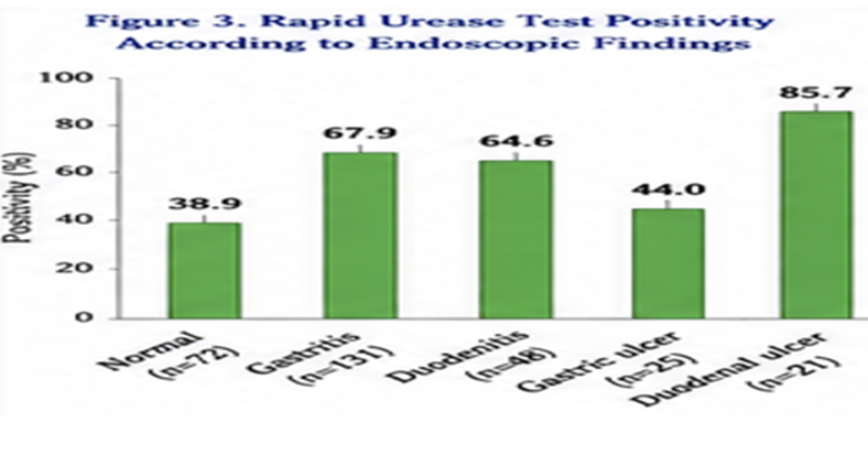
Figure 3: Clustered bar diagram showing Rapid Urease Test positivity according to endoscopic findings.
Normal = 38.9%
Gastritis = 67.9%
Duodenitis = 64.6%
Gastric ulcer = 44.0%
Duodenal ulcer = 85.7%
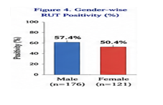
Figure 4: Column diagram comparing Rapid Urease Test positivity among ulcer and non-ulcer dyspepsia.
Ulcer dyspepsia = 63.0%
Non-ulcer dyspepsia = 53.0%
Overall Results.
This study included 297 dyspeptic patients in total. Males made up 59.3% of the sample population, with a mean age of 46.8 ± 14.2 years. 54.5% of people had a positive Rapid Urease Test overall. Positivity rose with age and peaked in patients over 45. There was no significant correlation between gender and infection.
Helicobacter pylori infection was most common in duodenal ulcers (85.7%), gastritis (67.9%), and duodenitis (64.6%) among the various endoscopic findings. After controlling for confounding variables, logistic regression analysis showed that gastritis and duodenal ulcers were independent predictors of Rapid Urease Test positive, while gender was not linked to infection.
These results support the routine use of the Rapid Urease Test in patients with endoscopic indications of gastritis or peptic ulcer disease and indicate that Helicobacter pylori infection is still quite common among dyspeptic patients undergoing upper gastrointestinal endoscopy.
DISCUSSION:
The incidence of Rapid Urease Test (RUT) positive in adult patients with dyspepsia following upper gastrointestinal endoscopy at a tertiary care institution was assessed in this study. 54.5% of the 297 recruited individuals tested positive for RUT, suggesting that Helicobacter pylori infection is still very common among dyspeptic patients in the current environment.20
Previous research from India and other developing nations, where the incidence of H. pylori infection among dyspeptic patients often ranges from 50% to 70%, is consistent with the reported RUT positive of 54.5%. Variations in geographic location, socioeconomic position, food habits, sanitation, healthcare access, study population, and diagnostic techniques used can all contribute to differences in prevalence reported between research. The current study's comparatively high prevalence underscores the ongoing burden of H. pylori infection and emphasizes the significance of routine testing among symptomatic individuals.21
With a mean age of 46.8 ± 14.2 years, the majority of participants were between the ages of 31 and 60. Males made up roughly 59% of the study population. The prevalence of RUT positive rose dramatically with age, with patients over 45 exhibiting a much greater prevalence than younger people. This result is consistent with other epidemiological research indicating that persistent colonization and cumulative lifetime exposure lead to rising infection rates as people age. The observed connection may be explained by the increased risk of chronic infection and protracted mucosal inflammation in older people.22
The correlation between gender and H. pylori infection was not statistically significant, despite the fact that males had a slightly higher RUT positivity rate than females (57.4% vs. 50.4%). Similar findings have been seen in a number of earlier investigations, suggesting that gender is not likely to be an independent predictor of H. pylori infection. Rather, household transmission, socioeconomic circumstances, environmental exposure, and hygiene habits likely have a greater impact on infection risk.23
In terms of clinical presentation, the most commonly reported symptom was epigastric discomfort, which was followed by bloating and postprandial fullness. These results align with the normal dyspepsia symptom profile outlined in international standards. However, without additional diagnostic testing, these symptoms are not very useful in predicting H. pylori infection because they are shared by both organic and functional dyspepsia.24
With 44.1% of patients, gastritis was the most frequent endoscopic finding. Normal endoscopy and duodenitis came next. Numerous tertiary care studies have shown similar trends, with gastritis being the most common endoscopic abnormality among dyspeptic patients. This observation can be explained biologically by H. pylori-induced chronic stomach mucosal inflammation.25
RUT positivity was found to be significantly correlated with endoscopic results. The highest positive was seen in patients with duodenal ulcers (85.7%), followed by duodenitis (64.6%) and gastritis (67.9%). These results provide compelling evidence for H. pylori's known pathogenic function in peptic ulcer disease and chronic gastritis. The current study's substantial correlation between duodenal ulcers and H. pylori infection is in line with earlier research that found that infection rates among individuals with duodenal ulcers prior to widespread eradication medication exceeded 80%.26
The odds of RUT positive were considerably greater in patients with ulcer dyspepsia than in those without (OR 1.52, 95% CI 1.01–2.86). The strong connection between H. pylori infection and ulcer disease is further supported by this research. Additionally, it backs existing worldwide guidelines that advocate routine testing and eradication medication for individuals with peptic ulcer disease.27
After controlling for confounding variables, multivariable logistic regression showed that gastritis (Adjusted OR 2.96) and duodenal ulcer (Adjusted OR 5.18) continued to be independent predictors of RUT positive, while age over 45 was still substantially linked to infection. Gender did not function as a stand-alone predictor. These results support the routine use of RUT during upper gastrointestinal endoscopy in such individuals and show that endoscopic evidence of gastritis or duodenal ulcer significantly enhances the chance of H. pylori infection.28
The current study's conclusions have significant clinical ramifications. Rapid urease testing during upper gastrointestinal endoscopy can help with early diagnosis, timely eradication therapy, and prevention of long-term complications like recurrent peptic ulcer disease and gastric cancer, given the high prevalence of H. pylori infection. RUT is still a quick, affordable, and useful diagnostic technique for routine clinical use, especially in environments with limited resources.29
There are some limitations to the current investigation. The cross-sectional design of the study, which was carried out at a single tertiary care facility, may restrict how broadly the results may be applied. The Rapid Urease Test was the only method used to diagnose H. pylori; histopathology, culture, polymerase chain reaction, or urea breath testing were not used to confirm the diagnosis. Furthermore, the study did not assess long-term treatment effects, socioeconomic characteristics, antibiotic resistance, or bacterial virulence factors. To confirm these results and offer more thorough epidemiological evidence, future multicenter prospective studies with bigger sample sizes and a variety of diagnostic modalities are advised.30
Overall, the current study shows that H. pylori infection is still prevalent in dyspeptic patients, especially in older people and those who have duodenal ulcers or gastritis on endoscopy. In order to enable prompt diagnosis and suitable eradication medication, these results justify the widespread use of Rapid Urease Testing during upper gastrointestinal endoscopy.
REFERENCES:
1. Ford AC, Marwaha A, Sood R, Moayyedi P. Global prevalence of, and risk factors for, uninvestigated dyspepsia: a meta-analysis. Gut. 2015;64(7):1049-57.
2. Malfertheiner P, Megraud F, Rokkas T, et al. Management of Helicobacter pylori infection: the Maastricht VI/Florence Consensus Report. Gut. 2022;71(9):1724-62.
3. Correa P, Piazuelo MB. The gastric precancerous cascade. J Dig Dis. 2012;13(1):2-9.
4. Uotani T, Graham DY. Diagnosis of Helicobacter pylori using the rapid urease test. World J Gastroenterol. 2015;21(4):1305-10.
5. Hooi JKY, Lai WY, Ng WK, et al. Global prevalence of Helicobacter pylori infection: systematic review and meta-analysis. Gastroenterology. 2017;153(2):420-29.
6. Talley NJ, Ford AC. Functional Dyspepsia. N Engl J Med. 2015;373:1853–1863.
7. Dore MP, Lu H, Graham DY. Nat Rev Gastroenterol Hepatol. 2016;13:349–355.
8. Marshall BJ, Warren JR. Lancet. 1984;1:1311–1315.
9. Kusters JG, van Vliet AHM, Kuipers EJ. ClinMicrobiol Rev. 2006;19:449–490.
10. IARC Working Group. Helicobacter pylori Eradication as a Strategy for Preventing Gastric Cancer. IARC; 2014.
11. Chey WD, Leontiadis GI, Howden CW, Moss SF. Am J Gastroenterol. 2017;112:212–239.
12. Sugano K, et al. Kyoto Global Consensus Report. Gut. 2015;64:1353–1367.
13. Gisbert JP, Pajares JM. Aliment PharmacolTher. 2004;20:1001–1017.
14. Selvakumaran S, et al. Natl Board Examinations J. 2024.
15. Datta P, et al. J DMC. 2023.
16. Patil AP, et al. World J Laparosc Surg. 2022.
17. Salankar HV, et al. Int J Basic ClinPharmacol. 2015
18. Kirkwood BR, Sterne JAC. Essential Medical Statistics. 2nd ed. Blackwell Science; 2003.
19. World Medical Association. World Medical Association Declaration of Helsinki: Ethical Principles for Medical Research Involving Human Subjects. JAMA.2013;310:2191-2194.
20. Malfertheiner P, Megraud F, Rokkas T, Gisbert JP, Liou JM, Schulz C, et al. Management of Helicobacter pylori infection: the Maastricht VI/Florence Consensus Report. Gut. 2022;71(9):1724-1762.
21. Chey WD, Leontiadis GI, Howden CW, Moss SF. ACG Clinical Guideline: Treatment of Helicobacter pylori infection. Am J Gastroenterol. 2017;112(2):212-239.
22. Hooi JKY, Lai WY, Ng WK, Suen MMY, Underwood FE, Tanyingoh D, et al. Global prevalence of Helicobacter pylori infection: systematic review and meta-analysis. Gastroenterology. 2017;153(2):420-429.
23. Ford AC, Marwaha A, Sood R, Moayyedi P. Global prevalence of, and risk factors for, uninvestigated dyspepsia: a meta-analysis. Gut. 2015;64(7):1049-1057.
24. Talley NJ, Ford AC. Functional dyspepsia. N Engl J Med. 2015;373(19):1853-1863.
25. Uotani T, Graham DY. Diagnosis of Helicobacter pylori using the rapid urease test. World J Gastroenterol. 2015;21(4):1305-1313.
26. Sugano K, Tack J, Kuipers EJ, Graham DY, El-Omar EM, Miura S, et al. Kyoto global consensus report on Helicobacter pylori gastritis. Gut. 2015;64(9):1353-1367.
27. Correa P, Piazuelo MB. The gastric precancerous cascade. J Dig Dis. 2012;13(1):2-9.
28. Gisbert JP, Pajares JM. Diagnosis of Helicobacter pylori infection by rapid urease test: a systematic review. Aliment PharmacolTher. 2004;20(10):1001-1017.
29. Dore MP, Lu H, Graham DY. Role of Helicobacter pylori in gastric diseases. Nat Rev GastroenterolHepatol. 2016;13(6):349-355.
30. Marshall BJ, Warren JR. Unidentified curved bacilli in the stomach of patients with gastritis and peptic ulceration. Lancet. 1984;323(8390):1311-1315.